. 2007 Jan;36(1):43-52.
doi: 10.1165/rcmb.2006-0197OC. Epub 2006 Aug 10.

Silica-directed mast cell activation is enhanced by scavenger receptors

Affiliations

Silica-directed mast cell activation is enhanced by scavenger receptors

Jared M Brown et al. Am J Respir Cell Mol Biol. 2007 Jan.

Abstract

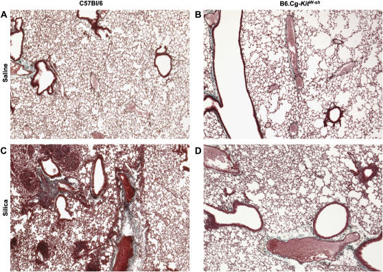
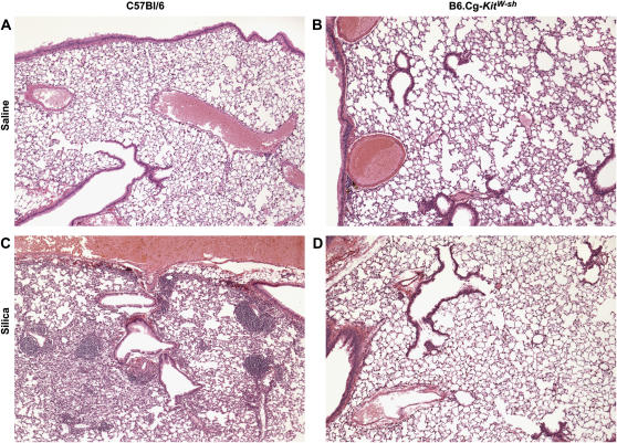
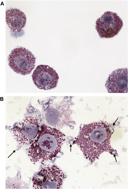
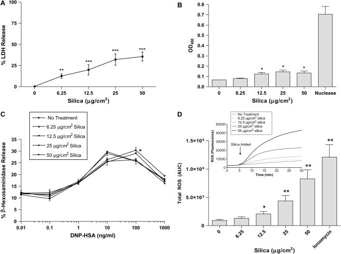
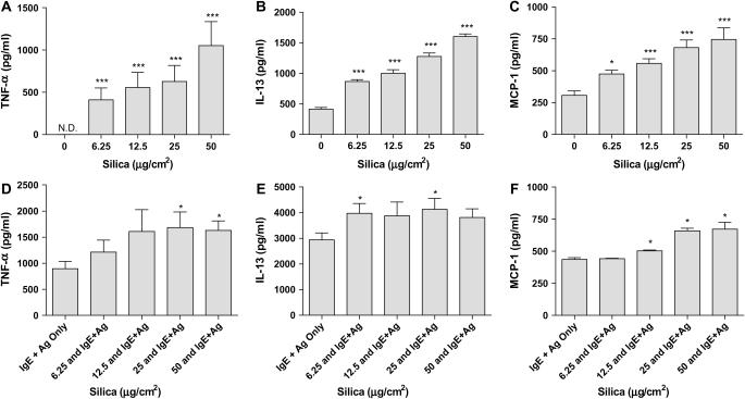
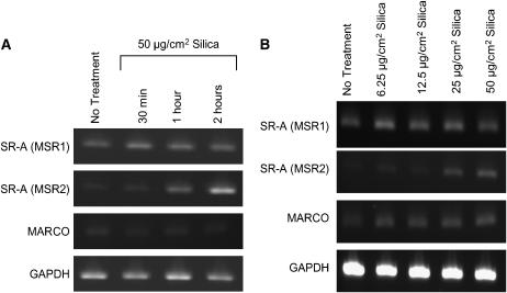
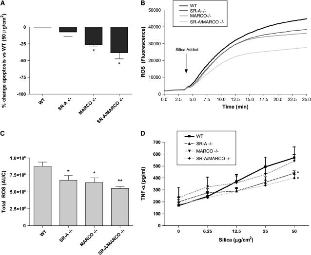
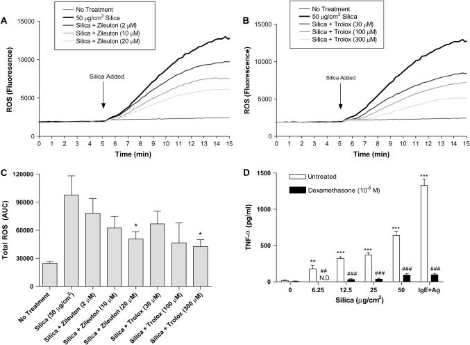
Inhalation of crystalline silica results in pulmonary fibrosis and silicosis. It has been suggested that mast cells play a role in these conditions. How mast cells would influence pathology is unknown. We thus explored mast cell interactions with silica in vitro and in B6.Cg-kit(W-sh) mast cell-deficient mice. B6.Cg-kit(W-sh) mice did not develop inflammation or significant collagen deposition after instillation of silica, while C57Bl/6 wild-type mice did have these findings. Given this supporting evidence of a role for mast cells in the development of silicosis, we examined the ability of silica to activate mouse bone marrow-derived mast cells (BMMC), including degranulation (beta-hexosaminidase release); production of reactive oxygen species (ROS) and inflammatory mediators; and the effects of silica on Fc epsilon RI-dependent activation. Silica did not induce mast cell degranulation. However, TNF-alpha, IL-13, monocyte chemotactic protein-1, protease activity, and production of ROS were dose-dependently increased after silica exposure, and production was enhanced after Fc epsilon RI stimulation. This mast cell activation was inhibited by anti-inflammatory compounds. As silica mediates some effects in macrophages through scavenger receptors (SRs), we first determined that mast cells express scavenger receptors; then explored the involvement of SR-A and macrophage receptor with colleagenous structure (MARCO). Silica-induced ROS formation, apoptosis, and TNF-alpha production were reduced in BMMC obtained from SR-A, MARCO, and SR-A/MARCO knockout mice. These findings demonstrate that silica directs mast cell production of inflammatory mediators, in part through SRs, providing insight into critical events in the pathogenesis and potential therapeutic targets in silicosis.


Figures

<b>Figure 1.</b>
Figure 1.
Silica induces collagen deposition in C57Bl/6 wild-type, but not B6.Cg-kitW-sh mast cell–deficient mouse lungs 3 mo after two intranasal instillations of 1 mg silica. C57Bl/6 mice were instilled with either saline (A) or silica (C). B6.Cg-kitW-sh received instillations of either saline (B) or silica (D). Lungs were stained with Gomori's trichrome stain to examine collagen deposition. Images were obtained at ×5 magnification and are representative of six mice per group.
<b>Figure 2.</b>
Figure 2.
Silica induces inflammation in C57Bl/6 wild-type, but not B6.Cg-kitW-sh mast cell–deficient mouse lungs 3 mo after two intranasal instillations of 1 mg silica. C57Bl/6 mice were instilled with either saline (A) or silica (C). B6.Cg-kitW-sh received instillations of either saline (B) or silica (D). Lungs were stained with H&E to examine inflammation. Images were obtained at ×5 magnification and are representative of six mice per group.
<b>Figure 3.</b>
Figure 3.
Silica interacts with and is internalized by BMMC. Images of (A) untreated or (B) BMMC exposed to 25 μg/cm2 silica for 24 h and stained with Wright-Giemsa (magnification: ×100). Arrows indicate silica particles.
<b>Figure 4.</b>
Figure 4.
Silica induces LDH release, apoptosis, and ROS production, but has little effect on β-hexosaminidase release in BMMC. (A) LDH release was measured in supernatant of silica-exposed BMMC. (B) Apoptosis was determined by DNA fragmentation in BMMC (1 × 10⁵/well) exposed to 0, 6.25, 12.5, 25, or 50 μg/cm2 silica for 24 h. (C) FcεRI-mediated β-hexosaminidase release, as a measure of degranulation, was determined 24 h after exposure of BMMC (50,000/well) to 0, 6.25, 12.5, 25, or 50 μg/cm2 silica. (D) ROS production in silica-exposed BMMC where silica was added to BMMC 5 min after addition of the fluorescent probe DCF, at doses of 0, 6.25, 12.5, 25, or 50 μg/cm2. Intracellular ROS production was measured up to 30 min (insert in D). Area under the curve of silica-induced ROS production with ionomycin as a positive control (D). Data are means ± SEM for three experiments. *P ⩽ 0.05, **P ⩽ 0.01, and ***P ⩽ 0.001 as compared with untreated BMMC.
<b>Figure 5.</b>
Figure 5.
Silica directs TNF-α, IL-13, and MCP-1 production and enhances FcεRI-mediated production of these mediators in BMMC. BMMC (2 × 10⁵/well) were exposed to 0, 6.25, 12.5, 25, or 50 μg/cm2 silica for 24 h before the measurement of TNF-α (A), IL-13 (B), and MCP-1 (C). In addition, BMMC sensitized with IgE anti-DNP and exposed overnight to 0, 6.25, 12.5, 25, or 50 μg/cm2 silica before challenge with DNP-HSA for 8 h were measured for production of TNF-α (D), IL-13 (E), and MCP-1 (F). Data are means ± SEM for three independent experiments. *P ⩽ 0.05 and ***P ⩽ 0.001 as compared with untreated BMMC.
<b>Figure 6.</b>
Figure 6.
Silica induces release of proteases from BMMC. Protease activity was measured in supernatants of BMMC exposed to 0, 6.25, 12.5, 25, or 50 μg/cm2 silica for 24 h. Data are means ± SEM for three independent experiments. *P ⩽ 0.05 and ***P ⩽ 0.001 as compared with untreated BMMC.
<b>Figure 7.</b>
Figure 7.
Demonstration that BMMC express mRNA for SR-A (MSR1 and MSR2) and MARCO. (A) BMMC (1 × 10⁶ cells/condition) were examined for mRNA expression of SR-A and MARCO in untreated or BMMC exposed to 50 μg/cm2 silica for 30 min, 1 h, or 2 h. (B) In addition, mRNA for SR-A and MARCO were examined in BMMC at 2 h after addition of 0, 6.25, 12.5, 25, or 50 μg/cm2 silica. GAPDH was used as a control for constitutive gene expression. Results are representative examples of three separate experiments.
<b>Figure 8.</b>
Figure 8.
Apoptosis, ROS, and TNF-α production are reduced in silica exposed BMMC cultured from SR-AI/II KO, MARCO KO, or SR-A/MARCO double KO mice. (A) DNA fragmentation was measured in BMMC obtained from scavenger receptor KO mice and exposed to 50 μg/cm2 silica for 24 h. Data are presented as the percentage decrease in OD values as compared with C57Bl/6 WT BMMC exposed to 50 μg/cm2 silica. (B) ROS production using the fluorescent probe DCF, following exposure to 50 μg/cm2 silica in SR-A KO, MARCO KO and SR-A/MARCO double KO BMMC is shown over 25 min. (C) ROS production was also analyzed as AUC in BMMC from scavenger receptor KO and WT mice exposed to 50 μg/cm2 silica. (D) TNF-α production measured by ELISA is shown 24 h after addition of 50 μg/cm2 silica in SR-A KO, MARCO KO, and SR-A/MARCO double KO BMMC (silica doses presented in a linear form for representation only). Data are presented as means ± SEM (n = 3). *P ⩽ 0.05 and **P ⩽ 0.01 as compared with wild-type BMMC exposed to the same dose of silica.
<b>Figure 9.</b>
Figure 9.
Treatment of silica exposed BMMC with Zileuton and Trolox reduces ROS formation and treatment with dexamethasone inhibits silica-directed TNF-α production. (A) Zileuton, a 5-lipoxygenase inhibitor, was added to BMMC at concentrations of 2, 10, and 20 μM 5 min before addition of 50 μg/cm2 silica to inhibit ROS formation. (B) In addition, the vitamin E derivative Trolox was added at concentrations of 30, 100, and 300 μM 5 min before addition of 50 μg/cm2 silica to inhibit intracellular ROS formation. (C) Area under the curve was calculated for the reduction of silica-induced ROS formation using Zileuton and Trolox. (D) TNF-α was measured in BMMC (2 × 10⁵/well) that were treated with dexamethasone (10−6 M) 1 h before the addition of 50 μg/cm2 silica for 24 h. BMMC that were sensitized overnight with IgE anti-DNP and challenged for 8 h with 100 ng/ml DNP-HSA were used as a control for the inhibition of TNF-α production by dexamethasone. Data are means ± SEM for three independent experiments. *P ⩽ 0.05, **P ⩽ 0.01, and ***P ⩽ 0.001 as compared with silica-exposed BMMC not treated with either Zileuton, Trolox, or dexamethasone. ##P ⩽ 0.01 and ###P ⩽ 0.001, silica-exposed BMMC treated with dexamethasone as compared with silica-exposed BMMC.

References

    1. American thoracic society committee of the scientific assembly on environmental and occupational health. Adverse effects of crystalline silica exposure. Am J Respir Crit Care Med 1997;155:761–768. - PubMed
    1. Bang KM, Mazurek JM, Attfield MD. Silicosis mortality, prevention, and control–United States, 1968–2002. MMWR Morb Mortal Wkly Rep 2005;54:401–405. - PubMed
    1. Kopinski P, Czunko P, Soja J, Lackowska B, Gil K, Jedynak U, Szczeklik J, Sladek K, Chlap Z. Pneumonol Alergol Pol 2000;68:109–119. (cytoimmunologic changes in material obtained from bronchoalveolar lavage (bal) in asymptomatic individuals chronically exposed to silica dust). - PubMed
    1. Hamada H, Vallyathan V, Cool CD, Barker E, Inoue Y, Newman LS. Mast cell basic fibroblast growth factor in silicosis. Am J Respir Crit Care Med 2000;161:2026–2034. - PubMed
    1. Suzuki H, Kurihara Y, Takeya M, Kamada N, Kataoka M, Jishage K, Ueda O, Sakaguchi H, Higashi T, Suzuki T, et al. A role for macrophage scavenger receptors in atherosclerosis and susceptibility to infection. Nature 1997;386:292–296. - PubMed

Publication types

MeSH terms