. 2006 Aug;3(8):e277.
doi: 10.1371/journal.pmed.0030277.

Mycobacterium tuberculosis induces interleukin-32 production through a caspase- 1/IL-18/interferon-gamma-dependent mechanism

Affiliations

Mycobacterium tuberculosis induces interleukin-32 production through a caspase- 1/IL-18/interferon-gamma-dependent mechanism

Mihai G Netea et al. PLoS Med. 2006 Aug.

Abstract

Background: Interleukin (IL)-32 is a newly described proinflammatory cytokine that seems likely to play a role in inflammation and host defense. Little is known about the regulation of IL-32 production by primary cells of the immune system.

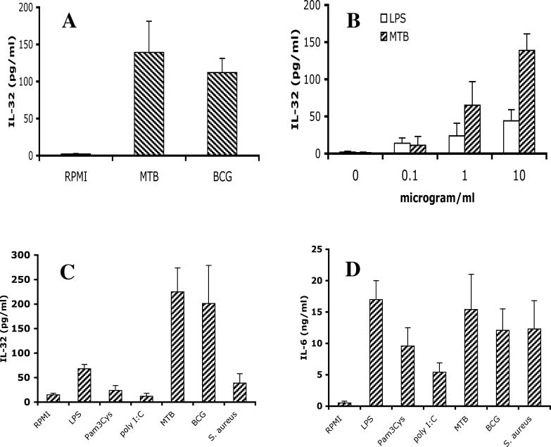
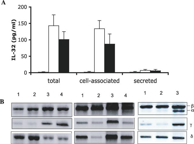
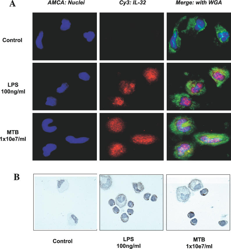
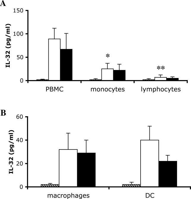
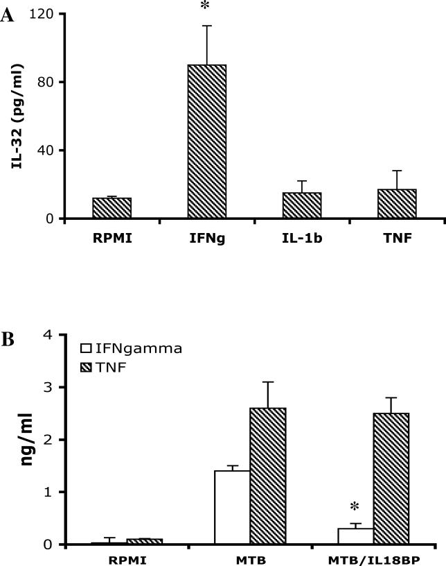
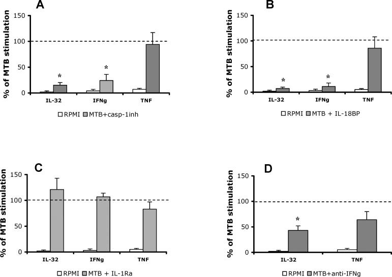
Methods and findings: In the present study, freshly obtained human peripheral blood mononuclear cells were stimulated with different Toll-like receptor (TLR) agonists, and gene expression and synthesis of IL-32 was determined. We demonstrate that the TLR4 agonist lipopolysaccharide induces moderate (4-fold) production of IL-32, whereas agonists of TLR2, TLR3, TLR5, or TLR9, each of which strongly induced tumor necrosis factor alpha and IL-6, did not stimulate IL-32 production. However, the greatest amount of IL-32 was induced by the mycobacteria Mycobacterium tuberculosis and M. bovis BCG (20-fold over unstimulated cells). IL-32-induced synthesis by either lipopolysaccharide or mycobacteria remains entirely cell-associated in monocytes; moreover, steady-state mRNA levels are present in unstimulated monocytes without translation into IL-32 protein, similar to other cytokines lacking a signal peptide. IL-32 production induced by M. tuberculosis is dependent on endogenous interferon-gamma (IFNgamma); endogenous IFNgamma is, in turn, dependent on M. tuberculosis-induced IL-18 via caspase-1.

Conclusions: In conclusion, IL-32 is a cell-associated proinflammatory cytokine, which is specifically stimulated by mycobacteria through a caspase-1- and IL-18-dependent production of IFNgamma.


Conflict of interest statement

Competing Interests: The authors have declared that no competing interests exist.

Figures

Figure 1
Figure 1. Stimulation of IL-32 by Mycobacteria
(A) Whole blood was diluted 1:4 with RPMI and stimulated with 10 μg/ml sonicated M. tuberculosis (MTB) or M. bovis BCG (BCG). Triton X-100 (0.5%) was added 24 h later, and IL-32 concentration was measured by ECL. (B) Stimulation of freshly isolated PBMCs with various concentrations of LPS or sonicated M. tuberculosis stimulated IL-32 synthesis in a dose-dependent manner. (C and D) Freshly isolated PBMCs were stimulated with TLR4 (LPS, 100 ng/ml), TLR2 (Pam3Cys, 10 μg/ml), TLR3 (poly I:C, 5 μg/ml), M. tuberculosis, M. bovis BCG, or S. aureus (all at 1 × 10⁷ microorganisms/milliliter, heat-killed). After 24 h of stimulation, LPS-, M. tuberculosis–, and M. bovis BCG–stimulated production of IL-32 (C) or IL-6 (D) was measured by ECL. Data are presented as means ± SEM (n = 8).
Figure 2
Figure 2. IL-32 Induced by Inflammatory Stimuli Is a Cell-Associated Cytokine
(A) Human PBMCs were stimulated with 10 μg/ml sonicated M. tuberculosis. After 24 h, supernatants and cell lysates were assayed for IL-32 (see Methods). (B) RT-PCR of the four IL-32 isoforms in freshly lysed PBMCs (lane1), PBMCs incubated in medium for 4 h (lane 2), stimulated with 100 ng/ml LPS (lane 3), or with M. tuberculosis (lane 4).
Figure 3
Figure 3. IL-32 Immunostaining of Human PBMCs Stimulated with LPS or M. tuberculosis
(A) Freshly isolated PBMCs were left to adhere, stimulated with either control medium (first row), LPS (second row), or M. tuberculosis (MTB; third row), and 24 h later stained for the presence of IL-32. Nuclei were stained with AMCA (blue, left panels), and IL-32 was stained with Cy3 (red, middle panels). The right panels show the merged pictures with WGA-stained cellular surface (green).The stained cells were examined by digital deconvoluted microscopy. (B) PBMCs stimulated with control medium (left panel), LPS (middle panel), or M. tuberculosis (right panel) were stained with an anti-IL-32 monoclonal antibody and counterstained with Mayer's hematoxylin for optical microscopy.
Figure 4
Figure 4. IL-32 Production by Monocytes, Macrophages, and DCs
(A) PBMCs were incubated for 2 h at 37 °C in 96-well plates, and non-adherent lymphocytes were transferred to a different well. The total PBMCs, the adherent monocytes, or the non-adherent lymphocytes were stimulated with RPMI (hatched bars), M. tuberculosis (open bars), or M. bovis BCG (solid bars) for an additional 24 h. After 24 h, IL-32 concentrations were measured. (B) Adherent monocytes were incubated for 5 d with RPMI and 20% human serum for macrophage differentiation, or a combination of 50 ng/ml GM-CSF and 20 ng/ml IL-4 for DC differentiation. The macrophages and DCs were thereafter stimulated for an additional 24 h with RPMI (hatched bars), M. tuberculosis (open bars), or M. bovis BCG (solid bars). IL-32 concentration was measured by ECL. Data are presented as means ± SEM (n = 8); *, p < 0.05; **, p < 0.01.
Figure 5
Figure 5. IFNγ Is a Stimulus of IL-32 Production
(A) PBMCs isolated from five volunteers were stimulated with IFNγ, IL-1β, or TNFα (all at a concentration of 10 ng/ml). IL-32 concentrations were measured by ECL 24 h later in cells lysed with Triton X-100. (B) PBMCs were stimulated for 24 h with RPMI, M. tuberculosis (MTB), or M. tuberculosis in combination with 10 μg/ml IL-18BP. IFNγ and TNFα concentrations were measured 24 h later. Data are presented as means ± SEM (n = 5); *, p < 0.05.
Figure 6
Figure 6. M. tuberculosis Stimulates IL-32 through a Caspase-1/IL-18/IFNγ-Dependent Mechanism
PBMCs were stimulated with RPMI, M. tuberculosis (MTB), or a combination of M. tuberculosis and cytokine inhibitors: 20 μM of a caspase-1 inhibitor (casp-1inh) (A), 10 μg/ml IL-18BP (B), IL-1Ra (C), or 10 μg/ml of a monoclonal mouse anti-human IFNγ antibody (D). As a control for the anti-IFNγ antibody, a similar concentration of a purified mouse IgG1 antibody was used in the control stimulation. After 24 h of incubation, IL-32, IFNγ, and TNF concentrations were measured by ECL. The dashed line represents the 100% stimulation induced by M. tuberculosis alone. The open bars represent the unstimulated cells, and the solid bars represent the cells stimulated with M. tuberculosis in the presence of the cytokine inhibitors, expressed as percent of M. tuberculosis production. Data are presented as means ± SEM (n = 8); *, p < 0.05.

References

    1. Kim SH, Han SY, Azam T, Yoon DY, Dinarello CA. Interleukin-32: A cytokine and inducer of TNF-alpha. Immunity. 2005;22:131–142. - PubMed
    1. Netea MG, Azam T, Ferwerda G, Girardin SE, Walsh M, et al. IL-32 synergizes with nucleotide oligomerization domain (NOD) 1 and NOD2 ligands for IL-1beta and IL-6 production through a caspase 1-dependent mechanism. Proc Natl Acad Sci U S A. 2005;102:16309–16314. - PMC - PubMed
    1. Ottenhoff TH, Verreck FAW, Hoeve MA, van de Vosse E. Control of human host immunity to mycobacteria. Tuberculosis. 2005;85:53–64. - PubMed
    1. Murray HW, Scavuzzo D, Jacobs JL, Kaplan MH, Libby DM, et al. In vitro and in vivo activation of human mononuclear phagocytes by interferon-g. J Immunol. 1987;138:2457–2462. - PubMed
    1. Perrusia B, Kobayashi M, Rossi ME, Anegon I, Trinchieri G. Immune interferon enhances functional properties of human granulocytes: Role of Fc receptors and effect of lymphotoxin, tumor necrosis factor, and granulocyte-macrophage colony-stimulating factor. J Immunol. 1987;138:765–774. - PubMed

Publication types

MeSH terms