. 2006 Aug;3(8):e310.
doi: 10.1371/journal.pmed.0030310.

Macrophages inhibit neovascularization in a murine model of age-related macular degeneration

Affiliations

Macrophages inhibit neovascularization in a murine model of age-related macular degeneration

Rajendra S Apte et al. PLoS Med. 2006 Aug.

Abstract

Background: Age-related macular degeneration (AMD) is the leading cause of blindness in people over 50 y of age in at least three continents. Choroidal neovascularization (CNV) is the process by which abnormal blood vessels develop underneath the retina. CNV develops in 10% of patients with AMD but accounts for up to 90% of the blindness from AMD. Although the precise etiology of CNV in AMD remains unknown, the macrophage component of the inflammatory response, which has been shown to promote tumor growth and support atherosclerotic plaque formation, is thought to stimulate aberrant angiogenesis in blinding eye diseases. The current theory is that macrophage infiltration promotes the development of neovascularization in CNV.

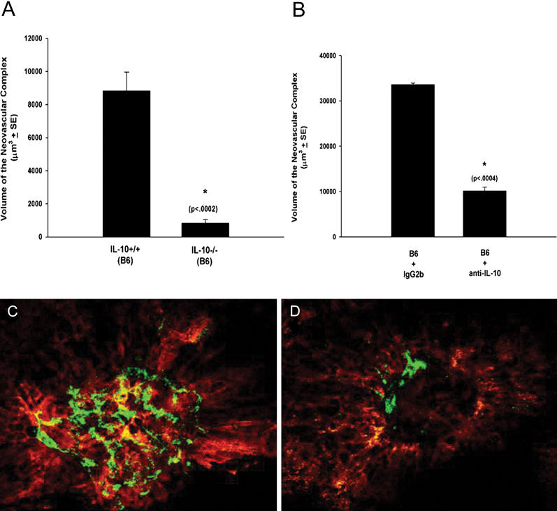
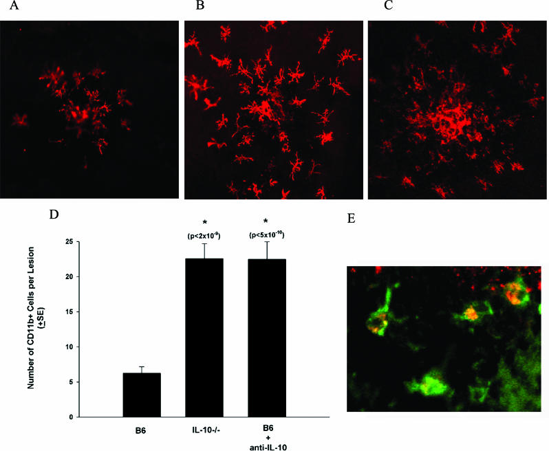
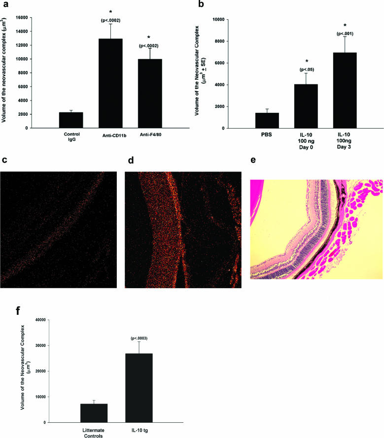
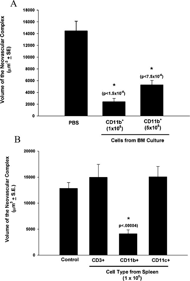
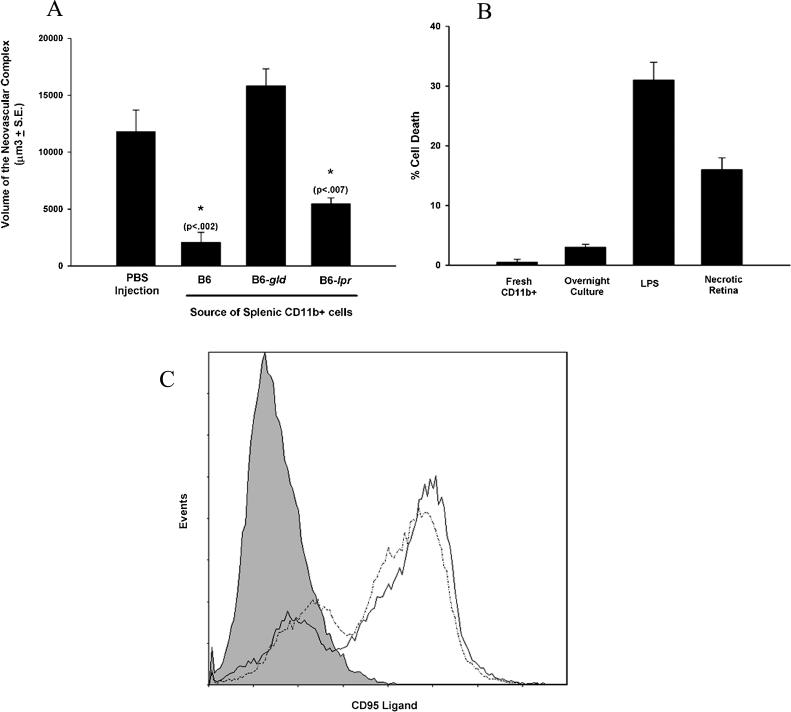
Methods and findings: We examined the role of macrophages in a mouse model of CNV. IL-10(-/-) mice, which have increased inflammation in response to diverse stimuli, have significantly reduced CNV with increased macrophage infiltrates compared to wild type. Prevention of macrophage entry into the eye promoted neovascularization while direct injection of macrophages significantly inhibited CNV. Inhibition by macrophages was mediated by the TNF family death molecule Fas ligand (CD95-ligand).

Conclusions: Immune vascular interactions can be highly complex. Normal macrophage function is critical in controlling pathologic neovascularization in the eye. IL-10 regulates macrophage activity in the eye and is an attractive therapeutic target in order to suppress or inhibit CNV in AMD that can otherwise lead to blindness.


Conflict of interest statement

Competing Interests: The authors have declared that no competing interests exist.

Figures

Figure 1
Figure 1. IL-10 and Neovascularization (CNV)
(A and B) CNV was induced in IL-10 −/− (volume of the neovascular complex: 829.6 ± 225.7 μm3) and B6 (IL-10 +/+; 8,830.7 ± 1,130.3 μm3) mice (A) and B6 mice injected on days 0, 3, and 5 with anti-IL-10 (10,115.3 ± 850.5 μm3) or isotype control antibody (IgG2b; 33,602.3 ± 317.4 μm3) (B). Asterisks indicate values significantly different from control; p-values given in parentheses are based on Student's t test performed as described [17,18]. (C and D) Seven days following laser treatment, the volumes of the neovascular complexes (green) were determined by confocal microscopy. Shown is representative CNV in a B6 eye (C) and an IL-10 −/− eye (D).
Figure 2
Figure 2. Cellular Infiltrates in CNV Lesions
(A–C) On day 7 following laser treatment, whole mount stains were performed to determine the cells present in the area of the neovascular complex. Stains for CD11b were performed on (A) B6, (B) IL-10 −/−, and (C) B6 mice treated with neutralizing anti-IL-10. Images were take by confocal microscopy (magnification 200×) centered on the laser lesion. (D) The number of CD11b+ cells per lesion was counted (200× high-power field centered on the laser lesion) (B6, 6.3 ± 0.9; IL10 −/−, 22.6 ± 2.1; B6 + anti-IL10, 22.5 ± 2.5). Asterisks indicate values significantly different from control. (E) Dual staining was performed on day 7 following laser treatment using FITC-conjugated anti-CD11b (green) and PE-conjugated anti-F4/80 (red) (magnification 400×).
Figure 3
Figure 3. Inhibition of Macrophage Infiltration Promotes CNV
(A) IL-10 −/− mice were injected with anti-CD11b (volume of the neovascular complex: 12,903.1 ± 2,171.3 μm3), anti-F4/80 (9,977.3 ± 1,572.7 μm3), or control IgG (2,262.3 ± 313.4 μm3) on days −1, 0, and +1. CNV volumes were determined on day 7. (B) IL-10 −/− mice were injected in the vitreous with PBS (1,394 ± 382.4 μm3) or 100 ng of rIL-10 on day 0 (the day of laser treatment; 4,033.6 ± 1,026.5 μm3) or day 3 (6,949.8 ± 1,475.5 μm3). (C and D) Cross-sections of the retinas of a control littermate (C) and a VMD2-IL-10 Tg mouse (D) stained with anti-IL-10 and examined by confocal microscopy. (E) Hematoxylin and eosin staining of the retina of a VMD2-IL-10 Tg mouse (magnification 200×). (F) VMD2-IL-10 Tg mice (24,428.6 ± 4,360.7 μm3) and littermate controls (6,830.1 ± 1,324.9 μm3) were subjected to laser treatment and CNV volumes were determined on day 7. Asterisks indicate values significantly different from control; p-values are given in parentheses.
Figure 4
Figure 4. Injection of Macrophages Inhibits CNV
Cells obtained from bone marrow (BM) cultures (A) or spleen (B) were purified by magnetic beads and injected into the vitreous cavity on the day of laser treatment. Seven days later, the volume of the neovascular complex was determined by confocal microscopy. Asterisks indicate values significantly different from control; p-values given in parentheses are based on Student's t test. (A) Volume of the neovascular complex for injection of PBS (14,470.9 ± 1,636.4 μm3), CD11b cells—1 × 10⁵ cells (2,431.2 ± 551.3 μm3), and CD11b cells—5 × 10⁵ cells (5,274.2 ± 772.5 μm3). (B) Volume of the neovascular complex for injection of PBS (12,857.9 ± 1,094.9 μm3), CD3 cells (14,960.5 ± 2,502.0 μm3), CD11b cells (4,135.1 ± 714.4 μm3), and CD11c cells (15,063.3 ± 1,982.4 μm3).
Figure 5
Figure 5. Macrophages Inhibit CNV via CD95L
(A) CD11b+ cells from B6, B6-lpr, and B6-gld mice were purified from spleen, and 1 × 10⁵ cells were injected into the vitreous cavity on the day of laser induction of CNV. Seven days later, the volume of the neovascular complex was determined by confocal microscopy for PBS (11,786.4 ± 1,907.3 μm3), B6 (2,084.6 ± 874.8 μm3), gld (15,824.9 ± 1,483.6 μm3), and lpr (5,443.9 ± 542.1 μm3). Asterisks indicate values significantly different from control. (B) Purified CD11b+ cells were cultured overnight with LPS or necrotic retina and tested for killing against L1210-Fas. (C) The expression of CD95L in CD11b cells was determined by flow cytometry following overnight culture with LPS (dotted line) or necrotic retina (solid line). The line with shading underneath represents untreated cells.

Comment in

References

    1. Nakao S, Kuwano T, Tsutsumi-Miyahara C, Ueda SI, Kimura YN, et al. Infiltration of COX-2-expressing macrophages is a prerequisite for IL-1beta-induced neovascularization and tumor growth. J Clin Invest. 2005;115:2979–2991. - PMC - PubMed
    1. Taylor PR, Martinez-Pomares L, Stacey M, Lin HH, Brown GD, et al. Macrophage receptors and immune recognition. Annu Rev Immunol. 2005;23:901–944. - PubMed
    1. Hansson GK. Inflammation, atherosclerosis, and coronary artery disease. N Engl J Med. 2005;352:1685–1695. - PubMed
    1. Sakurai E, Anand A, Ambati BK, van Rooijen N, Ambati J. Macrophage depletion inhibits experimental choroidal neovascularization. Invest Ophthalmol Vis Sci. 2003;44:3578–3585. - PubMed
    1. Espinosa-Heidmann DG, Suner IJ, Hernandez EP, Monroy D, Csaky KG, et al. Macrophage depletion diminishes lesion size and severity in experimental choroidal neovascularization. Invest Ophthalmol Vis Sci. 2003;44:3586–3592. - PubMed

Publication types

Associated data