Comparative Study
. 2006 Sep;4(9):e269.
doi: 10.1371/journal.pbio.0040269.

The structural basis for promoter -35 element recognition by the group IV sigma factors

Affiliations
Comparative Study

The structural basis for promoter -35 element recognition by the group IV sigma factors

William J Lane et al. PLoS Biol. 2006 Sep.

Abstract

The control of bacterial transcription initiation depends on a primary sigma factor for housekeeping functions, as well as alternative sigma factors that control regulons in response to environmental stresses. The largest and most diverse subgroup of alternative sigma factors, the group IV extracytoplasmic function sigma factors, directs the transcription of genes that regulate a wide variety of responses, including envelope stress and pathogenesis. We determined the 2.3-A resolution crystal structure of the -35 element recognition domain of a group IV sigma factor, Escherichia coli sigma(E)4, bound to its consensus -35 element, GGAACTT. Despite similar function and secondary structure, the primary and group IV sigma factors recognize their -35 elements using distinct mechanisms. Conserved sequence elements of the sigma(E) -35 element induce a DNA geometry characteristic of AA/TT-tract DNA, including a rigid, straight double-helical axis and a narrow minor groove. For this reason, the highly conserved AA in the middle of the GGAACTT motif is essential for -35 element recognition by sigma(E)4, despite the absence of direct protein-DNA interactions with these DNA bases. These principles of sigma(E)4/-35 element recognition can be applied to a wide range of other group IV sigma factors.


Conflict of interest statement

Competing interests. The authors have declared that no competing interests exist.

Figures

Figure 1
Figure 1. Overview of Ec σE 4/−35 Element DNA Structure
(A) Synthetic 12-mer oligonucleotides use for crystallization. The black numbers above the sequence denote the DNA position with respect to the transcription start site at +1. The −35 element is colored light green (nontemplate strand) and dark green (template strand). The flanking bases are colored light gray (nontemplate strand) and dark gray (template strand). (B) Two views of the Ec σE 4/−35 element DNA complex, related by a 90° rotation about the horizontal axis as shown. The protein is shown as an α-carbon backbone ribbon, with σE 4.1 colored yellow and σE 4.2 colored light blue. The DNA is color coded as in (A).
Figure 2
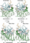
Figure 2. Ec σE 4/DNA Contacts; Structural View
Two stereo views (front and back) of the Ec σE 4/−35 element DNA complex, related by a 180° rotation about the vertical axis as shown. The protein is shown as an α-carbon backbone worm, with σE 4.1 colored yellow and σE 4.2 colored light blue. Side chains are shown for those residues that make protein–DNA contacts. Carbon atoms of the side chains are colored as the backbone, except atoms involved in polar contacts with the DNA are colored (nitrogen atoms, blue; oxygen atoms, red). The DNA is color-coded as in Figure 1A, except atoms involved in polar contacts with the protein are colored (nitrogen atoms, blue; oxygen atoms, red). Water molecules are indicated with red spheres. Dashed black lines indicate hydrogen bonds or salt bridges.
Figure 3
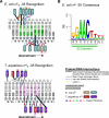
Figure 3. Ec σE 4/DNA Contacts; Schematic View
(A) Schematic representation of σ4–DNA interactions for Ec σE 4 (top) and Taq σA (bottom; [4]). The nontemplate/template strand DNA is colored light gray/dark gray (respectively), except the −35 element is colored light green/dark green (for Ec σE 4) or pink/magenta (for Taq σA). Colored boxes denote protein residues. Color-coding for the proteins, as well as the meaning of the lines indicating interactions, is explained in the legend (lower right). Double thick solid black lines indicate two hydrogen bonds with the same residue. Water molecules mediating protein–DNA contacts are shown as red circles. (B) Sequence logo denoting sequence conservation within the Ec σE 4 −35 element [17,51].
Figure 4
Figure 4. Ec σE −35 Element DNA Geometry
(A) Cartoon views of the DNA backbone geometry. The DNA was aligned using the template strand DNA from −35′ to −30′, giving an RMSD of 0.839 over 30 atoms for Ec σE 4/DNA and Taq σA 4/DNA. Straight B-form dsDNA is blue, Ec σE −35 element DNA is green, while Taq σA −35 element DNA is magenta. The paths of the DNA helical axes, calculated using Curves (http://www.ibpc.fr/UPR9080/Curindex.html), are also shown. (B) Graph showing the DNA minor groove width (calculated using 3DNA) for B-form DNA (blue), Ec σE 4 −35 element DNA (green), and Taq σA −35 element DNA (magenta; [49]). Minor groove width was calculated as the P-P distance minus 5.8 Å to take into account the radii of the phosphate groups. (C) View of the hydrogen bonds important in stabilizing the unique geometry of the downstream σE −35 element DNA. The waters participating in the spine of hydration are indicated by red spheres. Dashed black lines indicate water-mediated minor groove hydrogen bonds. Dashed blue lines indicate cross-strand hydrogen bonds formed between adjacent bases.
Figure 5
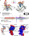
Figure 5. Structural Comparisons of Ec σE 4 and Taq σA 4 −35 Element Recognition
(A) Ec σE 4/−35 element DNA and Taq σA 4/−35 element DNA complexes were aligned using the template strand DNA from −35′ to −30′, giving an RMSD of 0.839 over 30 atoms. The two views are related by a 90° rotation about the horizontal axis as shown. Proteins are shown as α-carbon backbone worms, color-coded as shown. The Ec σE −35 element DNA is colored light green (nontemplate strand) and dark green (template strand). The Taq σA −35 element is colored pink (nontemplate strand) and magenta (template strand). (B) Comparison of the Ec σE 4 and Taq σA 4 protein–DNA interactions. The Cα-backbone of Ec σE 4 and Taq σA 4 were aligned using Ec σE 4 residues 137 to 150 and 155 to 182 with Taq σA 4 residues 375 to 388 and 397 to 424, giving an RMSD of 1.00 Å over 42 atoms. Protein residue numbering is shown between the sequences (Taq/Ec). Residues in σ4.1 are highlighted in red/yellow (Taq σA/Ec σE) and those in σ4.2 are colored purple/blue. Red dots denote protein residues that make base-specific DNA contacts. Colored dots denote protein residues that make DNA contacts. Black dots denote hydrogen bonds (less than 3.2 Å) or salt bridges (less than 4.0 Å) originating from the protein side chain. Magenta dots denote hydrogen bonds originating from the protein main chain. Blue dots denote van der Waals (hydrophobic) contacts (less than 4.0 Å). Yellow dots denote cation–π interactions. The positions along the DNA that are contacted by each residue are indicated above and below the contact circles. (C) The protein α-carbon backbones of Ec σE 4 and Taq σA 4 were aligned as described in (B). The superimposed proteins, shown as α-carbon backbone worms, are shown on the left, color-coded as in (A). The Ec σE 4/−35 element and Taq σA/−35 element complexes are shown separately (middle and left, respectively). In these views, the proteins are shown as molecular surfaces, color-coded according to electrostatic surface potential. The DNAs are shown as phosphate-backbone ribbons, with bases indicated schematically as sticks.
Figure 6
Figure 6. Correlation of σ4 and −35 Element Sequences for Several Group IV σ Factors
The top shows a sequence alignment of the proposed −35 element DNA binding region of several group IV σ factors. The residue positions that are important in −35 element DNA recognition in the Ec σE 4/−35 element DNA structure are highlighted green (similar to Ec σE) or red (dissimilar to Ec σE). The bottom shows the alignment of the known −35 consensus sequences from several group IV σ factors. The three −35 element regions are highlighted with the upstream G region (blue), the middle AAC motif (red), and the downstream T rich region (green). Lines connecting the two alignments indicate protein residue–DNA base interactions important for −35 element recognition in the Ec σE 4/DNA structure.

Comment in

References

    1. Darst SA. Bacterial RNA polymerase. Curr Opinion Struct Biol. 2001;11:155–162. - PubMed
    1. Gross CA, Chan C, Dombroski A, Gruber T, Sharp M, et al. The functional and regulatory roles of sigma factors in transcription. Cold Spring Harbor Symp Quant Biol. 1998;63:141–155. - PubMed
    1. Murakami K, Darst SA. Bacterial RNA polymerases: The wholo story. Curr Opin Struct Biol. 2003;13:31–39. - PubMed
    1. Campbell EA, Muzzin O, Chlenov M, Sun JL, Olson CA, et al. Structure of the bacterial RNA polymerase promoter specificity sigma factor. Mol Cell. 2002;9:527–539. - PubMed
    1. Murakami K, Masuda S, Campbell EA, Muzzin O, Darst SA. Structural basis of transcription initiation: An RNA polymerase holoenzyme/DNA complex. Science. 2002;296:1285–1290. - PubMed

Publication types