

Why stones break better at slow shockwave rates than at fast rates: in vitro study with a research electrohydraulic lithotripter
- PMID: 16903810
- PMCID: PMC2442574
- DOI: 10.1089/end.2006.20.537
Why stones break better at slow shockwave rates than at fast rates: in vitro study with a research electrohydraulic lithotripter
Abstract
Background and purpose: Stones break better when the rate of shockwave (SW) delivery is slowed. It has been hypothesized that the greater cavitation accompanying a fast rate shields pulse propagation, thus interfering with the delivery of SW energy to the stone. We tested this idea by correlating waveforms measured at the SW focus with cavitation viewed using high-speed imaging.
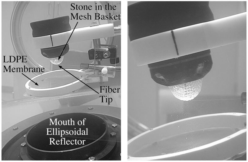
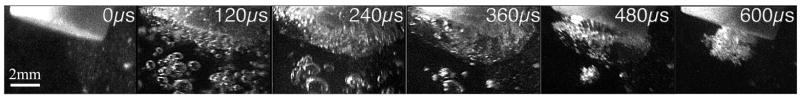
Materials and methods: A series of U30 gypsum stones held in a 2-mm mesh basket were exposed to 200 SWs at 30 or 120 SW/min from a research electrohydraulic lithotripter (HM3 clone). Waveforms were collected using a fiberoptic probe hydrophone. High-speed imaging was used to observe cavitation bubbles in the water and at the stone surface.
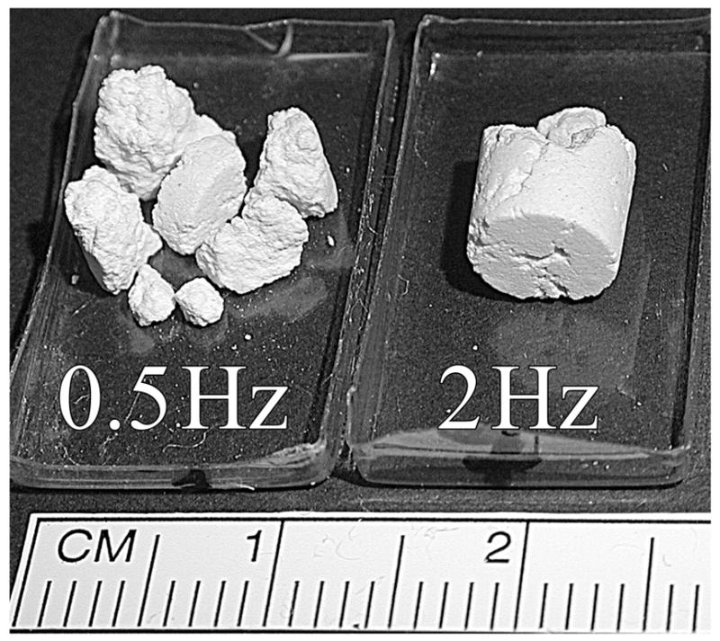
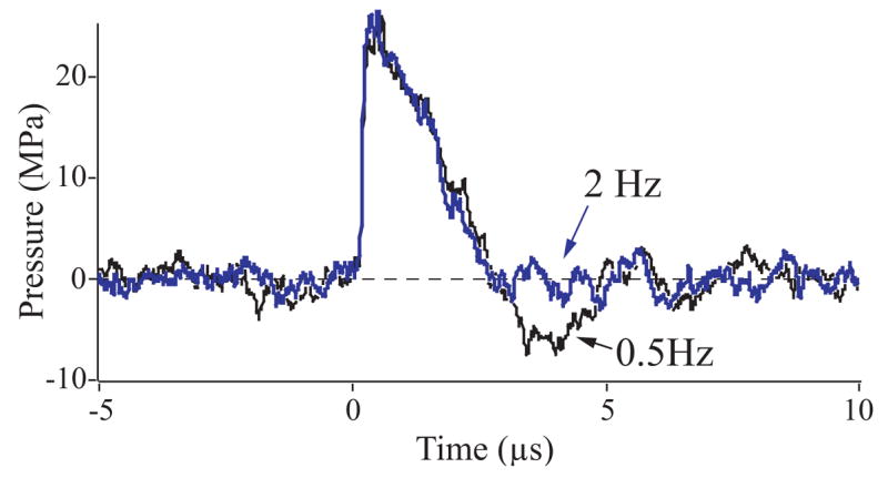
Results: Stone breakage was significantly better at 30 SW/min than at 120 SW/min. The rate had little effect on SW parameters in the water free field. In the presence of particulates released from stones, the positive pressure of the SW remained unaffected, but the trailing tensile phase of the pulse was significantly reduced at 120 SW/min.
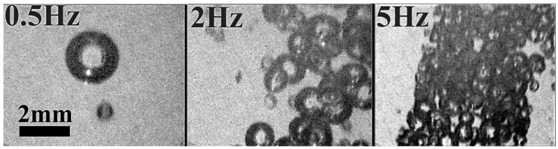
Conclusions: Cavitation bubbles do not persist between SWs. Thus, mature bubbles from one pulse do not interfere with the next pulse, even at 120 SW/min. However, cavitation nuclei carried by fine particles released from stones can persist between pulses. These nuclei have little effect on the compressive wave but seed cavitation under the influence of the tensile wave. Bubble growth draws energy from the negative-pressure phase of the SW, reducing its amplitude. This likely affects the dynamics of cavitation bubble clusters at the stone surface, reducing the effectiveness of bubble action in stone comminution.
Figures
References
-
- Vallancien G, Munoz R, Borghi M, et al. Relationship between the frequency of piezoelectric shock waves and the quality of renal stone fragmentation. In vitro study and clinical implications. European Urology. 1989;16:41–44. - PubMed
-
- Weir MJ, Tariq N, Honey RJ. Shockwave frequency affects fragmentation in a kidney stone model. J Endourol. 2000;14:547–550. - PubMed
-
- Greenstein A, Matzkin H. Does the rate of extracorporeal shock wave delivery affect stone fragmentation? Urology. 1999;54:430–432. - PubMed
-
- Léger P, Daudon M, Magnier M. Expérience ≪ in vitro≫ de lithotripsie pyézo-électrique à repérage ultra-sonique sur le lithotripteur EDAP LT 01. Journal d’Urologie. 1990;96:353–364. - PubMed
-
- Paterson RF, Lifshitz DA, Lingeman JE, et al. Stone fragmentation during shock wave lithotripsy is improved by slowing the shock wave rate: Studies with a new animal model. J Urol. 2002;168:2211–2215. - PubMed
Publication types
MeSH terms
Grants and funding
LinkOut - more resources
Full Text Sources
Other Literature Sources
