. 2006 Nov;119(3):393-403.
doi: 10.1111/j.1365-2567.2006.02448.x. Epub 2006 Aug 14.

Enhanced neutrophil motility by granulocyte colony-stimulating factor: the role of extracellular signal-regulated kinase and phosphatidylinositol 3-kinase

Affiliations

Enhanced neutrophil motility by granulocyte colony-stimulating factor: the role of extracellular signal-regulated kinase and phosphatidylinositol 3-kinase

Mika Nakamae-Akahori et al. Immunology. 2006 Nov.

Abstract

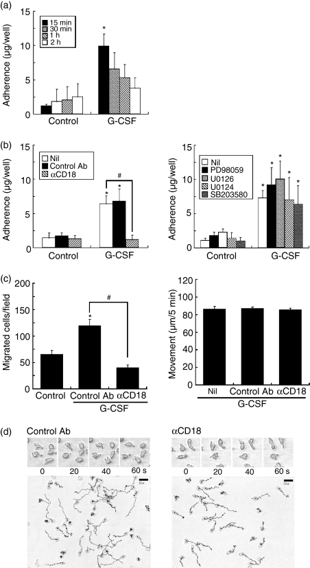
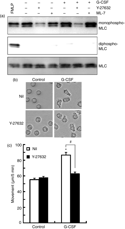
The effect of granulocyte colony-stimulating factor (G-CSF) on human neutrophil motility was studied using videomicroscopy. Stimulation of neutrophils with G-CSF resulted in enhanced motility with morphological change and increased adherence. Enhanced neutrophil motility was detected within 3-5 min after G-CSF stimulation, reached a maximum at 10 min, and was sustained for approximately 35 min. The maximum migration rate was 84.4 +/- 2.9 microm/5 min. A study using the Boyden chamber method revealed that G-CSF-stimulated neutrophils exhibited random migration but not chemotaxis. Enhanced neutrophil motility and morphological change were inhibited by MEK [mitogen-activated protein kinase (MAPK)/extracellular signal-regulated kinase (ERK) kinase] inhibitors (PD98059 and U0126), and a phosphatidylinositol 3-kinase (PI3K) inhibitor (wortmannin), but not by a p38 MAPK inhibitor (SB203580). These findings are consistent with the fact that G-CSF selectively activates MEK/ERK and PI3K, but not p38, in neutrophils. MEK/ERK activation was associated with G-CSF-induced redistribution of F-actin and phosphorylated myosin light chain. Enhanced neutrophil motility was observed even in the presence of neutralizing anti-CD18 antibody, which prevented cell adherence. These findings indicate that G-CSF induces human neutrophil migration via activation of MEK/ERK and PI3K.


Figures

Figure 1
Figure 1
Stimulation of human neutrophil migration by G-CSF. Neutrophils were stimulated with G-CSF (50 ng/ml), and cell migration was monitored under a videomicroscope. (a) The images at the indicated time points after G-CSF stimulation. (b) The migration rate, expressed as μm/5 min. The data are expressed as the mean ± SEM (n = 34 to n = 64 cells). *Significantly greater as compared with unstimulated control cells (P < 0·01).
Figure 2
Figure 2
Effects of MEK and p38 MAPK inhibitors on G-CSF-induced neutrophil migration and morphological change. Neutrophils were pretreated with PD98059 (50 μm), U0126 (10 μm), U0124 (10 μm), or SB203580 (10 μm) for 30 min, and thereafter stimulated with G-CSF (50 ng/ml) or FMLP (10−7m). (a) The images at 30–31 min after G-CSF stimulation. Each arrowhead in each series indicates the same cell. The images shown are representative of three to five independent experiments. (b) The tracks that neutrophils moved along at 10–15 min after G-CSF stimulation. Bars represent 20 μm. The images shown are representative of three to five independent experiments. (c) The migration rate that was determined at 10–15 min after G-CSF or FMLP stimulation. The data are expressed as the mean ± SEM (n = 40 to n = 93 cells). *Significantly greater as compared with unstimulated control cells (P < 0·01). #Significantly inhibited by PD98059 or U0126 (P < 0·01).
Figure 3
Figure 3
Effects of MEK and p38 MAPK inhibitors on G-CSF-induced actin reorganization and ERK1/2 phosphorylation. (a) Neutrophils were pretreated with U0126 (10 μm), or SB203580 (10 μm) for 30 min, and thereafter stimulated with G-CSF (50 ng/ml) for 20 min at 37°. Left panel of images: Nomarski images; right panel of images: F-actin distribution. The images shown are representative of three independent experiments. (b) Neutrophils were pretreated with PD98059 (50 μm), U0126 (10 μm), U0124 (10 μm), or SB203580 (10 μm) for 30 min, and thereafter stimulated with G-CSF (50 ng/ml) for 10 min at 37°. The immunoblotting was performed using antibodies against phosphorylated ERK1/2, phosphorylated p38 and p38. Results shown are representative of three independent experiments.
Figure 4
Figure 4
Analysis of G-CSF-induced neutrophil migration using Boyden chamber method. (a) G-CSF (50 ng/ml) was added to the upper and lower wells, or the lower well alone. As a positive control, FMLP (10−7 m) was added to the lower well. Neutrophil suspension (1 × 10⁶/ml) was added to the upper well. After incubation for 2 hr, cells migrating to the lower well were determined. The data are expressed as the mean ± SEM of five independent experiments. *Significantly greater as compared with control cells (P < 0·01). (b) Neutrophils were pretreated with PD98059 (50 μm), U0126 (10 μm), U0124 (10 μm), or SB203580 (10 μm) for 30 min, and thereafter stimulated with G-CSF (50 ng/ml). G-CSF and each inhibitor were added to the upper and lower wells. The data are expressed as the mean ± SEM of five independent experiments. *Significantly greater as compared with control cells (P < 0·01). #Significantly inhibited by PD98059 and U0126 (P < 0·01).
Figure 5
Figure 5
Effects of anti-CD18 antibody and MEK and p38 MAPK inhibitors on G-CSF-induced neutrophil adherence and migration. (a) Neutrophils were stimulated with G-CSF (50 ng/ml) for the indicated periods at 37°, and thereafter cell adherence was determined. The data are expressed as the mean ± SD of three independent experiments. *Significantly greater as compared with control cells (P < 0·05). (b) Neutrophils were pretreated with anti-CD18 antibody (10 μg/ml), isotype-matched control antibody (10 μg/ml), PD98059 (50 μm), U0126 (10 μm), U0124 (10 μm), or SB203580 (10 μm) for 30 min, and thereafter stimulated with G-CSF (50 ng/ml) for 15 min for determination of cell adherence. The data are expressed as the mean ± SD of four or five independent experiments. *Significantly greater as compared with control cells (P < 0·05). #Significantly inhibited by anti-CD18 antibody (P < 0·01). (c) Neutrophils were pretreated with anti-CD18 antibody (10 μg/ml), or isotype-matched control antibody (10 μg/ml) for 30 min, and thereafter G-CSF-induced neutrophil migration was determined with Boyden chamber method (left panel) and videomicroscopy (right panel). G-CSF (50 ng/ml) and each antibody were added to the upper and lower wells of Boyden chambers (left panel). The migration rate was determined at 10–15 min after G-CSF (50 ng/ml) stimulation (right panel). The data are expressed as the mean ± SD of five independent experiments. *Significantly greater as compared with control cells (P < 0·01). #Significantly inhibited by anti-CD18 antibody (P < 0·01). (d) Neutrophils were pretreated with anti-CD18 antibody (10 μg/ml), or isotype-matched control antibody (10 μg/ml) for 30 min, and thereafter G-CSF-induced neutrophil migration was determined with video microscopy. Upper panel: the images at 10–11 min after G-CSF stimulation. Lower panel: the tracks that neutrophils moved along at 10–15 min after G-CSF stimulation. Bars represent 20 μm. The images shown are representative of three to five independent experiments.
Figure 6
Figure 6
Effects of MEK and p38 MAPK inhibitors on G-CSF-induced redistribution of phosphorylated MLC. (a) Neutrophils were pretreated with U0126 (10 μm), or SB203580 (10 μm) for 30 min, and thereafter stimulated with G-CSF (50 ng/ml) for 20 min at 37°. The distribution of F-actin and phosphorylated MLC was analysed with confocal microscopy. The images shown are representative of three independent experiments. (b) Neutrophils were stimulated with G-CSF (50 ng/ml) for 10 min, or FMLP (10−7 m) for 1 min at 37°. The immunoblotting was performed using antibodies against Ser19-mono-phosphorylated MLC, Thr18/Ser19-di-phosphorylated MLC, and MLC. Results shown are representative of three independent experiments.
Figure 7
Figure 7
Effects of Y-27632 and ML-7 on G-CSF-induced neutrophil migration and phosphorylation of MLC. (a) Neutrophils were pretreated with Y-27632 (10 μm), or ML-7 (10 μm) for 30 min, and thereafter stimulated with G-CSF (50 ng/ml) for 10 min at 37°. As a positive control, neutrophils were stimulated with FMLP (10−7 m) for 1 min at 37°. The immunoblotting was performed using antibodies against Ser19-mono-phosphorylated MLC, Thr18/Ser19-di-phosphorylated MLC and MLC. Results shown are representative of three independent experiments. (b) Neutrophils were pretreated with Y-27632 (10 μm) for 30 min, and thereafter stimulated with G-CSF (50 ng/ml). The images at 10 min after G-CSF stimulation are shown. The images shown are representative of five independent experiments. (c) Neutrophils were pretreated with Y-27632 (10 μm) for 30 min, and thereafter stimulated with G-CSF (50 ng/ml). The migration rate was determined at 10–15 min after G-CSF stimulation. The data are expressed as the mean ± SEM (n = 40 to n = 60 cells). *Significantly greater as compared with unstimulated control cells (P < 0·01). #Significantly inhibited by Y-27632 (P < 0·01).
Figure 8
Figure 8
Effect of wortmannin on G-CSF-induced neutrophil migration and adherence. (a) Neutrophils were pretreated with wortmannin (100 nm) for 30 min, and thereafter G-CSF-induced neutrophil migration was determined with Boyden chamber method (left panel) and videomicroscopy (right panel). G-CSF (50 ng/ml) and wortmannin were added to the upper and lower wells of Boyden chambers (left panel). The migration rate was determined at 10–15 min after G-CSF (50 ng/ml) stimulation (right panel). The data are expressed as the mean ± SD of three independent experiments. *Significantly greater as compared with control cells (P < 0·01). #Significantly inhibited by wortmannin (P < 0·01). (b) Neutrophils were pretreated with wortmannin (100 nm) for 30 min, and thereafter stimulated with G-CSF (50 ng/ml) for 15 min for determination of cell adherence. The data are expressed as the mean ± SD of three independent experiments. *Significantly greater as compared with control cells (P < 0·01). #Significantly inhibited by wortmannin (P < 0·01). (c) Neutrophils were pretreated with wortmannin (100 nm) for 30 min, and thereafter G-CSF-induced neutrophil migration was determined with video microscopy. Upper panel: the images at 10–11 min after G-CSF stimulation. Lower panel: the tracks that neutrophils moved along at 10–15 min after G-CSF stimulation. Bars represent 20 μm. The images shown are representative of three to five independent experiments. (d) Neutrophils were pretreated with U0126 (10 μm), or wortmannin (100 nm) for 30 min, and thereafter stimulated with G-CSF (50 ng/ml) for 10 min at 37°. The immunoblotting was performed using antibodies against phosphorylated Akt, phosphorylated ERK1/2, and p38. Results shown are representative of three independent experiments.

References

    1. Yuo A, Kitagawa S, Ohsaka A, et al. Recombinant human granulocyte colony-stimulating factor as an activator of human granulocytes: potentiation of responses triggered by receptor-mediated agonists and stimulation of C3bi receptor expression and adherence. Blood. 1989;74:2144–9. - PubMed
    1. Ohsaka A, Kitagawa S, Sakamoto S, Miura Y, Takanashi N, Takaku F, Saito M. In vivo activation of human neutrophil functions by administration of recombinant human granulocyte colony-stimulating factor in patients with malignant lymphoma. Blood. 1989;74:2743–8. - PubMed
    1. Demetri GD, Griffin JD. Granulocyte colony-stimulating factor and its receptor. Blood. 1991;78:2791–808. - PubMed
    1. Hasegawa T, Suzuki K, Sakamoto C, Ohta K, Nishiki S, Hino M, Tatsumi N, Kitagawa S. Expression of the inhibitor of apoptosis (IAP) family members in human neutrophils: up-regulation of cIAP2 by granulocyte colony-stimulating factor and overexpression of cIAP2 in chronic neutrophilic leukemia. Blood. 2003;101:1164–71. - PubMed
    1. Suzuki K, Hino M, Hato F, Tatsumi N, Kitagawa S. Cytokine-specific activation of distinct mitogen-activated protein kinase subtype cascades in human neutrophils stimulated by granulocyte colony-stimulating factor, granulocyte-macrophage colony-stimulating factor, and tumor necrosis factor-α. Blood. 1999;93:341–9. - PubMed

Publication types

MeSH terms

Substances

LinkOut - more resources