. 2006 Aug 22;103(34):12813-8.
doi: 10.1073/pnas.0605270103. Epub 2006 Aug 11.

Bistable expression of WOR1, a master regulator of white-opaque switching in Candida albicans

Affiliations

Bistable expression of WOR1, a master regulator of white-opaque switching in Candida albicans

Guanghua Huang et al. Proc Natl Acad Sci U S A. .

Abstract

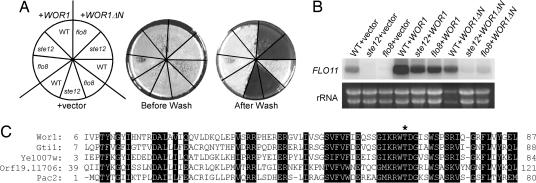
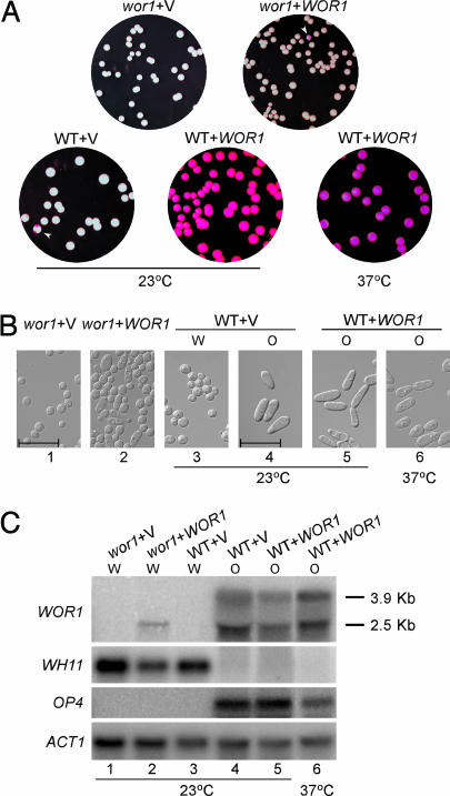
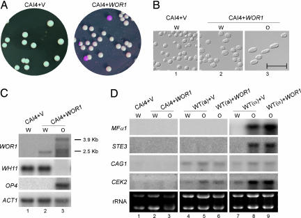
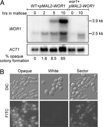
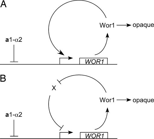
Candida albicans, a commensal organism and a pathogen of humans, can switch stochastically between a white phase and an opaque phase without an intermediate phase. The white and opaque phases have distinct cell shapes and gene expression programs. Once switched, each phase is stable for many cell divisions. White-opaque switching is under a1-alpha2 repression and therefore only happens in a or alpha cells. Mechanisms that control the switching are unknown. Here, we identify Wor1 (white-opaque regulator 1) as a master regulator of white-opaque switching. The deletion of WOR1 blocks opaque cell formation. The ectopic expression of WOR1 converts all cells to stable opaque cells in a or alpha cells. In addition, the ectopic expression of WOR1 in a/alpha cells is sufficient to induce opaque cell formation. Importantly, WOR1 expression displays an all-or-none pattern. It is undetectable in white cells, and it is highly expressed in opaque cells. The ectopic expression of Wor1 induces the transcription of WOR1 from the WOR1 locus, which correlates with the switch to opaque phase. We present genetic evidence for feedback regulation of WOR1 transcription. The feedback regulation explains the bistable and stochastic nature of white-opaque switching.


Conflict of interest statement

Conflict of interest statement: No conflicts declared.

Figures

Fig. 1.
Fig. 1.
Identification of Wor1 as a high-copy suppressor of a S. cerevisiae flo8 mutant in invasive growth. (A and B) Invasive assay (A) and FLO11 Northern blot (B) of a haploid S. cerevisiae WT strain (L5528), a flo8 mutant (HLY850) (31), and a ste12 mutant (HLY362) (32) carrying a vector (pVT102-U), WOR1 (pCF68), and WOR1ΔN (pVT-WOR1ΔN). Cells were grown at 30°C on YPD plates for 3 days for invasive growth and in liquid YPD for Northern blot analysis. (C) Protein sequence alignment of the Wor1 N-terminal domain with Sc. pombe Gti1, S. cerevisiae Yel007w, C. albicans Orf19.11706, and Sc. pombe Pac2. Conserved residues are shaded, and a putative protein kinase A phosphorylation site is marked with an asterisk.
Fig. 2.
Fig. 2.
Ectopic expression of WOR1 in WT MTLa strains converts all a cells into stable opaque phase. (A) Representative colonies of MTLa wor1 (CAH3) with vector (pACT1) or WOR1 (pACT1-WOR1), and WT MTLa (JYC5) with vector (pACT1) or WOR1 (pACT1-WOR1). Cells of each strain from 5-day-old white colonies on Lee’s medium plates were resuspended and plated on SC plus 5 μg/ml phloxine B plates for 7 days at 23°C or for 3 days at 37°C as indicated. The opaque sectors are marked with white arrowheads. (B) Photographs of cells grown from the white or opaque colonies in A under the indicated conditions. (Scale bars, 20 μm.) (C) Northern blot analysis of WOR1 and phase-specific genes of cells in B. Strains are as follows: lane 1, MTLa wor1/wor1 (CAH3) with vector (pACT1); lane 2, CAH3 with WOR1 (pACT1-WOR1); lanes 3 and 4, MTLa WT (JYC5) with vector from white or opaque colonies, respectively; lanes 5 and 6, JYC5 with WOR1 from opaque colonies. Cells in lanes 1–5 were grown in SC at 23°C overnight. Cells in lane 6 were grown in SC at 23°C overnight and then shifted to 37°C for 8 h. The white or opaque phase of each culture was determined by cell morphology and is indicated above each lane. WH11 and OP4 were used to detect white and opaque phase gene expression. ACT1 was used as a loading control. The exposure times for the probes were 6 h for WOR1, 4 h for OP4, 1 h for WH11, and 3 h for ACT1.
Fig. 3.
Fig. 3.
Ectopic expression of WOR1 promotes opaque cell formation in a/α cells. (A) Representative white and opaque colonies of CAI4 (MTLa/α) with vector (pACT1) (Left) or with WOR1 (pACT1-WOR1) (Right). (B) Cells from white and opaque colonies in A were grown in SC at 23°C overnight and photographed. (Scale bar, 20 μm.) (C) Logarithmic-phase cultures of white and opaque cells from CAI4 with vector or WOR1 were harvested, and RNA was extracted for Northern blot analysis. Lane 1, white cells of CAI4 with vector; lane 2, white cells of CAI4 with WOR1; lane 3, opaque cells of CAI4 with WOR1. WOR1, WH11, OP4, and ACT1 probes were used for hybridization as described in Materials and Methods. (D) Northern blot analysis of genes required for mating. Lanes 1–3, same as lanes 1–3 in C; lanes 4 and 5, MTLa WT (JYC5) with vector from white or opaque colonies; lane 6, JYC5 with WOR1; lanes 7 and 8, MTLα WT (CHY257) with vector from white or opaque colonies; lane 9, CHY257 with WOR1. WOR1, WH11, OP4, MFα1, STE3, CAG1, CEK2, and ACT1 probes were used for hybridization as described in Materials and Methods.
Fig. 4.
Fig. 4.
Wor1 induces its own transcription and bistable expression of WOR1 in single cells. (A) Expression of Wor1 from the inducible MAL2 promoter promotes the expression of WOR1 from its own promoter and the transition to opaque phase. MTLa cells (JYC5) transformed with the pMAL2-WOR1-HA construct from a fresh white colony were grown to logarithmic phase in SD, washed five times, and resuspended into SC/maltose at 25°C. Cells were harvested at the indicated times for Northern blot analysis and plated to determine the percentages of opaque colony formation. The percentage of opaque colonies on SD plates with 5 μg/ml phloxine B is shown for each time point. MTLa wor1 transformed with the pMAL2-WOR1-HA construct was included to show the WOR1 transcribed from the MAL2 promoter. (B) Expression of WOR1 shows an on-and-off pattern in single cells. Differential interference contrast microscopy (DIC) and FITC images were taken with cells of MTLa pWOR1-GFP (JYC1 plus pWOR1-GFP) collected from a white colony, an opaque colony, and a sector with both white and opaque cells. GFP expression is under the control of the WOR1 promoter in this strain.
Fig. 5.
Fig. 5.
Model of feedback loops for WOR1 expression and white–opaque switching. A simplified diagram depicts the idea that either a positive feedback loop (A) or a double-negative feedback loop (B) can establish the bistable expression pattern of WOR1. Once expressed, Wor1 activates opaque cell formation.

Comment in

References

    1. Soll D. R., Lockhart S. R., Zhao R. Eukaryot. Cell. 2003;2:390–397. - PMC - PubMed
    1. Slutsky B., Staebell M., Anderson J., Risen L., Pfaller M., Soll D. R. J. Bacteriol. 1987;169:189–197. - PMC - PubMed
    1. Bennett R. J., Johnson A. D. Annu. Rev. Microbiol. 2005;59:233–255. - PubMed
    1. Rikkerink E. H., Magee B. B., Magee P. T. J. Bacteriol. 1988;170:895–899. - PMC - PubMed
    1. Srikantha T., Soll D. R. Gene. 1993;131:53–60. - PubMed

Publication types

Substances

Associated data

LinkOut - more resources