. 2006 Sep;7(9):933-9.
doi: 10.1038/sj.embor.7400773. Epub 2006 Aug 11.

Cytochrome c-d regulates developmental apoptosis in the Drosophila retina

Affiliations

Cytochrome c-d regulates developmental apoptosis in the Drosophila retina

César S Mendes et al. EMBO Rep. 2006 Sep.

Abstract

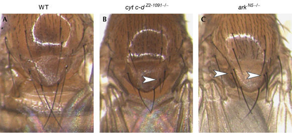
The role of cytochrome c (Cyt c) in caspase activation has largely been established from mammalian cell-culture studies, but much remains to be learned about its physiological relevance in situ. The role of Cyt c in invertebrates has been subject to considerable controversy. The Drosophila genome contains distinct cyt c genes: cyt c-p and cyt c-d. Loss of cyt c-p function causes embryonic lethality owing to a requirement of the gene for mitochondrial respiration. By contrast, cyt c-d mutants are viable but male sterile. Here, we show that cyt c-d regulates developmental apoptosis in the pupal eye. cyt c-d mutant retinas show a profound delay in the apoptosis of superfluous interommatidial cells and perimeter ommatidial cells. Furthermore, there is no apoptosis in mutant retinal tissues for the Drosophila homologues of apoptotic protease-activating factor 1 (Ark) and caspase 9 (Dronc). In addition, we found that cyt c-d--as with ark and dronc-regulates scutellar bristle number, which is known to depend on caspase activity. Collectively, our results indicate a role of Cyt c in caspase regulation of Drosophila somatic cells.


Figures

Figure 1
Figure 1
Interommatidial cell and perimeter ommatidial cell death are delayed in cyt c-d mutant retina. (A,B,DG) Pupal retinas were stained with Arm protein to visualize retinal cell membranes. (A,B) Extra IOCs are marked red in staged 42 h APF animals. (A) Wild-type (WT) ommatidia contain a definite number of IOCs (that is, six secondary and three tertiary cells). The yellow hexagon is a defined area used to count IOCs (see the Interommatidial cell counts section). (B) cyt c-dZ2-1091−/− retinas show extra IOCs with the normal shape and position of secondary or tertiary cells (see Table 1 for values). (C) Graph showing the difference in the number of IOCs per ommatidium between WT and cyt c-dZ2-1091−/− at 22 and 42 h APF. The number of extra IOCs is significantly higher at 22 h than at 42 h APF (t-test, ***P<0.001). (DG) Staged 38 and 40 h APF retinas stained against Arm (white). (D,E) At 38 h APF, extra ommatidial clusters (yellow arrows) are visualized in (D) WT and (E) cyt c-dZ2-1091−/− retinas in a thick layer of IOCs (yellow bracket). (F) In WT retinas at 40 h APF, POC death is almost complete and only occasional extra ommatidial clusters remain at the edge in a thin layer of IOCs. (G) In cyt c-dZ2-1091−/− 40 h APF retinas, many extra ommatidial clusters (yellow arrows) are still visible in a thick layer of IOCs. (H) Reverse transcription–PCR analysis of cyt c-d expression at 22 h APF. As a negative control, we used bln1 mutant flies in which no cyt c-d transcript is detected (Arama et al, 2003). APF, after puparium formation; Arm, Armadillo; IOC, interommatidial cell; POC, perimeter ommatidial cell.
Figure 2
Figure 2
Loss of cyt c-d has no effect on ATP levels and retinal development. (A) ATP levels in wild-type (WT; CS, OR and yw), cyt c-dZ2-1091−/+ and cyt c-dZ2-1091−/− adult fly heads (1 day after eclosion). Each value shows the mean±s.e.m. of three independent experiments. (BD) WT and (EG) cyt c-dZ2-1091−/− mutant eye discs were stained with various differentiation markers. (B,E) Posterior is to the right. Third-instar larval eye discs stained with antibodies against the photoreceptor cell marker, Elav (green), and R8 cell marker, Boss (red), show similar pattern in (B) WT and (E) cyt c-dZ2-1091−/−. 42 h APF (C,D) WT and (F,G) cyt c-dZ2-1091−/− retinas stained with Arm (apical view; C,F) and the secondary and tertiary pigment marker, Hth (basal view; D,G), in two distinct focal planes of the retina. In cyt c-dZ2-1091−/−, extra secondary or tertiary cells are visualized with Arm staining and their nuclei express Hth (an extra yellow arrow compared with WT retina). APF, after puparium formation; Arm, Armadillo; Hth, Homothorax.
Figure 3
Figure 3
Ark and Dronc are required for apoptosis in the pupal retina. (A,B) ark and dronc mutant clones are visualized in 42 h APF retinas by the absence of green fluorescent protein (GFP; green) and the membrane outlined with Arm antibody (red). Numerous extra IOCs are observed in the clonal area of (A) (ey-FLP; FRT42D arkL46/FRT42D Ubi-GFP) and (B) (ey-FLP;; droncI29 FRT80B/Ubi-GFP FRT80B). (A′) and (B′) show the corresponding schematic ommatidium, in which the yellow line represents the clonal boundaries and extra IOCs are marked in red. (CF) Retinal edges in ark and dronc mutant clones at 42 h APF are visualized by the absence of GFP. (C,D) POCs stained with Arm antibody (red) in (C) arkL46 and (D) droncI29 mutant clones show extra POCs (white arrows) compared with wild-type retina. (E,F) Retinal edges stained with TdT-mediated dUTP nick end labelling (TUNEL; red) in (E) arkL46 and (F) droncI29 mutant clones show the absence of TUNEL staining compared with the non-mutant tissue (GFP positive). (E′) and (F′) are the corresponding panels without GFP staining, in which a white line marks the boundary of the clone. (G) Comparison of the number of IOCs in arkL46, droncI29 mutant clones and retinas expressing GMR-p35 at 42 h APF. APF, after puparium formation; Arm, Armadillo; IOC, interommatidial cell; POC, perimeter ommatidial cell.
Figure 4
Figure 4
Adult flies with the mutation cyt c-d−/− have an extra bristle in the scutellum. (A) Wild-type (WT; CS) flies show four bristles. (B) cyt c-dZ2-1091−/− mutant showing an extra bristle (arrowhead). (C) arkN5−/− fly with two extra bristles (arrowheads). See supplementary Table S1 online for statistical analysis.

References

    1. Akdemir F et al. (2006) Autophagy occurs upstream or parallel to the apoptosome during histolytic cell death. Development 133: 1457–1465 - PubMed
    1. Arama E, Agapite J, Steller H (2003) Caspase activity and a specific cytochrome c are required for sperm differentiation in Drosophila. Dev Cell 4: 687–697 - PubMed
    1. Arama E, Bader M, Srivastava M, Bergmann A, Steller H (2006) The two Drosophila cytochrome c proteins can function in both respiration and caspase activation. EMBO J 25: 232–243 - PMC - PubMed
    1. Cagan RL, Ready DF (1989) The emergence of order in the Drosophila pupal retina. Dev Biol 136: 346–362 - PubMed
    1. Chew SK, Akdemir F, Chen P, Lu WJ, Mills K, Daish T, Kumar S, Rodriguez A, Abrams JM (2004) The apical caspase dronc governs programmed and unprogrammed cell death in Drosophila. Dev Cell 7: 897–907 - PubMed

Publication types