. 2006 Sep;2(9):494-502.
doi: 10.1038/nchembio811. Epub 2006 Aug 13.

Biosynthesis of Dictyostelium discoideum differentiation-inducing factor by a hybrid type I fatty acid-type III polyketide synthase

Affiliations

Biosynthesis of Dictyostelium discoideum differentiation-inducing factor by a hybrid type I fatty acid-type III polyketide synthase

Michael B Austin et al. Nat Chem Biol. 2006 Sep.

Abstract

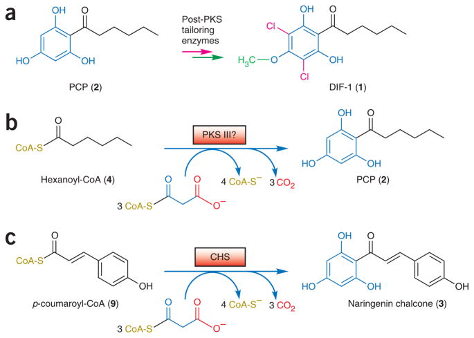
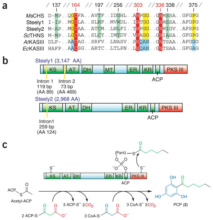
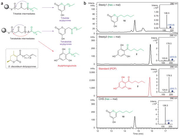
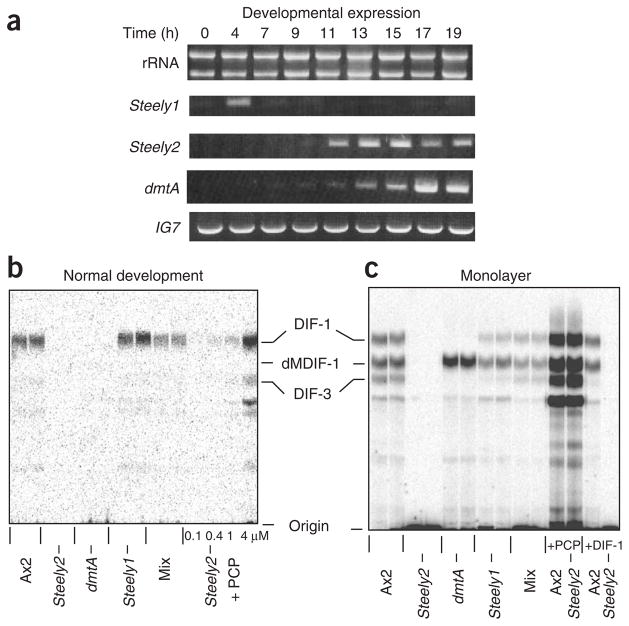
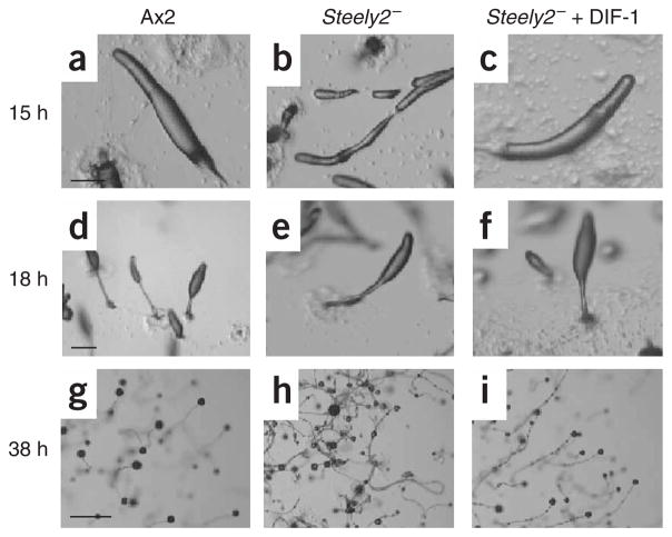
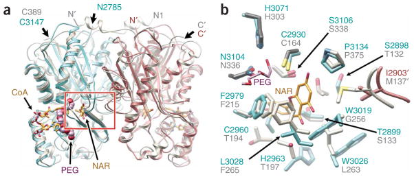
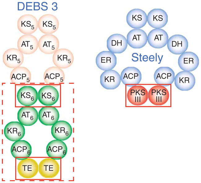
Differentiation-inducing factors (DIFs) are well known to modulate formation of distinct communal cell types from identical Dictyostelium discoideum amoebas, but DIF biosynthesis remains obscure. We report complimentary in vivo and in vitro experiments identifying one of two approximately 3,000-residue D. discoideum proteins, termed 'steely', as responsible for biosynthesis of the DIF acylphloroglucinol scaffold. Steely proteins possess six catalytic domains homologous to metazoan type I fatty acid synthases (FASs) but feature an iterative type III polyketide synthase (PKS) in place of the expected FAS C-terminal thioesterase used to off load fatty acid products. This new domain arrangement likely facilitates covalent transfer of steely N-terminal acyl products directly to the C-terminal type III PKS active sites, which catalyze both iterative polyketide extension and cyclization. The crystal structure of a steely C-terminal domain confirms conservation of the homodimeric type III PKS fold. These findings suggest new bioengineering strategies for expanding the scope of fatty acid and polyketide biosynthesis.


Conflict of interest statement

COMPETING INTERESTS STATEMENT

The authors declare that they have no competing financial interests.

Figures

Figure 1
Figure 1
Biosynthesis of D. discoideum DIF-1. (a) DIF-1 results from chlorination (red) and O-methylation (green) of PCP. The presence of saturated (black) and hydroxylated (blue) portions of PCP suggests two stages of biosynthesis. (b) Hypothetical type III PKS biosynthesis of PCP from hexanoyl-CoA, suggested by similarity to the phloroglucinol core of chalcone. (c) In plants, CHS transfers p-coumaroyl from CoA to a catalytic cysteine, then catalyzes sequential decarboxylative condensations with three malonyl-CoA molecules. Chalcone is formed and off loaded by an intramolecular Claisen cyclization of the resulting linear tetraketide.
Figure 2
Figure 2
D. discoideum type III PKSs occur as C-terminal domains of steely FAS-PKS hybrids. (a) Active site and signature sequence comparison of steely type III PKSs with plant and bacterial type III PKSs (Medicago sativa (alfalfa) CHS and Streptomyces coelicolor 1,3,6,8-tetrahydroxynaphthalene synthase, THNS) and related type II (plant and bacterial) FAS enzymes (Arabidopsis thaliana and E. coli KAS III enzymes). Signature sequences are highlighted in yellow or blue, red marks the conserved catalytic triad, and green indicates type III PKS specificity-determining positions. Numbering is for alfalfa CHS2. (b) Predicted enzymatic domains of D. discoideum steely proteins, including N-terminal type I FAS-PKS and C-terminal type III PKS domains. Type I FAS-PKS intermediates are tethered by a thioester linkage to either a KS cysteine or the prosthetic Ppant arm of the ACP-like domain. AA (amino acid) labels indicate the total number of residues in each steely protein or the amino acid position of intron boundaries. (c) Proposed PCP biosynthesis by a steely FAS I–PKS III hybrid. Direct transfer of a hexanoyl intermediate to the type III PKS domain based on analogous off loading of conventional type I FAS-PKS products via activity of thioesterase (TE) domains (Supplementary Fig. 1).
Figure 3
Figure 3
Hexanoyl-primed in vitro product specificity of steely C-terminal type III PKS domains. (a) Polyketide cyclization routes leading to acylpyrones (blue arrows) and acylphloroglucinols (red arrows). Carbons 1, 5 and 6 are involved in cyclization. Sphere represents CoA or active site cysteine. Starter-derived moieties are green; n = 3 and n = 2 for hexanoyl and pentanoyl moieties (respectively) of known D. discoideum acylphloroglucinols, and n = 3 and n = 1 for hexanoyl- and butanoyl-CoA substrates (respectively) tested here (see b and Supplementary Figs. 3 and 4). Conversely, dictyopyrone biosynthesis may involve condensation of a diketide (black) with another small molecule (gold). (b) Acylphloroglucinol (PCP) biosynthesis by Steely2, but not Steely1. Main enzymatic products of hexanoyl-CoA–primed in vitro type III PKS assays with malonyl-CoA as determined by negative-mode LC-MS-MS (insets). Parent (MS) masses for each MS-MS spectrum are given in blue. We also analyzed minor hexanoyl-primed enzymatic products and all butanoyl-primed enzymatic products (see Supplementary Figs. 3 and 4).
Figure 4
Figure 4
Steely gene expression and DIF-1 synthesis by steely null mutants. (a) Expression of the steely genes during development of wild-type Ax2 cells determined by reverse transcription PCR. Hours of development are given; rRNA and the IG7 gene are shown as loading and PCR controls. (b,c) DIF-1 synthesis detected by labeling with 36Cl during normal development on an agar surface or development as a submerged monolayer of cells in tissue culture dishes. Cells: Ax2 is parental control; HM1154 is Steely2 (stlB); HM1030 is dmtA, blocked in the last step of DIF-1 biosynthesis; HM1157 is Steely1 (stlA); mix is a 1:1 mix of HM1154 and HM1030 cells, which develop intermingled with each other and can therefore exchange membrane-permeable metabolites. Additions: (b) PCP added to the medium at the concentrations indicated; (c) 4 μM PCP and 100 nM DIF-1 added to the medium as indicated. Markers: dMDIF-1 is the immediate precursor of DIF-1; DIF-3 is the first breakdown product of DIF-1; the other labeled compounds are likely to be more derived metabolites. Incorporation of radioactivity (36Cl) into DIF-1 by Ax2 cells: (b) 6 c.p.m. by 108 cells per lane; (c) 40 c.p.m. by 4 × 10⁷ cells per lane.
Figure 5
Figure 5
Development of Steely2 mutant and rescue by DIF-1. (ac) Migratory slugs; those of the mutant are long and thin (and they break apart) but are restored by 100 nM DIF-1 in the agar. (df) Early culminates. (gi) Mature fruiting bodies; those of the wild type each have a spore mass supported by a stalk, but the mutant stalks lay down on the surface and the spores slip down to their base, giving an untidy appearance, which is again restored by DIF-1. Cells used are Ax2 (parental control) and HM1154 (Steely2). Each horizontal set of panels is at the same magnification; scale bars used are (a,d) 0.2 mm, (g) 1 mm.
Figure 6
Figure 6
Crystal structure of Steely1 C-terminal domain confirms conservation of the PKS III fold, homodimeric assembly and internal active site cavity. (a) Structure of D. discoideum Steely1 homodimeric C-terminal domain (cyan and rose ribbons) overlaid with alfalfa CHS2 (gray ribbons, PDB ID 1CJK). A PEG molecule (space-filling lavender and red) is bound in the Steely1 pantetheine-binding tunnel, illustrated here by CHS-bound CoA (gold stick, superimposed from PDB ID 1BQ6), which presents thioester substrates to the internal active site cavity, delineated here by CHS-bound naringenin (NAR). The red box indicates area detailed in b. Apostrophes designate the dyad-related protein chain. (b) Active site comparison confirms homology-predicted assignments of important active site residues (Fig. 2a and text), but with subtle conformational changes. One end of the Steely1-bound PEG molecule (shown here as a stick model) is positioned in the ‘oxyanion hole’ of the conserved catalytic triad.
Figure 7
Figure 7
Steely1 type III PKS homodimer suggests functional similarity of steely hybridization interface to modular type I PKS interfaces. Domain interaction schematic (adapted from ref. 14) showing a current model for the parallel dimeric assembly of modular type I PKS proteins. Shown here is the the final polypeptide chain of DEBS, chain 3, which is composed of modules 5 and 6 (numbered subscripts). Solid red boxes compare the steely ACP–PKS III interface to DEBS intramodular (ACP-TE) and intermodular (ACP-KS) domain interactions. Dashed box highlights functional equivalence of steely PKS III with an entire PKS I module.

Comment in

  • A type III PKS makes the DIFference.
    Sankaranarayanan R. Sankaranarayanan R. Nat Chem Biol. 2006 Sep;2(9):451-2. doi: 10.1038/nchembio0906-451. Nat Chem Biol. 2006. PMID: 16921353 No abstract available.

References

    1. Kessin RH. Dictyostelium. Cambridge University Press; Cambridge, UK: 2001.
    1. Morris HR, Taylor GW, Masento MS, Jermyn KA, Kay RR. Chemical structure of the morphogen differentiation inducing factor from Dictyostelium discoideum. Nature. 1987;328:811–814. - PubMed
    1. Thompson CR, Kay RR. The role of DIF-1 signaling in Dictyostelium development. Mol Cell. 2000;6:1509–1514. - PubMed
    1. Gokan N, et al. Structural requirements of Dictyostelium differentiation-inducing factors for their stalk-cell-inducing activity in Dictyostelium cells and anti-proliferative activity in K562 human leukemic cells. Biochem Pharmacol. 2005;70:676–685. - PubMed
    1. Kay RR. The biosynthesis of differentiation-inducing factor, a chlorinated signal molecule regulating Dictyostelium development. J Biol Chem. 1998;273:2669–2675. - PubMed

Publication types

MeSH terms