. 2006 Oct;112(4):389-404.
doi: 10.1007/s00401-006-0127-z. Epub 2006 Aug 12.

Staging of Alzheimer disease-associated neurofibrillary pathology using paraffin sections and immunocytochemistry

Affiliations

Staging of Alzheimer disease-associated neurofibrillary pathology using paraffin sections and immunocytochemistry

Heiko Braak et al. Acta Neuropathol. 2006 Oct.

Abstract

Assessment of Alzheimer's disease (AD)-related neurofibrillary pathology requires a procedure that permits a sufficient differentiation between initial, intermediate, and late stages. The gradual deposition of a hyperphosphorylated tau protein within select neuronal types in specific nuclei or areas is central to the disease process. The staging of AD-related neurofibrillary pathology originally described in 1991 was performed on unconventionally thick sections (100 mum) using a modern silver technique and reflected the progress of the disease process based chiefly on the topographic expansion of the lesions. To better meet the demands of routine laboratories this procedure is revised here by adapting tissue selection and processing to the needs of paraffin-embedded sections (5-15 mum) and by introducing a robust immunoreaction (AT8) for hyperphosphorylated tau protein that can be processed on an automated basis. It is anticipated that this revised methodological protocol will enable a more uniform application of the staging procedure.


Figures

Fig. 1
Fig. 1
Scheme showing the location of the three blocks of tissue required for staging of AD-related neurofibrillary changes. The first block at the far left (a) includes anteromedial portions of the temporal lobe. It is cut at the mid-uncal or amygdala level (frontal section at the level of the mamillary bodies) and includes the parahippocampal and adjoining occipito-temporal gyri (see enlarged insert below a). The cutting line runs through the rhinal sulcus. The second block comes from the same level and includes part of the medial and superior temporal gyri (a). The third block at the far right (c) is removed from basal portions of the occipital lobe. The cut is oriented perpendicular to the calcarine fissure. The block includes the neocortex covering the lower bank of the calcarine fissure and the adjoining basal occipital gyri. It thus shows portions of the peristriate region as well as of the parastriate and striate areas (see enlarged insert below c). (b) This block provides the classical view of the hippocampal formation and is removed at the level of the lateral geniculate nucleus. It is routinely dissected for diverse diagnostic purposes of the hippocampal formation. The cutting line runs through the collateral sulcus (d). The parahippocampal gyrus at this latitude abuts posteriorly on the lingual gyrus and contains either posterior portions of the entorhinal and transentorhinal regions or lingual neocortex. Insofar as the first temporal block at mid-uncal level is essential for the evaluation of the transentorhinal and entorhinal regions (diagnosis of AD stages I–III), the classical hippocampus section is not absolutely required for staging. The middle drawing in the second row indicated by a double frame shows the anatomical landmarks of the entorhinal region seen basally. Note the wart-like elevations in anterior portions of the parahippocampal gyrus roughly outlining the extent of the entorhinal allocortex. The lower schemata highlight the lamination pattern of the areas that need to be evaluated for staging purposes. The various allocortical and neocortical laminae are indicated across the upper margins. 17, 18, 19 striate area, parastriate area, peristriate region. Abbreviations: CA1 first sector of the Ammon’s horn, ent entorhinal region, parasubic parasubiculum, presubic presubiculum, temp. neocortex temporal neocortex, tre transentorhinal region (mesocortex), transentorhin. transentorhinal
Fig. 2
Fig. 2
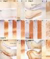
Comparison of Gallyas silver- and AT8-immunostaining of cortical neurofibrillary pathology as seen in adjacent serial 100 μm polyethylene glycol-embedded sections. The distribution pattern of the lesions throughout the various cortical fields that are necessary for staging purposes basically corresponds in both methods. It is possible with either technique to assess the progress of the neurofibrillary pathology. In the revised staging procedure, however, the greater emphasis on the presence of abnormal plexuses, which also include non-argyrophilic pretangle material in AT8-ir sections, facilitates rapid diagnostic assessment of the stages. a–d stage I: Mild involvement is confined to the transentorhinal region. Note that the plexus of AT8-ir nerve cell processes (b and d) is more conspicuous than that of argyrophilic neuropil threads (a and c). Sections originate from a non-demented 62-year-old male. e–h stage II: Lesional density increases and the pathology extends into the entorhinal region. Layer pre-α gradually sinks into a deeper position at the border between entorhinal and transentorhinal region (arrowhead). Note the greater breadth of the ir-plexus in comparison to silverstained nerve cell processes (compare f and h with e and g). Immunoreactions begin to show the deep entorhinal plexus (pri-α). The sections were obtained from a non-demented 78-year-old male. i–n stage III: The pathology in the outer and inner entorhinal (i, j) and transentorhinal (k, l) cellular layers worsens, and lesions extend into the adjoining neocortical association areas of the fusiform (occipito-temporal) gyrus (m, n). The sections originate from an 85-year-old female. o–t stage IV: The density of the lesions increases in both the entorhinal region (o, p) and fusiform gyrus (q–r) with a gradual decrease of the pallid lines (lamina dissecans in p and outer line of Baillarger in r). The neurofibrillary pathology now extends up to the medial temporal gyrus (s, t). Sections were taken from an 80-year-old female. u–x stage V: The lesions extend widely into the occipital lobe and appear in the peristriate region. Note the presence of a deep plexus in AT8-immunoreactions (v, x). Sections were obtained from a 66-year-old demented female. y-z′ stage VI: Lesions are visible even in the parastriate and striate areas of the occipital neocortex. Note the clear-cut line in layer V of the striate area (z′ and z″). The sections originate from a demented 75-year-old male. Scale bar in a applies to all overviews and that in c to all micrographs of cortical areas
Fig. 3
Fig. 3
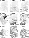
Stages I–VI of cortical neurofibrillary pathology in 100 μm polyethylene glycol-embedded hemisphere sections immunostained for hyperphosphorylated tau (AT8, Innogenetics). a stage I: Involvement is slight and all but confined to the transentorhinal region (part of the temporal mesocortex), located on the medial surface of the rhinal sulcus. The section originates from a non-demented 80-year-old female. b stage II: Additional immunoreactivity occurs in layer pre-α or layer II of the entorhinal region. The layer gradually sinks into a deeper position in the transentorhinal region (arrow). The border between the entorhinal and transentorhinal regions is clearly recognizable in these early stages (arrowhead). Furthermore, the lesions make headway into the hippocampus (arrow). The section was obtained from a non-demented 80-year-old male. c stage III: The lesions in the hippocampal formation worsen. Entorhinal layers pre-α and, additionally, pri-α of the deep layers become strongly involved. Lesions extend through the transentorhinal region into the adjoining high order sensory association areas of the temporal neocortex. The lesions generally do not extend beyond the occipito-temporal gyrus laterally (arrow) and lingual gyrus posteriorly. The section originates from a 90-year-old female. d stage III: A flat section through the entire basal surface of the temporal lobe reveals the severe involvement of the entorhinal cortex (anterior portions of the parahippocampal gyrus) at stage III and shows the tendency of the pathology to extend from there into the adjacent neocortex, i.e., occipito-temporal gyrus laterally (arrow) and lingual gyrus posteriorly (arrow). e stage IV: The third and fourth sectors of the Ammon’s horn and a large portion of the insular cortex (arrow) become affected. The involvement of the neocortical high order sensory association cortex of the temporal lobe now extends up to the medial temporal gyrus and stops short of the superior temporal gyrus (arrow). The primary fields of the neocortex (see transverse gyrus of Heschl) and, to a large extent, also the premotor and first order sensory association areas of the neocortex remain intact. This section was taken from an 82-year-old demented female. f stage V: In addition to the presence of AD-related lesions in all of the regions involved in stage IV, pathological changes appear in the superior temporal gyrus and even encroach to a mild degree upon the premotor and first order sensory association areas of the neocortex. g stage V: In the occipital lobe, the peristriate region shows varying degrees of affection, and lesions occasionally can even be seen in the parastriate area. Stage V sections were obtained from a 90-year-old female with dementia. h–i stage VI: Strong immunoreactivity can be detected even in the first order sensory association areas (e.g., the parastriate area) and the primary areas of the neocortex (e.g., the striate area) of the occipital neocortex. Compare the superior temporal gyrus and transverse gyrus of Heschl at stage V with the same structures at stage VI. Both stage VI sections originate from a severely demented 70-year-old female Alzheimer patient. Scale bar applies to all thick sections
Fig. 4
Fig. 4
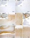
Progress of cortical neurofibrillary pathology, as seen in paraffin sections immunostained for hyperphosphorylated tau (AT8, Innogenetics). a, b Control case displaying no AT8-ir intraneuronal changes. Note that even the transentorhinal region is devoid of immunoreactivity. c, d stage I: The first AT8-ir pyramidal cells often are more easily detected in thick sections than in paraffin material. Closer inspection of the predilection site (transentorhinal region in d, framed area in c), however, reveals the initial lesions. The meshwork of ir-neurites is as yet not well developed. e–g stage II: Many AT8-ir projection neurons are seen in the transentorhinal region accompanied by a well-developed plexus of ir-neurites (f, g). The pathology also extends into the entorhinal layers pre-α and pri-α (arrowheads in e). h–l stage III: The transentorhinal and entorhinal regions are more severely involved than in the preceeding stage, and the pathology now extends into the adjoining temporal neocortex of the occipito-temporal and lingual gyri (h, j). The middle temporal gyrus remains uninvolved (i). Scale bar in a, c, e, h is also valid for i and Fig. 5 a, e, f, j, k, o below
Fig. 5
Fig. 5
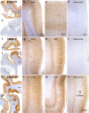
Progress of cortical neurofibrillary pathology, as seen in paraffin sections immunostained for hyperphosphorylated tau (AT8, Innogenetics). a–e stage IV: The disease process extends into the high order sensory association neocortex of the temporal lobe (temp.) and includes the medial temporal gyrus (a–c). The peristriate region as well as the parastriate field and striate area of the occipital lobe (occ.) still lack the neurofibrillary pathology (e, d). f–j stage V: Large numbers of neuritic plaques appear in the neocortex (g, h). Pathological changes now encroach to a mild degree upon premotor areas and first order sensory association fields. In the occipital lobe (j), it is chiefly the peristriate region (h) that shows varying degrees of affection, and lesions occasionally even develop in the parastriate area. The striate area remains uninvolved (i, j). k–o stage VI: Drastic affection of the neocortex is seen at stage VI with involvement of almost all areas. Strong immunoreactivity can be recognized even in premotor areas and first order sensory association areas (e.g., the parastriate area m), as well as in primary neocortical areas (e.g., the striate area n). The borderline between the striate and the parastriate areas is drawn easily and—owing to the sudden cessation of the line of Gennari (plexus of myelinated fibers)—usually can be detected with the unaided eye. A key feature of stage VI is the involvement of the striate area (n), characterized by a dense neuritic mesh in layer V with sharply drawn upper and lower boundaries. Note that the myelin-rich line of Gennari (layer IVb n) is virtually devoid of neurofibrillary pathology. Scale bar in n also applies to g, h, i, l, m

References

    1. Alafuzoff I, Pikkarainen M, Al-Sarraj S, Arzberger T, Bell J, Bodi I, Bogdanovic N, Budka H, Bugiani O, Ferrer I, Gelpi E, Gaiccone G, Graeber MB, Hauw JJ, Kamphorst W, King A, Kopp N, Korkolopolou P, Kovacs GG, Meyronet D, Marchi P, Patsouris E, Preusser M, Ravid R, Roggendorf W, Seilhean D, Streichneberger N, Thal DR, BNE consortium, Kretzschmar H (2006) Inter-laboratory comparison of assessments of AD-related lesions. A study of the BrainNet Europe consortium. J Neuropathol Exp Neurol 65 (in press) - PubMed
    1. Alzheimer A. Über einen eigenartigen schweren Erkrankungsprozeß der Hirnrinde. Neurolog Centralbl. 1906;23:1129–1136.
    1. Alzheimer A. Über eigenartige Krankheitsfälle des späteren Alters. Z ges Neurol Psychiatr. 1911;4:356–385.
    1. Arnold SE, Hyman BT, Flory J, Damasio AR, van Hoesen GW. The topographical and neuroanatomical distribution of neurofibrillary tangles and neuritic plaques in the cerebral cortex of patients with Alzheimer’s disease. Cereb Cortex. 1991;1:103–116. - PubMed
    1. Arriagada PV, Growdon J, Hedley-Whyte E, Hyman BT. Neurofibrillary tangles but not senile plaques parallel duration and severity of Alzheimer’s disease. Neurology. 1992a;42:631–639. - PubMed

Publication types

MeSH terms