

Different inflammatory cell pattern and macrophage phenotype in chronic obstructive pulmonary disease patients, smokers and non-smokers
- PMID: 16907910
- PMCID: PMC1809704
- DOI: 10.1111/j.1365-2249.2006.03154.x
Different inflammatory cell pattern and macrophage phenotype in chronic obstructive pulmonary disease patients, smokers and non-smokers
Abstract
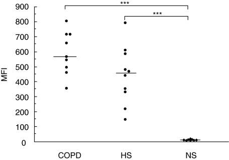
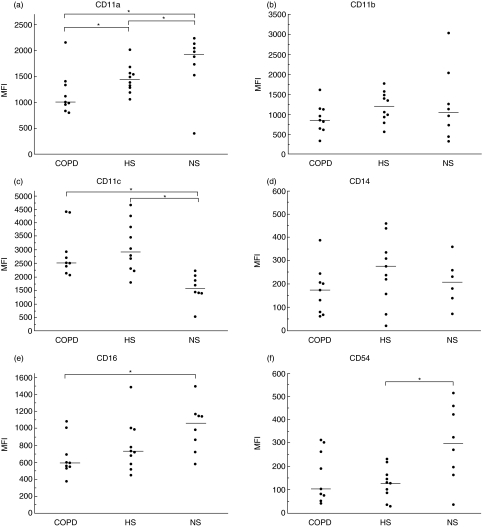
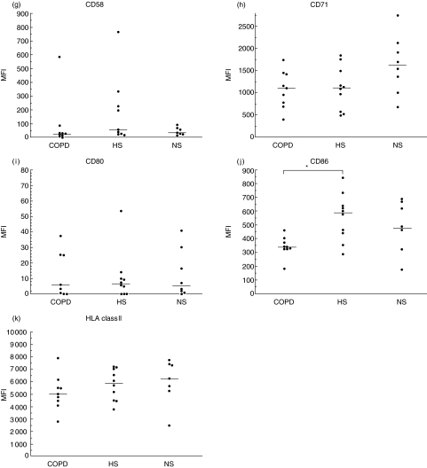
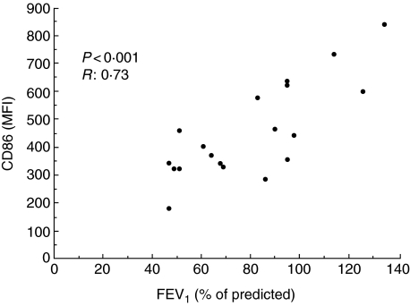
Smokers exhibit airway inflammation and increased number of alveolar macrophages (AM), but not all develop chronic obstructive pulmonary disease (COPD). We hypothesized that AMs in COPD patients have an altered functional capacity mirrored in a different phenotype. Sixteen steroid-naive COPD patients [forced expiratory volume in 1 s (FEV(1)) < 70% of predicted] underwent bronchoalveolar lavage (BAL). Age- and smoking-matched non-obstructive smokers (n = 10) and healthy non-smokers (n = 9) served as controls. Nine COPD patients had a BAL cell yield sufficient for flow cytometry analysis, where expression of AM cell surface markers reflecting various functions was determined. AMs from COPD patients showed decreased expression of CD86 (co-stimulation) and CD11a (adhesion) compared to smokers' AMs (P < 0.05). Furthermore, smokers' AMs showed lower (P < 0.05) expression of CD11a compared to non-smokers. AM expression of CD11c was higher in the COPD and smokers groups compared to non-smokers (P < 0.05). The expression of CD54 (adhesion) was lower in smokers' AMs compared to non-smokers (P < 0.05), whereas CD16 was lower (P < 0.05) in COPD patients compared to non-smokers. The AM expression of CD11b, CD14, CD58, CD71, CD80 and human leucocyte antigen (HLA) Class II did not differ between the three groups. The AM phenotype is altered in COPD and further research may develop disease markers. The lower AM expression of CD86 and CD11a in COPD implies a reduced antigen-presenting function. Some alterations were found in smokers compared to non-smokers, thus indicating that changes in AM phenotype may be associated with smoking per se. The functional relevance of our findings remains to be elucidated.
Figures
Similar articles
-
Toll-like receptor 2 expression is decreased on alveolar macrophages in cigarette smokers and COPD patients.Respir Res. 2005 Jul 8;6(1):68. doi: 10.1186/1465-9921-6-68. Respir Res. 2005. PMID: 16004610 Free PMC article. Clinical Trial.
-
A novel insight into adaptive immunity in chronic obstructive pulmonary disease: B cell activating factor belonging to the tumor necrosis factor family.Am J Respir Crit Care Med. 2010 Oct 15;182(8):1011-9. doi: 10.1164/rccm.200911-1700OC. Epub 2010 Jun 25. Am J Respir Crit Care Med. 2010. PMID: 20581172
-
The phenotype of alveolar macrophages and its correlation with immune cells in bronchoalveolar lavage.Eur Respir J. 1993 Oct;6(9):1287-94. Eur Respir J. 1993. PMID: 7904572
-
Altered macrophage function in chronic obstructive pulmonary disease.Ann Am Thorac Soc. 2013 Dec;10 Suppl:S180-5. doi: 10.1513/AnnalsATS.201305-123AW. Ann Am Thorac Soc. 2013. PMID: 24313770 Review.
-
Immune response in chronic obstructive pulmonary disease.Expert Rev Clin Immunol. 2013 Sep;9(9):821-33. doi: 10.1586/1744666X.2013.828875. Expert Rev Clin Immunol. 2013. PMID: 24070046 Review.
Cited by
-
Expansion of Phenotypically Altered Dendritic Cell Populations in the Small Airways and Alveolar Parenchyma in Patients with Chronic Obstructive Pulmonary Disease.J Innate Immun. 2023;15(1):188-203. doi: 10.1159/000526080. Epub 2022 Aug 23. J Innate Immun. 2023. PMID: 35998572 Free PMC article.
-
Cigarette smoke exposure and alveolar macrophages: mechanisms for lung disease.Thorax. 2022 Jan;77(1):94-101. doi: 10.1136/thoraxjnl-2020-216296. Epub 2021 May 13. Thorax. 2022. PMID: 33986144 Free PMC article. Review.
-
CD11b immunophenotyping identifies inflammatory profiles in the mouse and human lungs.Mucosal Immunol. 2016 Mar;9(2):550-63. doi: 10.1038/mi.2015.84. Epub 2015 Sep 30. Mucosal Immunol. 2016. PMID: 26422753 Free PMC article.
-
Smoking-dependent reprogramming of alveolar macrophage polarization: implication for pathogenesis of chronic obstructive pulmonary disease.J Immunol. 2009 Aug 15;183(4):2867-83. doi: 10.4049/jimmunol.0900473. Epub 2009 Jul 27. J Immunol. 2009. PMID: 19635926 Free PMC article.
-
Beyond Smoking: Emerging Drivers of COPD and Their Clinical Implications in Low- and Middle-Income Countries: A Narrative Review.J Clin Med. 2025 Jun 30;14(13):4633. doi: 10.3390/jcm14134633. J Clin Med. 2025. PMID: 40649006 Free PMC article. Review.
References
-
- Di Stefano A, Caramori G, Ricciardolo FL, Capelli A, Adcock IM, Donner CF. Cellular and molecular mechanisms in chronic obstructive pulmonary disease: an overview. Clin Exp Allergy. 2004;34:1156–67. - PubMed
-
- Thompson AB, Daughton D, Robbins RA, Ghafouri MA, Oehlerking M, Rennard SI. Intraluminal airway inflammation in chronic bronchitis. Characterization and correlation with clinical parameters. Am Rev Respir Dis. 1989;140:1527–37. - PubMed
-
- Linden M, Rasmussen JB, Piitulainen E, et al. Airway inflammation in smokers with nonobstructive and obstructive chronic bronchitis. Am Rev Respir Dis. 1993;148:1226–32. - PubMed
-
- Tetley TD. Macrophages and the pathogenesis of COPD. Chest. 2002;121:156S–159S. - PubMed
Publication types
MeSH terms
Substances
LinkOut - more resources
Full Text Sources
Medical
Research Materials
