

Comparison of RCAS1 and metallothionein expression and the presence and activity of immune cells in human ovarian and abdominal wall endometriomas
- PMID: 16907986
- PMCID: PMC1574328
- DOI: 10.1186/1477-7827-4-41
Comparison of RCAS1 and metallothionein expression and the presence and activity of immune cells in human ovarian and abdominal wall endometriomas
Abstract
Background: The coexistence of endometrial and immune cells during decidualization is preserved by the ability of endometrial cells to regulate the cytotoxic immune activity and their capability to be resistant to immune-mediated apoptosis. These phenomena enable the survival of endometrial ectopic cells. RCAS1 is responsible for regulation of cytotoxic activity. Metallothionein expression seems to protect endometrial cells against apoptosis. The aim of the present study was to evaluate RCAS1 and metallothionein expression in human ovarian and scar endometriomas in relation to the presence of immune cells and their activity.
Methods: Metallothionein, RCAS1, CD25, CD69, CD56, CD16, CD68 antigen expression was assessed by immunohistochemistry in ovarian and scar endometriomas tissue samples which were obtained from 33 patients. The secretory endometrium was used as a control group (15 patients).
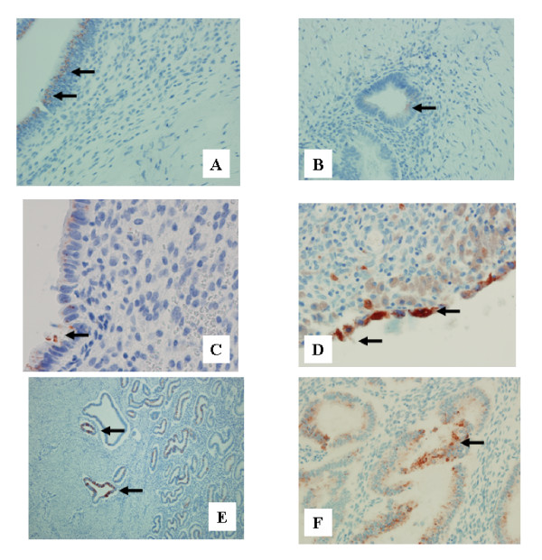
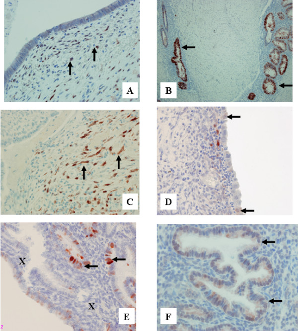
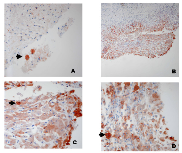
Results: The lowest metallothionein expression was revealed in ovarian endometriomas in comparison to scar endometriomas and to the control group. RCAS1 expression was at the highest level in the secretory endometrium and it was at comparable levels in ovarian and scar endometriomas. Similarly, the number of CD56-positive cells was lower in scar and ovarian endometriomas than in the secretory endometrium. The highest number of macrophages was found in ovarian endometriomas. RCAS1-positive macrophages were observed only in ovarian endometriomas. CD25 and CD69 antigen expression was higher in scar and ovarian endometriomas than in the control group.
Conclusion: The expression of RCAS1 and metallothionein by endometrial cells may favor the persistence of these cells in ectopic localization both in scar following cesarean section and in ovarian endometriosis.
Figures
Similar articles
-
Metallothionein and RCAS1 expression in comparison to immunological cells activity in endometriosis, endometrial adenocarcinoma and endometrium according to menstrual cycle changes.Gynecol Oncol. 2005 Dec;99(3):622-30. doi: 10.1016/j.ygyno.2005.07.003. Epub 2005 Aug 19. Gynecol Oncol. 2005. PMID: 16112719
-
Cycle dependent RCAS1 expression with respect to the immune cells presence and activity.Neuro Endocrinol Lett. 2006 Oct;27(5):645-50. Neuro Endocrinol Lett. 2006. PMID: 17159823
-
Analysis of metallothionein, RCAS1 immunoreactivity regarding immune cell concentration in the endometrium and tubal mucosa in ectopic pregnancy during the course of tubal rupture.Gynecol Obstet Invest. 2008;65(1):52-61. doi: 10.1159/000107649. Epub 2007 Aug 24. Gynecol Obstet Invest. 2008. PMID: 17717421
-
Characteristics of follicular fluid in ovaries with endometriomas.Eur J Obstet Gynecol Reprod Biol. 2017 Feb;209:34-38. doi: 10.1016/j.ejogrb.2016.01.032. Epub 2016 Feb 8. Eur J Obstet Gynecol Reprod Biol. 2017. PMID: 26895700 Review.
-
RCAS1, MT, and vimentin as potential markers of tumor microenvironment remodeling.Am J Reprod Immunol. 2010 Mar 1;63(3):181-8. doi: 10.1111/j.1600-0897.2009.00803.x. Epub 2010 Jan 19. Am J Reprod Immunol. 2010. PMID: 20085563 Review.
Cited by
-
Abdominal scar endometriosis.Indian J Surg. 2013 Jun;75(Suppl 1):217-9. doi: 10.1007/s12262-012-0567-8. Epub 2012 Jul 4. Indian J Surg. 2013. PMID: 24426570 Free PMC article.
-
Increased Expression Levels of Metalloprotease, Tissue Inhibitor of Metalloprotease, Metallothionein, and p63 in Ectopic Endometrium: An Animal Experimental Study.Rev Bras Ginecol Obstet. 2018 Nov;40(11):705-712. doi: 10.1055/s-0038-1675612. Epub 2018 Nov 28. Rev Bras Ginecol Obstet. 2018. PMID: 30485900 Free PMC article.
-
Scar endometrioma following obstetric surgical incisions: retrospective study on 33 cases and review of the literature.Sao Paulo Med J. 2009 Sep;127(5):270-7. doi: 10.1590/s1516-31802009000500005. Sao Paulo Med J. 2009. PMID: 20169275 Free PMC article. Review.
-
Abdominal scar endometriosis.Indian J Surg. 2008 Aug;70(4):184-7. doi: 10.1007/s12262-008-0050-8. Epub 2008 Sep 16. Indian J Surg. 2008. PMID: 23133054 Free PMC article.
References
-
- Scott RB, Telinde RW. Clinical external endometriosis: probable viability of menstrually shed fragments of endometrium. Obstet Gynecol. 1954;4:502–510. - PubMed
-
- Sampson JA. Endometrial carcinoma of the ovary, arising in endometrial tissue of that organ. Arch Surg. 1925;10:1–72.
-
- Fazleabas AT. A baboon model for inducing endometriosis. Methods Mol Med. 2006;121:95–99. - PubMed
-
- Nishida M, Nasu K, Fukuda J, Kawano Y, Narahara H, Miyakawa I. Down-regulation of Interleukin-1 receptor type 1 expression causes the dysregulated expression of CXC chemokines in endometriotic stromal cells: a possible mechanism for altered immunological functions in endometriosis. J Clin Endocrinol Metab. 2004;89:5094–5100. doi: 10.1210/jc.2004-0354. - DOI - PubMed
Publication types
MeSH terms
Substances
LinkOut - more resources
Full Text Sources
Medical
Research Materials
