Comparative Study
. 2006 Aug 17;51(4):431-40.
doi: 10.1016/j.neuron.2006.07.011.

NMDA receptor-nitric oxide transmission mediates neuronal iron homeostasis via the GTPase Dexras1

Affiliations
Comparative Study

NMDA receptor-nitric oxide transmission mediates neuronal iron homeostasis via the GTPase Dexras1

Jaime H Cheah et al. Neuron. .

Abstract

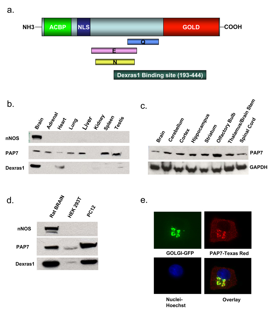
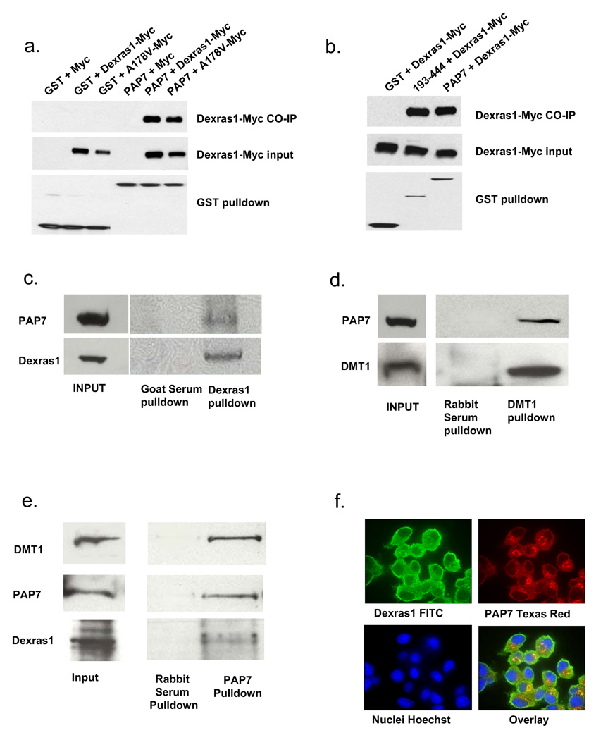
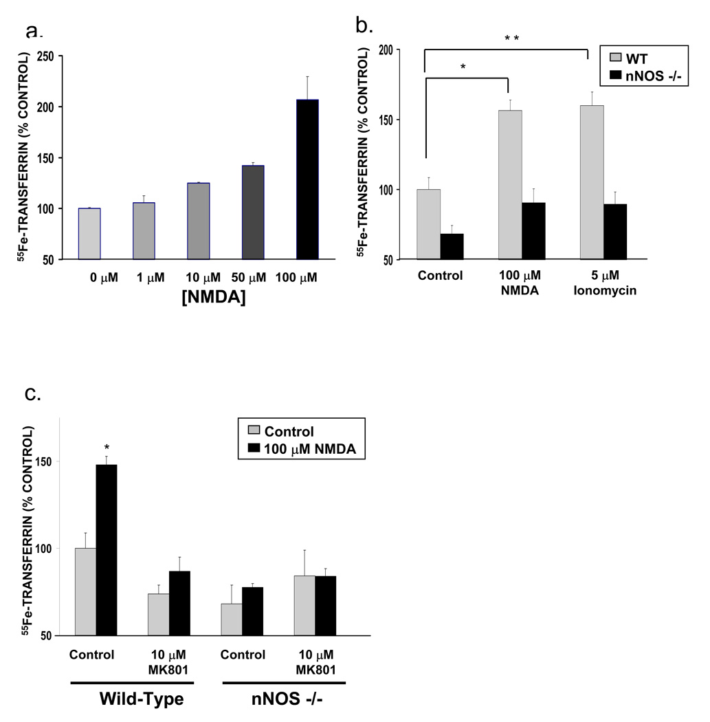
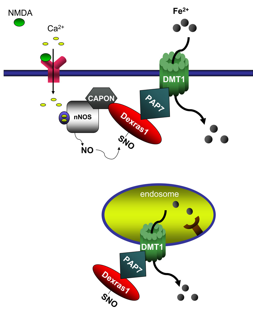
Dexras1 is a 30 kDa G protein in the Ras subfamily whose discovery was based on its pronounced inducibility by the glucocorticoid dexamethasone. It binds to neuronal nitric oxide synthase (nNOS) via the adaptor protein CAPON, eliciting S-nitrosylation and activation of Dexras1. We report that Dexras1 binds to the peripheral benzodiazepine receptor-associated protein (PAP7), a protein of unknown function that binds to cyclic AMP-dependent protein kinase and the peripheral benzodiazepine receptor. PAP7 in turn binds to the divalent metal transporter (DMT1), an iron import channel. We have identified a signaling cascade in neurons whereby stimulation of NMDA receptors activates nNOS, leading to S-nitrosylation and activation of Dexras1, which, via PAP7 and DMT1, physiologically induces iron uptake. As selective iron chelation prevents NMDA neurotoxicity in cortical cultures, the NMDA-NO-Dexras1-PAP7-DMT1-iron uptake signaling cascade also appears to mediate NMDA neurotoxicity.


Figures

Figure 1
Figure 1. Characterization of PAP7
(a) Schematic diagram of PAP7. An Acyl-CoA Binding Protein Domain (ACBP) and bipartite nuclear localization signal (NLS) are located at the N-terminus, while a Golgi Dynamics (GOLD) Domain resides at the C-terminus. The middle of the protein contains asparagine (N), glutamate (E) and glutamine (Q)-rich domains. The Dexras1 binding domain is located between amino acids 193–444 as determined by yeast two-hybrid analysis. (b) PAP7 is occurs in all major tissues except kidney, while nNOS is brain-specific, and Dexras1 is predominantly in the brain, with moderate levels in the heart and testis (c) PAP7 is present at similar levels in different regions of the brain. (d) Dexras1 and PAP7 are present in high levels in PC12 cells, with small levels in HEK 293T cells. (e) Undifferentiated PC12 cells were transfected with a Golgi marker and stained for endogenous PAP7, revealing that PAP7 is localized to both the Golgi apparatus and the cytosol.
Figure 2
Figure 2. Dexras1, PAP7 and DMT1 physiologically interact
(a) HEK 293T cells were transfected either with GST or GST-PAP7 and a Myc-tagged Dexras1 construct. A178V is a constitutively active form of Dexras1. Dexras1 and A178V specifically interact with PAP7. (b) HEK 293T cells were transfected either with GST or GST-PAP7 or GST-PAP7-193–444 and a Myc-tagged Dexras1 construct, showing that Dexras1 is able to bind to PAP7-193–444, as originally shown by yeast-two-hybrid analysis. (c) Immunoprecipitation experiments were performed using undifferentiated PC12 cells with a Dexras1 antibody, showing co-precipitation of PAP7. (d) Immunoprecipitation experiments were performed using undifferentiated PC12 cells with a DMT1 antibody, showing co-precipitation of PAP7. (e) Immunoprecipitation experiments were performed using mouse brain lysate with a PAP7 antibody, showing simultaneous immunoprecipitation of Dexras1 and DMT1. (f) Undifferentiated PC12 cells were fixed and stained for endogenous Dexras1 and PAP7. Dexras1 is shown to be localized to the cytosol and plasma membrane, while PAP7 is localized to the cytosol and Golgi apparatus.
Figure 3
Figure 3. Dexras1 is S-nitrosylated and NTBI uptake is increased in PC12 cells after treatment with NO donors
(a) HEK 293T cells were transfected with Dexras1 or Dexras1-A178V in the presence or absence of PAP7. NTBI uptake was measured as described in the Materials and Methods. (*, p<0.05, **, p<0.01) (b) Dexras1 in undifferentiated PC12 cells is S-nitrosylated by GSNO in a concentration-dependent manner (c) GSNO treatment for 3h leads to a concentration-dependent increase NTBI uptake in undifferentiated PC12 cells, as does (d) SNP for 1h and (e) DETA NONOate for 4h. (All NTBI uptake experiments were repeated three times, each sample in triplicate. Comparison with two-tailed student’s t-test; error bars represent SEM).
Figure 4
Figure 4. NO-mediated NTBI uptake is mediated by S-nitrosylation of Dexras1
(a) HEK 293T cells were transfected with GST or GST-PAP7 Dexras1-C11S-myc, a nitrosylation-dead mutant, which is able to bind specifically to PAP7. (b) HEK 293T cells were transfected with PAP7 and either wild-type Dexras1 or the C11S mutant. After transfection, the cells were treated with 100 µM GSNO for 3h and NTBI uptake was measured (*, p<0.01). GSNO treatment up-regulates NTBI uptake in cells containing wild-type Dexras1, but not the C11S mutant. (c) Undifferentiated PC12 cells were transfected with either control RNAi or Dexras1 RNAi (mock is untransfected). Dexras1 protein levels was fully depleted after transfection with Dexras1 RNAi, but not in control or mock-transfected cells (d) Undifferentiated PC12 cells were transfected with either control RNAi or Dexras1 RNAi, treated with 100 µM GSNO for 3h and NTBI uptake was measured (*, p<0.01). GSNO treatment up-regulates NTBI uptake in control cells, but not in cells depleted of Dexras1. (All NTBI uptake experiments were repeated three times, each sample in triplicate. Comparison with two-tailed student’s t-test; error bars represent SEM).
Figure 5
Figure 5. Glutamate-NMDA neurotransmission increases NTBI uptake, which is abolished in nNOS knockout mice
(a) NMDA stimulation increases NTBI uptake in primary cortical neurons in a concentration-dependent manner. (b) NTBI uptake in primary cortical neurons is increased by both NMDA stimulation and ionomycin treatment, a calcium ionophore. This up-regulation is not seen in primary cortical neurons from nNOS knockout mice (*, p<0.05, **, p<0.01) and (c) is abolished by pretreatment with MK801, an NMDA receptor antagonist (*, p<0.005). (All NTBI uptake experiments were repeated three times, each sample in triplicate. Comparison with two-tailed student’s t-test; error bars represent SEM).
Figure 6
Figure 6. Glutamate-NMDA neurotransmission increases Tf-iron uptake, which is abolished in nNOS knockout mice
(a) Tf-mediated iron uptake is increased in a dose-dependent manner with NMDA treatment in primary cortical neurons. (b) Tf-mediated iron uptake is increased by both NMDA and ionomycin (*, p<0.01, **, p<0.01). This up-regulation is not seen in primary cortical neurons from nNOS knockout mice and (c) is abolished by pre-treatment with MK801 (*, p<0.005). No increase in uptake is seen in neurons from nNOS knockout mice. (All Tf uptake experiments were repeated three times, each sample in triplicate. Comparison with two-tailed student’s t-test; error bars represent SEM).
Figure 7
Figure 7. Physiological and Pathophysiological Relevance of Iron Uptake
(a) Rate of heme biosynthesis was measured using incorporation of 55Fe into heme and was increased after NMDA treatment (*, p<0.01). (b) NTBI uptake is significantly increased after treatment with an excitotoxic concentration of NMDA. (c) An increase in HPF fluorescence was observed after treatment with NMDA, indicating an increase in ROS formation. This increase was abolished with pre-treatment of the neurons with SIH. (d) Treatment of primary cortical neurons with 300 µM NMDA elicits a high level of neurotoxicity, which is attenuated by the pre-treatment with either 10 µM MK801, an NMDA receptor antagonist, or SIH, an iron chelator.
Figure 8
Figure 8. Dexras1, PAP7 and DMT1 mediate both NTBI and Tf-mediated iron uptake
A model of a signaling cascade whereby glutamate-NMDA neurotransmission regulates cellular iron homeostasis. Glutamate-NMDA stimulation leads to the activation of nNOS and, via the scaffolding protein CAPON, to S-nitrosylation of Dexras1, which interacts with PAP7 and DMT1 both at the endosome and the plasma membrane, thus influencing cellular iron uptake in the cell.

References

    1. Blumer JB, Cismowski MJ, Sato M, Lanier SM. AGS proteins: receptor-independent activators of G-protein signaling. Trends Pharmacol. Sci. 2005;26:470–476. - PubMed
    1. Bredt DS, Snyder SH. Nitric oxide: a physiologic messenger molecule. Annu. Rev. Biochem. 1994;63:175–195. - PubMed
    1. Brenman JE, Chao DS, Gee SH, McGee AW, Craven SE, Santillano DR, Wu Z, Huang F, Xia H, Peters MF, Froehner SC, Bredt DS. Interaction of nitric oxide synthase with the postsynaptic density protein PSD-95 and alpha1-syntrophin mediated by PDZ domains. Cell. 1996;84:757–767. - PubMed
    1. Brenman JE, Christopherson KS, Craven SE, McGee AW, Bredt DS. Cloning and characterization of postsynaptic density 93, a nitric oxide synthase interacting protein. J. Neurosci. 1996;16:7407–7415. - PMC - PubMed
    1. Cellier M, Prive G, Belouchi A, Kwan T, Rodrigues V, Chia W, Gros P. Nramp defines a family of membrane proteins. Proc. Natl. Acad. Sci. U. S. A. 1995;92:10089–10093. - PMC - PubMed

Publication types

MeSH terms

Substances