. 2006 Dec;80(6):1224-30.
doi: 10.1189/jlb.0406287. Epub 2006 Aug 14.

Francisella tularensis LVS evades killing by human neutrophils via inhibition of the respiratory burst and phagosome escape

Affiliations

Francisella tularensis LVS evades killing by human neutrophils via inhibition of the respiratory burst and phagosome escape

Ramona L McCaffrey et al. J Leukoc Biol. 2006 Dec.

Abstract

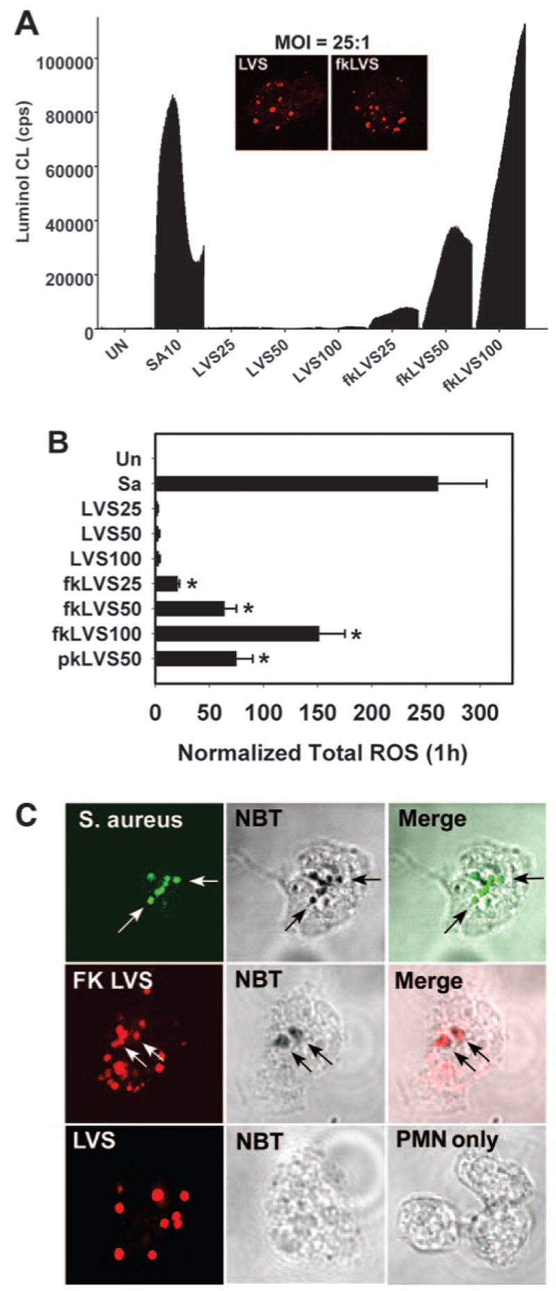
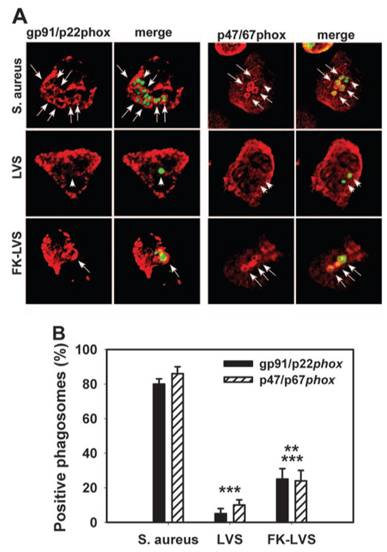
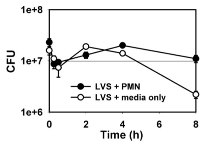
Francisella tularensis is a Gram-negative bacterium and the causative agent of tularemia. Recent data indicate that F. tularensis replicates inside macrophages, but its fate in other cell types, including human neutrophils, is unclear. We now show that F. tularensis live vaccine strain (LVS), opsonized with normal human serum, was rapidly ingested by neutrophils but was not eliminated. Moreover, evasion of intracellular killing can be explained, in part, by disruption of the respiratory burst. As judged by luminol-enhanced chemiluminescence and nitroblue tetrazolium staining, neutrophils infected with live F. tularensis did not generate reactive oxygen species. Confocal microscopy demonstrated that NADPH oxidase assembly was disrupted, and LVS phagosomes did not acquire gp91/p22(phox) or p47/p67(phox). At the same time, F. tularensis also impaired neutrophil activation by heterologous stimuli such as phorbol esters and opsonized zymosan particles. Later in infection, LVS escaped the phagosome, and live organisms persisted in the neutrophil cytosol for at least 12 h. To our knowledge, our data are the first demonstration of a facultative intracellular pathogen, which disrupts the oxidative burst and escapes the phagosome to evade elimination inside neutrophils, and as such, our data define a novel mechanism of virulence.


Figures

Fig. 1
Fig. 1
LVS does not trigger a respiratory burst in PMN. (A) Representative experiment depicting the total PMN oxidative burst over 1 h, as measured by luminol-ECL of uninfected cells (UN), PMN exposed to S. aureus MOI 10:1 (SA10), or live or formalin-killed (fk) LVS at MOI 25:1, 50:1, or 100:1. Data indicate CL as counts per second (cps) of triplicate samples from a representative experiment. (Inset) Confocal sections demonstrate equivalent phagocytosis of live and and fkLVS (both in red). (B) Total PMN ROS measured using luminol. Data are the mean ± SEM of three independent experiments performed in triplicate. pkLVS, Periodate-killed LVS. *, P < 0.05, versus LVS. (C) NBT staining of resting PMN or cells infected with S. aureus (green, MOI 5:1), LVS, or FK-LVS (red, both MOI 35:1) for 1 h. Arrows indicate formazan-positive phagosomes.
Fig. 2
Fig. 2
LVS phagosomes do not acquire NADPH oxidase subunits. (A) Confocal sections of PMN infected for 15 min show S. aureus, LVS, and formalin-killed (FK)-LVS in green and NAPDH oxidase components in red. Arrows and arrowheads indicate NADPH oxidase-positive and negative phagosomes, respectively. Comparable data were obtained at 5 min and 30 min. (B) Fraction of S. aureus, LVS, or FK-LVS phagosomes, which acquired gp91/p22phox or p47/67phox. Data are the average ± SEM from at least three independent experiments. **, P < 0.010, versus LVS; ***, P ≤ 0.001, versus S. aureus.
Fig. 3
Fig. 3
LVS evades killing inside PMN. Viability of LVS in media alone (RPMI 1640+10% AS) versus bacteria inside PMN (MOI 20:1). Data are the mean ± SEM for triplicate samples from one experiment representative of three independent determinations.
Fig. 4
Fig. 4
LVS survival is not impaired in PMA-activated PMN. (A) Resting and PMA-activated PMN (200 nM PMA, 5 min) were infected with LVS at MOI 20:1, and viable intracellular bacteria were quantified at the indicated time-points. Data are the mean ± SEM for triplicate samples from one experiment representative of three determinations. (B) LVS phagosomes (arrowheads) do not acquire p47/67phox in PMA-activated PMN. Data shown are representative of three independent experiments. (C) LVS impairs cell activation by PMA. PMN in luminol-containing buffer (B; 1st stimulus) were left untreated (B–B) or exposed to LVS (MOI 50:1; B–LVS) or 200 nM PMA (B–PMA) as indicated. Alternatively, cells were activated with PMA 10 min before (PMA–LVS) or 10 min after infection with LVS (LVS–PMA). Data are mean CL ± SD of triplicate samples from one experiment representative of four. (D) Same as C except formalin-killed (FK) LVS was used instead of live bacteria. Data are the mean ± SD of triplicate samples from one representative experiment.
Fig. 5
Fig. 5
LVS inhibits PMN activation by OpZ. (A) PMN were left in buffer or infected with LVS (MOI 50:1) and/or OpZ (ZYM, MOI 4:1) as indicated. Data are the mean ± SD of triplicate samples from one experiment representative of three. (B) PMN were left in buffer or activated with formalin-killed LVS (FK) and/or OpZ (ZYM) as indicated. Data are the mean ± SD of triplicate samples from a representative experiment.
Fig. 6
Fig. 6
LVS escapes PMN phagosomes. TEM images depict LVS entry into PMN (A) as well as bacteria at 15 min (B) and 4 (C), 6 (D and E), 9 (F and G), and 12 h postinfection (hpi; H). Arrow in A indicates a forming phagosome. Other black arrows indicate intact phagosomes. Black arrowheads indicate disrupted phagosome membranes. White arrows indicate bacteria free in the cytosol. (I) Overall PMN morphology at 9 hpi. (J) Time course of phagosome escape. Data are the mean ± SEM (n=60) for each time-point.

Similar articles

Cited by

References

    1. Nauseef WM. Assembly of the phagocyte NADPH oxidase. Histochem Cell Biol. 2004;122:277–291. - PubMed
    1. Curnutte JT. Chronic granulomatous disease: the solving of a clinical riddle at the molecular level. Clin Immunol Immunopathol. 1993;67:S2–S15. - PubMed
    1. Oyston PCF, Sjostedt A, Titball RW. Tularemia: bioterrorism defence renews interest in Francisella tularensis. Nat Rev Microbiol. 2004;2:967–978. - PubMed
    1. Santic M, Molmeret M, Klose KE, Abu Kwaik Y. Francisella tularensis travels a novel, twisted road within macrophages. Trends Microbiol. 2006;14:37–44. - PubMed
    1. Proctor RA, White JD, Ayala E, Canonico PG. Phagocytosis of Francisella tularensis by rhesus monkey peripheral leukocytes. Infect Immun. 1975;11:146–152. - PMC - PubMed

Publication types

MeSH terms