. 2006 Oct;26(20):7707-18.
doi: 10.1128/MCB.00849-06. Epub 2006 Aug 14.

Syntenin-1 is a new component of tetraspanin-enriched microdomains: mechanisms and consequences of the interaction of syntenin-1 with CD63

Affiliations

Syntenin-1 is a new component of tetraspanin-enriched microdomains: mechanisms and consequences of the interaction of syntenin-1 with CD63

Nadya Latysheva et al. Mol Cell Biol. 2006 Oct.

Abstract

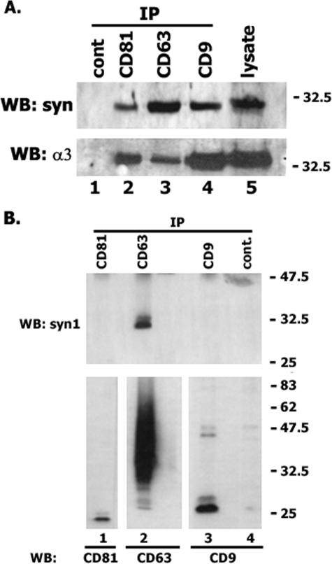
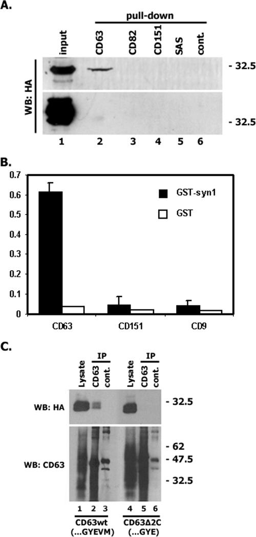
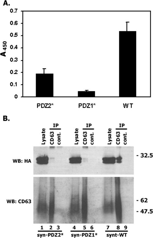
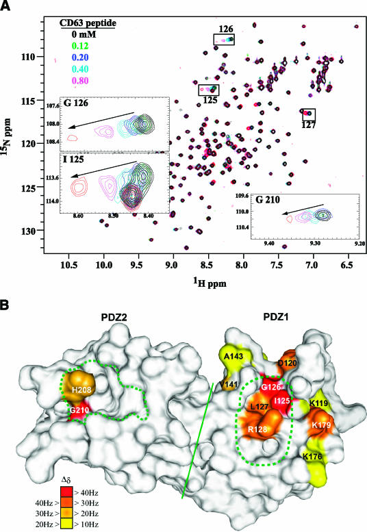
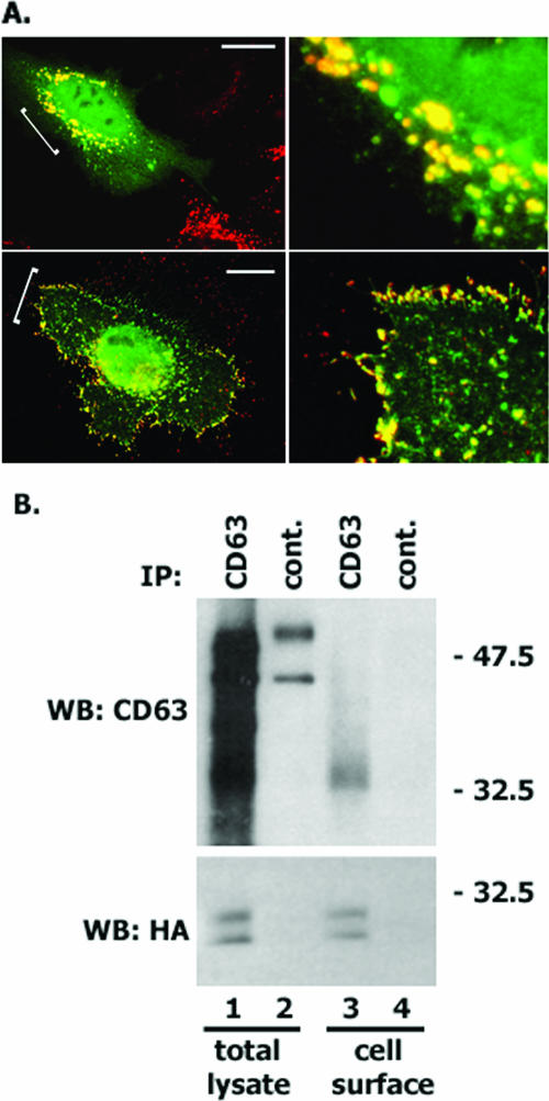
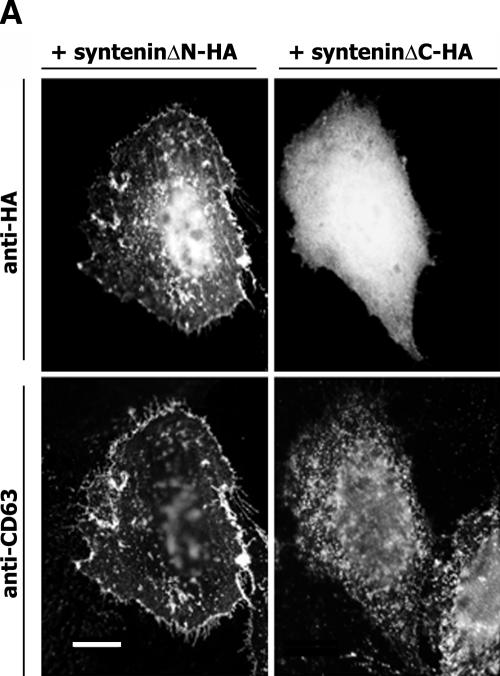
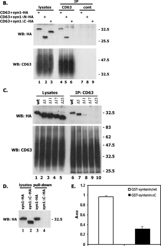
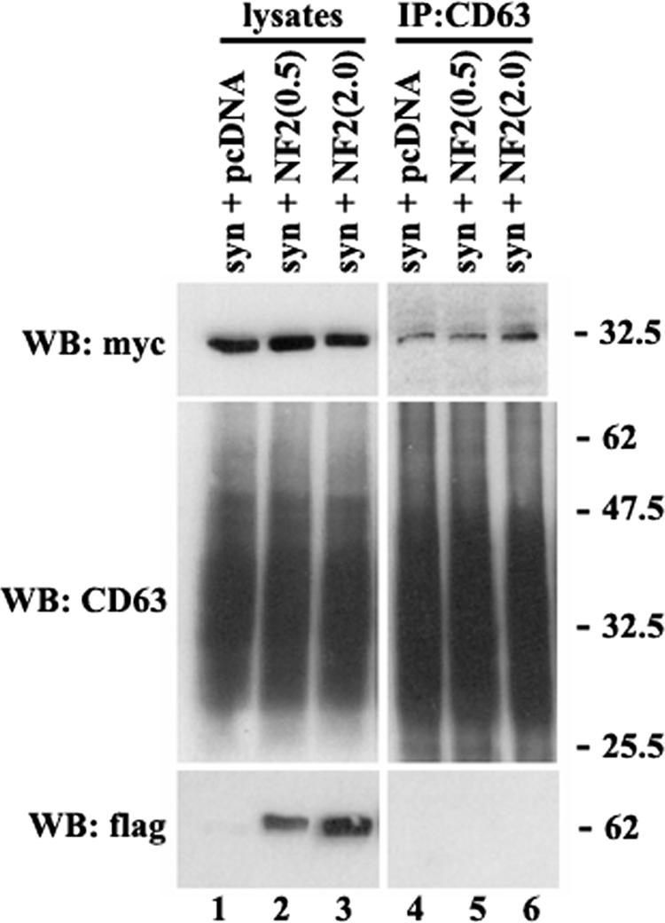
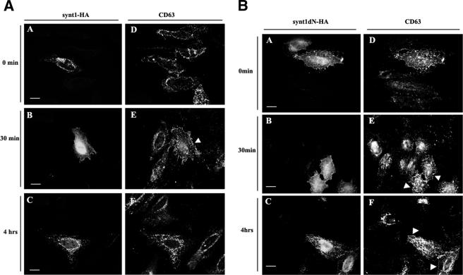
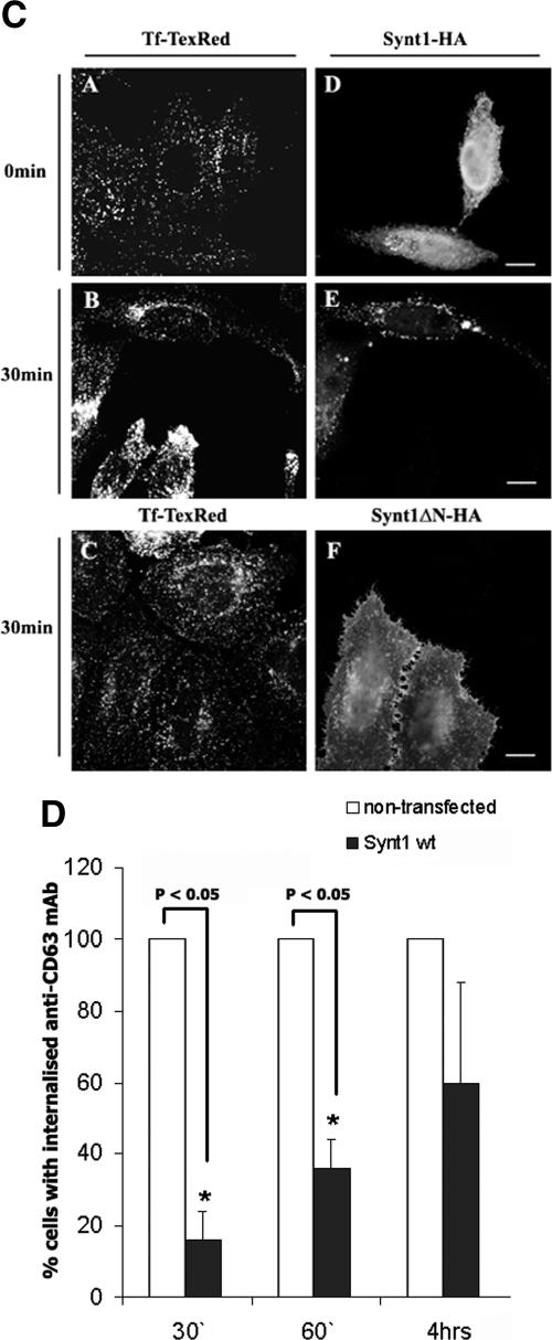
Tetraspanins are clustered in specific microdomains (named tetraspanin-enriched microdomains, or TERM) in the plasma membrane and regulate the functions of associated transmembrane receptors, including integrins and receptor tyrosine kinases. We have identified syntenin-1, a PDZ domain-containing protein, as a new component of TERM and show that syntenin-1 specifically interacts with the tetraspanin CD63. Detailed biochemical and heteronuclear magnetic resonance spectroscopy (NMR) studies have demonstrated that the interaction is mediated by the C-terminal cytoplasmic region of the tetraspanin and the PDZ domains of syntenin-1. Upon interaction, NMR chemical shift perturbations were predominantly localized to residues around the binding pocket of PDZ1, indicating a specific mode of recognition of the cytoplasmic tail of CD63. In addition, the C terminus of syntenin-1 has a stabilizing role in the CD63-syntenin-1 association, as deletion of the last 17 amino acids abolished the interaction. The CD63-syntenin-1 complex is abundant on the plasma membrane, and the elevated expression of the wild-type syntenin-1 slows down constitutive internalization of the tetraspanin. Furthermore, internalization of CD63 was completely blocked in cells expressing a syntenin-1 mutant lacking the first 100 amino acids. Previous results have shown that CD63 is internalized via AP-2-dependent mechanisms. Hence, our data indicate that syntenin-1 can counteract the AP-2-dependent internalization and identify this tandem PDZ protein as a new regulator of endocytosis.


Figures

FIG. 1.
FIG. 1.
Syntenin-1 is associated with the tetraspanin-enriched microdomains. MDA-MB-231 cells were lysed in the buffer containing either 0.8% Brij-0.2% Triton X-100 (A) or 0.5% Brij-0.5% Triton X-100 (B). Protein complexes were immunoprecipitated with the anti-CD9 MAb Syb1 (lane 2), anti-CD63 MAb 6H1 (lane 3), or anti-CD81 MAb M38 (lane 4). An irrelevant MAb (3E1) was used as a negative control (lane 1). The protein lysate (lane 5) was used as a positive control. Immunocomplexes were separated in 12% SDS-PAGE and transferred to nitrocellulose membranes. Membranes were developed with the polyclonal Abs to syntenin-1, the α3 integrin subunit, or MAbs to CD9 (C9-BB), CD81 (JS-64), or CD63 (1B5). WB, Western blot.
FIG. 2.
FIG. 2.
The C-terminal cytoplasmic region mediates the interaction of CD63 with syntenin-1. (A) 293T cells were transiently transfected with the plasmids encoding syntenin-1-HA (or syntenin-2-HA) and 48 h later lysed in 0.8% Brij-0.2% Triton X-100. Retention of syntenins by immobilized tetraspanin peptides was analyzed by Western blotting (WB). (B) Interaction of biotinylated peptides (0.25 μg) with the immobilized GST-syntenin-1 (or GST) was analyzed by ELISA as described in Materials and Methods. (C) CHO cells were transiently transfected with the plasmids encoding syntenin-1-HA and human CD63 (CD63h) constructs. After 48 h, cells were lysed in 0.8% Brij-0.2% Triton X-100. Protein complexes were immunoprecipitated (IP) with the anti-CD63 MAb (6H1) or an irrelevant MAb (3E1). The protein lysates (lanes 1 and 4) were used as positive controls. Immunocomplexes were separated in 12% SDS-PAGE and transferred on nitrocellulose membranes. Membranes were probed with the anti-HA polyclonal Abs or anti-CD63 MAb (1B5). wt, wild type.
FIG. 3.
FIG. 3.
Both PDZ domains contribute to binding of CD63 with syntenin-1. (A) The interaction of biotinylated CD63 peptide (0.25 μg) with the immobilized GST-tagged syntenins was analyzed by ELISA as described in Materials and Methods. WT, wild type. (B) CHO cells were transiently transfected with the plasmids encoding human CD63 and syntenin-1-HA (or PDZ domain mutants of syntenin-1). The association between the proteins was analyzed as described in the legend to Fig. 2C. syn-PDZ1* and syn-PDZ2* indicate the Gly126→Asp and Gly210→Glu mutations in the interacting loops of PDZ1 and PDZ2, respectively.
FIG. 4.
FIG. 4.
CD63 interacts specifically with the PDZ1 domain of syntenin-1. (A) Superposition of six two-dimensional 1H-15N-HSQC spectra of uniformly 15N-labeled syntenin-1 PDZ12 (200 μΜ), which are color coded, according to the concentration of titrated CD63 C-terminal peptide. Shown in the insets are traces for residues exhibiting chemical shift changes greater than 40 Hz. (B) The locations of PDZ12 residues involved in binding to the C-terminal peptide of CD63, based on chemical shift perturbation, are indicated on the surface of syntenin-1 PDZ12 monomer (1N99.pdb). The residues displaying combined chemical shift changes (Δδ) greater than the mean value plus one (10 Hz) are color coded as indicated. The canonical peptide binding pocket in PDZ2 (traced from 1W9E.pdb) and a corresponding putative pocket in PDZ1 are represented within green dotted lines. The bulk of the two domains are separated by a solid line.
FIG. 5.
FIG. 5.
Codistribution of CD63 with syntenin-1. CD63 is associated with the cell surface pool of syntenin-1. (A) CD63 and syntenin-1 are colocalized at the plasma membrane and vesicular organelles. Upper panels. HeLa cells were transiently transfected with the plasmid encoding syntenin-HA and 48 h later processed for immunofluorescence staining. The MAbs 6H1 (IgG1) and F7 (IgG2a) were used to examine the distribution of CD63 and HA-tagged syntenin-1. Staining was visualized using Alexa 594-conjugated goat anti-mouse IgG1 Ab and Alexa 488-conjugated goat anti-mouse IgG2a. The scale bar represents 10 μΜ. The right panel represents a magnified image of the area marked with a bracket on the left image. Note, some of the syntenin-1-positive vesicular organelles are devoid of CD63. Lower panels. Transfected HeLa cells were processed for immunofluorescence staining as described above except that they were preincubated with the anti-CD63 MAb for 1 h at 4°C prior to fixation and permeabilization. (B) CHO cells were transiently transfected with the plasmids encoding syntenin-HA and human CD63 and 48 h later were preincubated with the anti-CD63 MAb for 1 h at 4°C. After removal of unbound MAb, the association between CD63 and syntenin-1 was analyzed as described in the legend to Fig. 2C.
FIG.6.
FIG.6.
The C-terminal part of syntenin-1 stabilizes its association with CD63. (A) Distribution of deletion mutants of syntenin-1. HeLa cells were transiently transfected with the plasmids encoding syntenin-1ΔC-HA or syntenin-1ΔN-HA and 48 h later processed for immunofluorescence staining. The MAbs 6H1 (IgG1) and F7 (IgG2a) were used to examine the distribution of CD63 and HA-tagged syntenin-1. Staining was visualized using Alexa 594-conjugated goat anti-mouse IgG1 Ab and Alexa 488-conjugated goat anti-mouse IgG2a. The scale bar represents 10 μM. (B and C) Cos7 cells were transiently transfected with the plasmids encoding various HA-tagged syntenin-1 constructs and human CD63. After 48 h, cells were lysed in 0.5% Brij-0.5% Triton X-100. Protein complexes were immunoprecipitated (IP) with the anti-CD63 MAb (6H1) or an irrelevant MAb (3E1). The protein lysates (lanes 1 to 3 in panel B and lanes 1 to 5 in panel C) were used as positive controls. Immunocomplexes were separated in 11 SDS-PAGE and transferred on nitrocellulose membranes. Membranes were probed with the anti-HA polyclonal Abs or anti-CD63 MAb (1B5). (D) 293T cells were transiently transfected with the plasmids encoding syntenin-1-HA and syntenin-1ΔC-HA and 48 h later lysed in 0.5% Brij-0.5% Triton X-100. Retention of syntenins by immobilized CD63-C peptides was analyzed by Western blotting (WB) with the anti-HA polyclonal Abs. (E). Interaction of biotinylated CD63-C peptide (0.25 μg) with the immobilized GST-tagged syntenins was analyzed by ELISA as described in Materials and Methods. wt, wild type.
FIG.6.
FIG.6.
The C-terminal part of syntenin-1 stabilizes its association with CD63. (A) Distribution of deletion mutants of syntenin-1. HeLa cells were transiently transfected with the plasmids encoding syntenin-1ΔC-HA or syntenin-1ΔN-HA and 48 h later processed for immunofluorescence staining. The MAbs 6H1 (IgG1) and F7 (IgG2a) were used to examine the distribution of CD63 and HA-tagged syntenin-1. Staining was visualized using Alexa 594-conjugated goat anti-mouse IgG1 Ab and Alexa 488-conjugated goat anti-mouse IgG2a. The scale bar represents 10 μM. (B and C) Cos7 cells were transiently transfected with the plasmids encoding various HA-tagged syntenin-1 constructs and human CD63. After 48 h, cells were lysed in 0.5% Brij-0.5% Triton X-100. Protein complexes were immunoprecipitated (IP) with the anti-CD63 MAb (6H1) or an irrelevant MAb (3E1). The protein lysates (lanes 1 to 3 in panel B and lanes 1 to 5 in panel C) were used as positive controls. Immunocomplexes were separated in 11 SDS-PAGE and transferred on nitrocellulose membranes. Membranes were probed with the anti-HA polyclonal Abs or anti-CD63 MAb (1B5). (D) 293T cells were transiently transfected with the plasmids encoding syntenin-1-HA and syntenin-1ΔC-HA and 48 h later lysed in 0.5% Brij-0.5% Triton X-100. Retention of syntenins by immobilized CD63-C peptides was analyzed by Western blotting (WB) with the anti-HA polyclonal Abs. (E). Interaction of biotinylated CD63-C peptide (0.25 μg) with the immobilized GST-tagged syntenins was analyzed by ELISA as described in Materials and Methods. wt, wild type.
FIG. 7.
FIG. 7.
Elevated expression of NF-2/merlin does not interfere with the assembly of the CD63-syntenin-1 complex. Cos7 cells were transiently transfected with the plasmid encoding syntenin-1-myc (0.5 μg) and indicated amounts of the plasmid encoding Flag-tagged human NF-2/merlin. After 48 h, cells were lysed in 0.5% Brij-0.5% Triton X-100. Protein complexes were immunoprecipitated (IP) with the anti-CD63 MAb (6H1). The protein lysates (lanes 1 to 3) were used as positive controls. Immunocomplexes were separated in 11 to 12% SDS-PAGE and transferred on nitrocellulose membranes. Membranes were probed with the anti-myc and anti-Flag polyclonal Abs or anti-CD63 MAb (1B5). WB, Western blot.
FIG.8.
FIG.8.
Syntenin-1 regulates internalization of CD63. HeLa cells were transiently transfected with the plasmids encoding HA-tagged variants of syntenin-1 (A to C). Forty-eight hours later, cells were surface labeled with the anti-CD63 MAb (A and B) or Texas Red-labeled transferrin (Tf-TexRed) (C) for 1 h at 4°C and then placed to 37°C for indicated time intervals. Cells were subsequently processed for the immunofluorescence staining as described in the legend for Fig. 5A, upper panels. Arrowheads point to cells expressing syntenin-1 constructs. Note the distinct peripheral staining with anti-CD63 MAb. The scale bar represents 10 μM. (D) Quantitative analysis of three independent internalization experiments with 70 to 80 transfected cells counted in each experiment. Shown are the results of the experiments with cells expressing wild-type (wt) syntenin-1. Expression of the syntenin-1ΔN mutant completely blocked internalization of the anti-CD63 MAb. Error bars represent standard errors of the means.
FIG.8.
FIG.8.
Syntenin-1 regulates internalization of CD63. HeLa cells were transiently transfected with the plasmids encoding HA-tagged variants of syntenin-1 (A to C). Forty-eight hours later, cells were surface labeled with the anti-CD63 MAb (A and B) or Texas Red-labeled transferrin (Tf-TexRed) (C) for 1 h at 4°C and then placed to 37°C for indicated time intervals. Cells were subsequently processed for the immunofluorescence staining as described in the legend for Fig. 5A, upper panels. Arrowheads point to cells expressing syntenin-1 constructs. Note the distinct peripheral staining with anti-CD63 MAb. The scale bar represents 10 μM. (D) Quantitative analysis of three independent internalization experiments with 70 to 80 transfected cells counted in each experiment. Shown are the results of the experiments with cells expressing wild-type (wt) syntenin-1. Expression of the syntenin-1ΔN mutant completely blocked internalization of the anti-CD63 MAb. Error bars represent standard errors of the means.

References

    1. Adey, N. B., L. Huang, P. A. Ormonde, M. L. Baumgard, R. Pero, D. V. Byreddy, S. V. Tavtigian, and P. L. Bartel. 2000. Threonine phosphorylation of the MMAC1/PTEN PDZ binding domain both inhibits and stimulates PDZ binding. Cancer Res. 60:35-37. - PubMed
    1. Amezcua, C. A., S. M. Harper, J. Rutter, and K. H. Gardner. 2002. Structure and interactions of PAS kinase N-terminal PAS domain: model for intramolecular kinase regulation. Structure 10:1349-1361. - PubMed
    1. Berditchevski, F. 2001. Complexes of tetraspanins with integrins: more than meets the eye. J. Cell Sci. 115:4143-4151. - PubMed
    1. Berditchevski, F., G. Bazzoni, and M. E. Hemler. 1995. Specific association of CD63 with the VLA-3 and VLA-6 integrins. J. Biol. Chem. 270:17784-17790. - PubMed
    1. Berditchevski, F., S. Chang, J. Bodorova, and M. E. Hemler. 1997. Generation of monoclonal antibodies to integrin-associated proteins. Evidence that alpha3beta1 complexes with EMMPRIN/basigin/OX47/M6. J. Biol. Chem. 272:29174-29180. - PubMed

Publication types

MeSH terms

LinkOut - more resources