

Temporal horn index and volume of medial temporal lobe atrophy using a new semiautomated method for rapid and precise assessment
- PMID: 16908557
- PMCID: PMC7977513
Temporal horn index and volume of medial temporal lobe atrophy using a new semiautomated method for rapid and precise assessment
Abstract
Background and purpose: Quantitative markers of Alzheimer disease (AD), particularly in the early stages, are needed for clinical assessment and monitoring. We have evaluated a novel method to segment and visualize the ventricular system and obtain volumetric measures thereof. The temporal horn volume (THV) and index in patients with mild cognitive impairment (MCI) and in those with AD were evaluated.
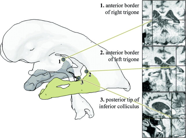
Methods: High-resolution T1-weighted volume imaging was performed in 52 subjects (21 patients with MCI, 10 with AD, and 21 healthy control subjects). An interactive watershed transformation and semiautomated histogram analysis were implemented to produce segmented THV and temporal horn indices (THI) (ratio of THV to lateral ventricular volume).
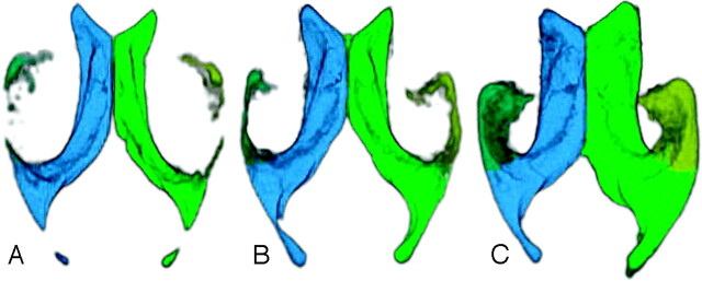
Results: Cerebral ventricular and temporal horn size could be semiautomatically quantified from all 52 datasets. The method was fast and rater-independent. Qualitative ventricular inspections using surface rendering shading could uncover atrophic process with enlargement of the whole and especially temporal horn volume. Both THV and THI of patients with AD were significantly larger than those of patients with MCI or control subjects (P < .005). There was no significant difference in THV and THI between patients with MCI or control subjects (P > .05). There was a significant correlation between the neuropsychologic performance and both THI and THV across groups (P < .01).
Conclusion: THV and THI could be used as markers of AD in the clinical environment and are expected to be helpful in monitoring therapeutic intervention.
Figures

Similar articles
-
Medial temporal lobe atrophy on MRI scans and the diagnosis of Alzheimer disease.Neurology. 2008 Dec 9;71(24):1986-92. doi: 10.1212/01.wnl.0000336925.79704.9f. Neurology. 2008. PMID: 19064880 Free PMC article.
-
Volumes of lateral temporal and parietal structures distinguish between healthy aging, mild cognitive impairment, and Alzheimer's disease.J Alzheimers Dis. 2011;26(4):719-34. doi: 10.3233/JAD-2011-101260. J Alzheimers Dis. 2011. PMID: 21709375
-
Medial temporal lobe atrophy and memory dysfunction as predictors for dementia in subjects with mild cognitive impairment.J Neurol. 1999 Jun;246(6):477-85. doi: 10.1007/s004150050387. J Neurol. 1999. PMID: 10431775
-
High-throughput, fully automated volumetry for prediction of MMSE and CDR decline in mild cognitive impairment.Alzheimer Dis Assoc Disord. 2009 Apr-Jun;23(2):139-45. doi: 10.1097/WAD.0b013e318192e745. Alzheimer Dis Assoc Disord. 2009. PMID: 19474571 Free PMC article.
-
Challenges of High-resolution Diffusion Imaging of the Human Medial Temporal Lobe in Alzheimer Disease.Top Magn Reson Imaging. 2010 Dec;21(6):355-65. doi: 10.1097/RMR.0b013e31823f6413. Top Magn Reson Imaging. 2010. PMID: 22158129 Free PMC article. Review.
Cited by
-
Hippocampo-Horn Percentage and Parietal Atrophy Score for Easy Visual Assessment of Brain Atrophy on Magnetic Resonance Imaging in Early- and Late-Onset Alzheimer's Disease.J Alzheimers Dis. 2021;84(3):1259-1266. doi: 10.3233/JAD-210372. J Alzheimers Dis. 2021. PMID: 34633317 Free PMC article.
-
Dementia in Down's syndrome: an MRI comparison with Alzheimer's disease in the general population.J Neurodev Disord. 2013 Aug 20;5(1):19. doi: 10.1186/1866-1955-5-19. J Neurodev Disord. 2013. PMID: 23962297 Free PMC article.
-
Cortical and Subcortical Grey and White Matter Atrophy in Myotonic Dystrophies Type 1 and 2 Is Associated with Cognitive Impairment, Depression and Daytime Sleepiness.PLoS One. 2015 Jun 26;10(6):e0130352. doi: 10.1371/journal.pone.0130352. eCollection 2015. PLoS One. 2015. PMID: 26114298 Free PMC article. Clinical Trial.
-
Influence of brain-derived neurotrophic factor and apolipoprotein E genetic variants on hemispheric and lateral ventricular volume of young healthy adults.Acta Neuropsychiatr. 2011 Jun;23(3):132-8. doi: 10.1111/j.1601-5215.2011.00546.x. Acta Neuropsychiatr. 2011. PMID: 21701702 Free PMC article.
-
Predicting the need for cerebrospinal fluid shunt implantation after spontaneous intracerebral hemorrhage: a challenging task.Front Neurol. 2023 Dec 7;14:1255477. doi: 10.3389/fneur.2023.1255477. eCollection 2023. Front Neurol. 2023. PMID: 38187155 Free PMC article.
References
-
- McKhann G, Drachman D, Folstein M, et al. Clinical diagnosis of Alzheimer’s disease: report of the NINCDS-ADRDA Work Group under the auspices of Department of Health and Human Services Task Force on Alzheimer’s Disease. Neurology 1984;34:939–44 - PubMed
-
- Kazee AM, Eskin TA, Lapham LW, et al. Clinicopathologic correlates in Alzheimer disease: assessment of clinical and pathologic diagnostic criteria. Alzheimer Dis Assoc Disord 1993;7:152–64 - PubMed
-
- Tierney MC, Fisher RH, Lewis AJ, et al. The NINCDS-ADRDA Work Group criteria for the clinical diagnosis of probable Alzheimer’s disease: a clinicopathologic study of 57 cases. Neurology 1988;38:359–64 - PubMed
-
- Seab JP, Jagust WJ, Wong ST, et al. Quantitative NMR measurements of hippocampal atrophy in Alzheimer’s disease. Magn Reson Med 1988;8:200–08 - PubMed
-
- Kesslak JP, Nalcioglu O, Cotman CW. Quantification of magnetic resonance scans for hippocampal and parahippocampal atrophy in Alzheimer’s disease. Neurology 1991;41:51–54 - PubMed
MeSH terms
LinkOut - more resources
Full Text Sources
Medical