

Creutzfeldt-Jakob disease: comparative analysis of MR imaging sequences
- PMID: 16908558
- PMCID: PMC7977534
Creutzfeldt-Jakob disease: comparative analysis of MR imaging sequences
Abstract
Background and purpose: MR imaging has played an increasingly important role in the diagnosis of Creutzfeldt-Jakob disease (CJD) since basal ganglia abnormalities on T2-weighted images have been described; thus, the aim of our study was to compare the value of different MR images in the diagnosis of CJD.
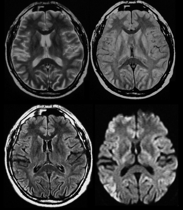
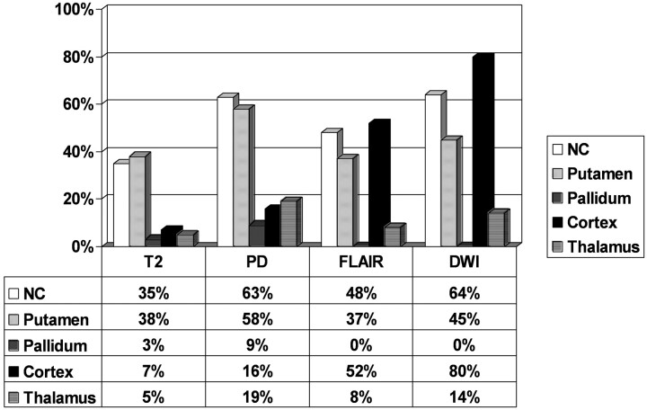
Methods: One hundred fifty-seven patients with CJD underwent MR imaging examinations. Ninety-two patients were neuropathologically confirmed, and 65 were clinically classified as having CJD through the CJD Surveillance Unit (probability of 95%). There was no standardized MR imaging protocol; thus, the examinations included 143 T2-weighted, 43 proton attenuation (PD)-weighted, 84 fluid-attenuated inversion recovery (FLAIR), and 44 diffusion-weighted images (DWI). The MR images were reviewed for pathologic changes of the basal ganglia, thalamus, and cerebral cortex.
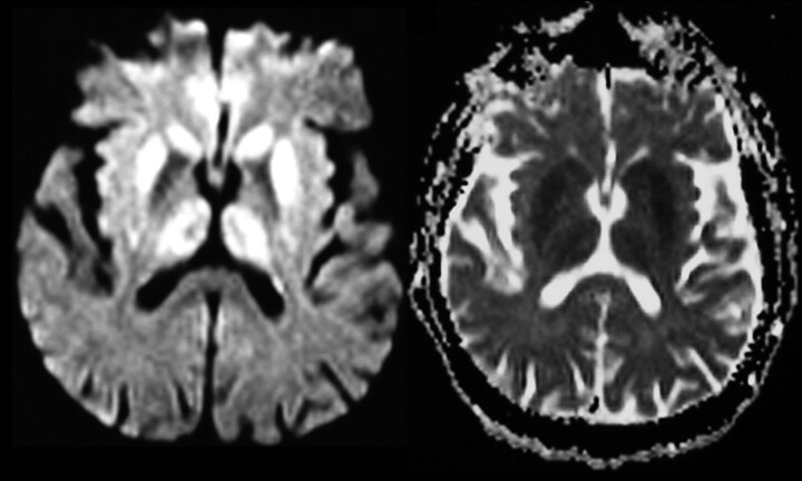
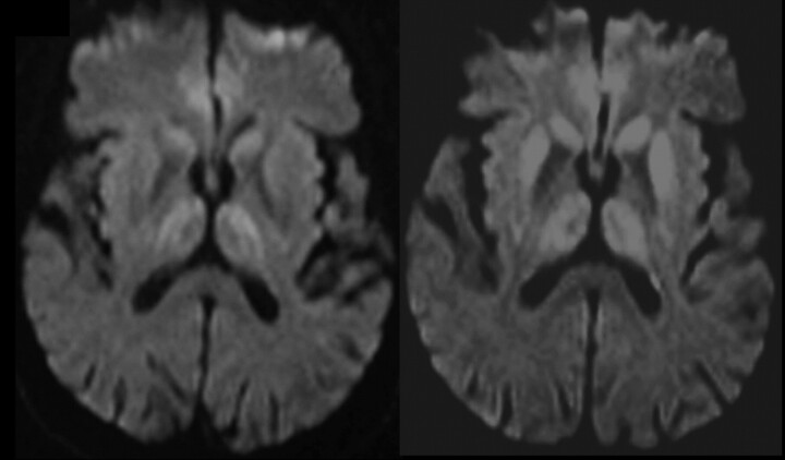
Results: Cortical abnormalities were present in 70 patients (45%) and were visible in 80% (35/44) of all available DWI examinations. The basal ganglia were affected in 94 patients (60%), in particular in the caudate nucleus; the most sensitive sequences were DWI (64%) and PD-weighted (63%). A thalamic involvement was more frequently diagnosed on PD-weighted images (19%) and DWI (14%) than on FLAIR or T2-weighted images.
Conclusion: PD-weighted images and DWI showed better results in the diagnosis of signal intensity changes in the basal ganglia compared with T2-weighted or FLAIR images; however, in the diagnosis of cortical changes, DWI was clearly superior. Our data suggest that DWI is the most sensitive MR imaging technique in the diagnosis of CJD.
Figures
References
-
- Masters CL, Harris JO, Gajdusek DC, et al. Creutzfeldt-Jakob disease: patterns of worldwide occurrence and the significance of familial and sporadic clustering. Ann Neurol 1979;5:177–88 - PubMed
-
- Kretzschmar HA, Ironside JW, DeArmond SJ, et al. Diagnostic criteria for sporadic Creutzfeldt-Jakob disease. Arch Neurol 1996;53:913–20 - PubMed
-
- Finkenstaedt M, Szudra A, Zerr I, et al. MR imaging of Creutzfeldt-Jakob disease. Radiology 1996;199:793–98 - PubMed
-
- Schroter A, Zerr I, Henkel K, et al. Magnetic resonance imaging in the clinical diagnosis of Creutzfeldt-Jakob disease. Arch Neurol 2000;57:1751–57 - PubMed
Publication types
MeSH terms
LinkOut - more resources
Full Text Sources
Medical