

Case Reports
Brain abscess formation: a delayed complication of carotid blowout syndrome treated by self-expandable stent-graft
Affiliations
- PMID: 16908577
- PMCID: PMC7977548
Item in Clipboard
Case Reports
Brain abscess formation: a delayed complication of carotid blowout syndrome treated by self-expandable stent-graft
AJNR Am J Neuroradiol.
2006 Aug.
Abstract
A patient with hypopharyngeal cancer developed carotid blowout syndrome (CBS) treated by self-expandable stent-graft in the left carotid artery. CT scan for progressive right hemiparesis 4 months later showed multiple left cerebral abscesses and left carotid thrombosis. Although deployment of stent-grafts for CBS can achieve initial hemostasis in patients with head-and-neck cancer, the placement of a stent-graft in a field of necrosis and infection is associated with poor long-term outcome. We recommend the use of prophylactic antibiotics if endovascular foreign materials are placed in a contaminated field.
Figures
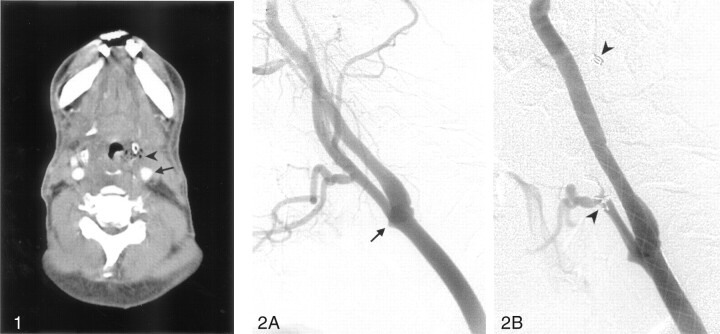
Axial CT of the neck with contrast medium shows slightly irregular axial contour of the left carotid bifurcation (arrow). An area of necrosis with gas bubbles is seen in the left pyriform sinus (arrowhead), adjacent to the left carotid artery.
Selective angiograms of the left carotid artery, with a lateral view. A, A pseudoaneurysm in the right distal common carotid artery is noted (arrow). B, A self-expandable Wallgraft stent is deployed from the right internal carotid artery to right common carotid artery. Three fiber coils in the main trunk and proximal branches of the left external carotid artery are also found (arrowheads).
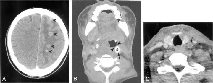
Axial CT scans of the brain and neck performed with contrast medium. A, Multiple brain abscesses are noted in the left centrum semiovale and the left high frontal region (arrowheads), in the junctional zone of the left anterior and middle cerebral arteries. B, Thrombosis with gas collection within the stent-graft is found in the left carotid artery (arrow). The stent has been extruded and exposed to the hypopharyngeal wall (arrowheads). C, Thrombosis of the left common carotid artery proximal to the stent is found (arrow).
References
-
- Macdonald S, Gan J, Mckay AJ, et al. Endovascular treatment of acute carotid blowout syndrome. J Vasc Interv Radiol 2000;11:1184–88 - PubMed
-
- Cohe J, Rad L. Contemporary management of carotid blowout. Curr Opin Otolaryngol Head Neck Surg 2004;12:110–15 - PubMed
-
- Warren FM, Cohen JI, Nesbit GM, et al. Management of carotid “blowout” with endovascular stent grafts. Laryngoscope 2002;112:428–33 - PubMed
Publication types
MeSH terms
LinkOut - more resources
Full Text Sources
Medical