. 2006 Aug 22;103(34):12769-74.
doi: 10.1073/pnas.0601990103. Epub 2006 Aug 14.

Efficient generation of retinal progenitor cells from human embryonic stem cells

Affiliations

Efficient generation of retinal progenitor cells from human embryonic stem cells

Deepak A Lamba et al. Proc Natl Acad Sci U S A. .

Abstract

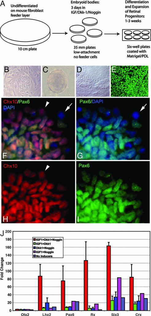
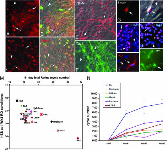
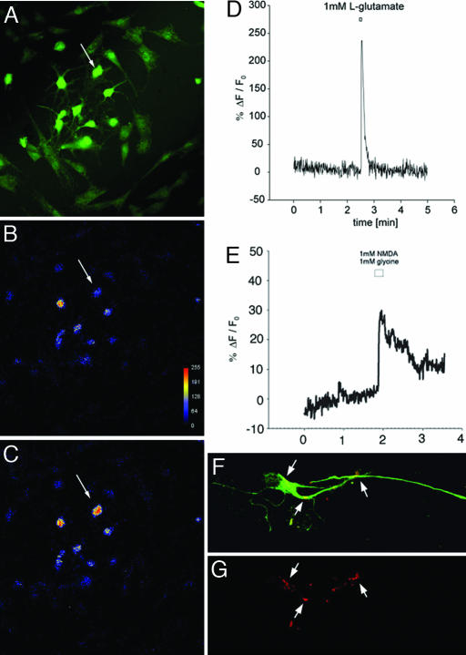
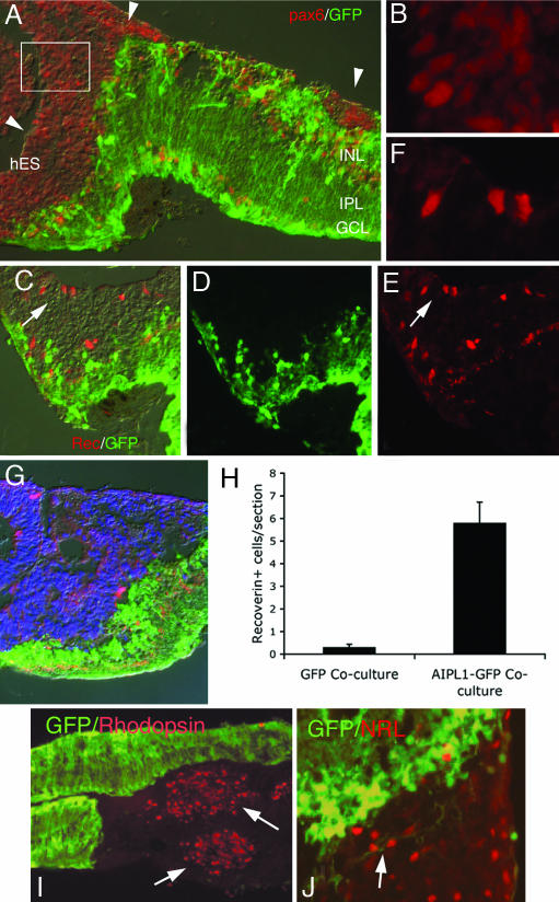
The retina is subject to degenerative conditions, leading to blindness. Although retinal regeneration is robust in lower vertebrates, regeneration does not occur in the adult mammalian retina. Thus, we have developed efficient methods for deriving retinal neurons from human embryonic stem (hES) cells. Under appropriate culture conditions, up to 80% of the H1 line can be directed to the retinal progenitor fate, and express a gene expression profile similar to progenitors derived from human fetal retina. The hES cell-derived progenitors differentiate primarily into inner retinal neurons (ganglion and amacrine cells), with functional glutamate receptors. Upon coculture with retinas derived from a mouse model of retinal degeneration, the hES cell derived retinal progenitors integrate with the degenerated mouse retina and increase in their expression of photoreceptor-specific markers. These results demonstrate that human ES cells can be selectively directed to a neural retinal cell fate and thus may be useful in the treatment of retinal degenerations.


Conflict of interest statement

Conflict of interest statement: No conflicts declared.

Figures

Fig. 1.
Fig. 1.
Efficient Retinal Induction of hES cells. (A) A schematic of the 3-week retinal determination protocol. (BD) Bright-field phase images of the progression of undifferentiated hES cells (B) through embryoid body (C) to formation of neural rosettes (D). At the end of 3 weeks, ≈80% cells show immunoreactivity to retinal progenitor markers like Pax6 (E, G, and I) and Chx10 (F and H). (FI) Coexpression of Pax6 (green) and Chx10 (red) by a group of cells labeled by DAPI in blue. Arrows in F and G point to a cell that expresses neither proteins, whereas arrowhead s in F and H point to a cell expressing Chx10 but not Pax6. For maximal induction of the eye-field transcription factors Pax6, Six3, and Rx, IGF1, Dkk1, and noggin are required. The experiment was performed by using the protocol and concentrations as described except that either IGF1, Dkk1, Noggin, or all three were omitted from the media. At the end of 1 week, comparison of the gene expression for the eye field transcription factors in each of the five cases (IGF1 + Dkk1+ Noggin, Dkk1+ noggin, IGF1 + noggin, IGF1 + Dkk1 and No inducers) was done by using quantitative RT-PCR (J). QPCR analysis of EFTFs at the end of 1 week under retinal determination conditions (n = 3; mean ± SEM) shows a 6–8 cycle (≈75- to 165-fold) increase in various retinal stem cell markers (Pax6, Lhx2, Rx, and Six3). None of the other conditions has a comparable inductive effect.
Fig. 2.
Fig. 2.
Multilineage differentiation of hES cell-derived retinal progenitors. (A) Two levels of Pax6 expression in these cells. Progenitors express lower levels of Pax6, whereas differentiating ganglion and amacrine cells express high levels of Pax6 and coexpress Hu C/D (green, B). (C and D) Similar pattern of Pax6 and Hu C/D expression in dissociated 78-day human fetal retinal cultures for comparison. (E and F) Human ES cells in RD conditions label for neurofilament-M (E) and Tuj-1 (F). (G and H) A cell expressing S-opsin in UV and Nomarski optics, respectively. (L) Cells labeled with the rod photoreceptor marker Rho-4D2 (rhodopsin). Many cells express the rod photoreceptor specific transcription factor, Nrl (red, I) or pan-photoreceptor marker, Crx (red, J); nuclei are also labeled with DAPI (blue). (K) Cell labeled with bipolar cell marker PKCα (red) with the nuclei labeled with DAPI (blue). In AK, an arrow marks and labels cell in the field, whereas an arrowhead indicates a cell not expressing that protein. (M) Comparative QPCR analysis of expression of various genes between a 91 day human fetal retina and hES cells after 3 weeks under RD conditions, showing the correlation between hES cell derived retinal cells and retinal cells isolated from fetal human retina. (N) QPCR analysis of retinal differentiation genes (n = 3). The graph shows a steady increase in retinal neuronal markers Crx, Math5, S-opsin, rhodopsin, recoverin, and PDE-β over the 3 weeks of induction.
Fig. 3.
Fig. 3.
Glutamate and NMDA induced calcium changes in hES cell-derived retinal neurons. (A) Oregon Green BAPTA-1 a.m. loading of the hES derived neurons. (B) The same cells at baseline using a rainbow LUT palette. (C) The same field of cells is shown immediately following application of 1 mM glutamate. Arrows in AC indicate a cell with large calcium transient after application. (D) The calcium change in the same cell expressed as a pseudoratio of fluorescence change expressed as a % ΔF/F0 over time. (E) A similar calcium change in a cell stimulated with 1 mM NMDA in the presence of 1 mM glycine, again expressed as a pseudoratio of fluorescence change. In each case at least four different preparations were analyzed (n = 4). (F and G) Cells with neuronal morphology and expressing internexin (green) show punctuate labeling with synaptophysin (red) antibody, a protein expressed in synapses.
Fig. 4.
Fig. 4.
Explant coculture of hES-derived retinal progenitors with retinas of Aipl1−/− GFP mice. (A) The mouse retina in green (GFP) with the outer nuclear layer missing (degenerated). Many of the hES cells express Pax6. (B) An enlarged view of boxed area in a. (CF) Recoverin expression, a marker of photoreceptors, in the cocultures. (C) A merged view of D and E, where D is showing mouse cells expressing GFP and E shows recoverin expression in a number of hES cell-derived neurons. Some mouse bipolars show recoverin and GFP co-expression. (F) A higher magnification of the region identified by the arrow in CE showing recoverin expressing hES cell-derived retinal neurons. (G) Confirmation of the identity of the recoverin (red) expressing cells in hES region using colabeling with human specific nuclear marker (blue). (H) Graph showing number of recoverin expressing hES cell-derived neurons per section (n = 3; mean ± SEM) from cocultures with wild-type mice expressing GFP and Aipl1−/− GFP mice. (I) Transplanted human cells expressing rod photoreceptor marker, Rho-4D2. (J) Transplanted human cells expressing another rod photoreceptor specific marker, Nrl.

References

    1. Moshiri A., Close J., Reh T. A. Int. J. Dev. Biol. 2004;48:1003–1014. - PubMed
    1. Meyer J. S, Katz M. L, Maruniak J. A., Kirk M. D. Stem Cells. 2006;24:274–283. - PMC - PubMed
    1. Ikeda H., Osakada F., Watanabe K., Mizuseki K., Haraguchi T., Miyoshi H., Kamiya D., Honda Y., Sasai N., Yoshimura N., et al. Proc. Natl. Acad. Sci. USA. 2005;102:11331–11336. - PMC - PubMed
    1. Bjorklund L. M., Sanchez-Pernaute R., Chung S., Andersson T., Chen I. Y., McNaught K. S., Brownell A. L., Jenkins B. G., Wahlestedt C., Kim K. S., Isacson O. Proc. Natl. Acad. Sci. USA. 2002;99:2344–2349. - PMC - PubMed
    1. Kim J. H., Auerbach J. M., Rodriguez-Gomez J. A., Velasco I., Gavin D., Lumelsky N., Lee S. H., Nguyen J., Sanchez-Pernaute R., Bankiewicz K., McKay R. Nature. 2002;418:50–56. - PubMed

Publication types