. 2006 Sep;79(3):469-80.
doi: 10.1086/507132. Epub 2006 Jul 7.

Mitochondrial DNA-deletion mutations accumulate intracellularly to detrimental levels in aged human skeletal muscle fibers

Affiliations

Mitochondrial DNA-deletion mutations accumulate intracellularly to detrimental levels in aged human skeletal muscle fibers

Entela Bua et al. Am J Hum Genet. 2006 Sep.

Abstract

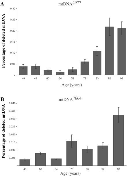
Skeletal muscle-mass loss with age has severe health consequences, yet the molecular basis of the loss remains obscure. Although mitochondrial DNA (mtDNA)-deletion mutations have been shown to accumulate with age, for these aberrant genomes to be physiologically relevant, they must accumulate to high levels intracellularly and be present in a significant number of cells. We examined mtDNA-deletion mutations in vastus lateralis (VL) muscle of human subjects aged 49-93 years, using both histologic and polymerase-chain-reaction (PCR) analyses, to determine the physiological and genomic integrity of mitochondria in aging human muscle. The number of VL muscle fibers exhibiting mitochondrial electron-transport-system (ETS) abnormalities increased from an estimated 6% at age 49 years to 31% at age 92 years. We analyzed the mitochondrial genotype of 48 single ETS-abnormal, cytochrome c oxidase-negative/succinate dehydrogenase-hyperreactive (COX-/SDH++) fibers from normal aging human subjects and identified mtDNA-deletion mutations in all abnormal fibers. Deletion mutations were clonal within a fiber and concomitant to the COX-/SDH++ region. Quantitative PCR analysis of wild-type and deletion-containing mtDNA genomes within ETS-abnormal regions of single fibers demonstrated that these deletion mutations accumulate to detrimental levels (>90% of the total mtDNA).


Figures

Figure  1.
Figure 1.
Detection of mtDNA-deletion mutations by major arc mtDNA-genome amplification. Total DNA from microdissected COX/SDH++ fibers was subjected to long-extension PCR. mtDNA-specific primers (F4840–R766) that amplify ∼13 kb of the full-length genome, including two origins of replication, were used. Full-length amplification products were detected in ETS-normal fibers (n). Smaller amplification products were identified in ETS-abnormal fibers (8, 11, 28, 31, and 34). Amplification of total mtDNA from tissue homogenates (T) detected both full-length and smaller amplification products. M = DNA molecular-weight standards.
Figure  2.
Figure 2.
Schematic representation of the human mtDNA-deletion mutations obtained from 48 COX/SDH++ fibers. mtDNA-deletion mutations were detected in all 48 ETS-abnormal fibers, and breakpoints were determined by DNA sequence analysis. Arcs represent the deleted regions of the genome. The location of the light-strand (OL) and the heavy-strand (OH) origins of replication are indicated. Cyt = cytochrome.
Figure  3.
Figure 3.
Log10 of mtDNA wild-type and partially deleted genomes along the length of fibers 19 (A), 36 (B), 37 (C), and 23 (D). Unblackened circles represent wild-type mtDNA, and blackened circles represent deletion-containing genomes.
Figure  4.
Figure 4.
mtDNA deletion–mutation abundance along the length of COX/SDH++ fibers. LCM was used to isolate 10-μm thick tissue sections at numerous sites along the length of single muscle fibers. Quantitative PCR defined the abundance of full-length and deleted mtDNA. A, B, and C, Percentage of deleted mtDNA7664 in the abnormal region of fibers 19, 36, and 37, respectively. D, Percentage of deleted mtDNA4989 in the abnormal region of fiber 23. a, Photomicrographs of tissue cross-sections sequentially double stained for COX and SDH activity. The individual fiber (arrow) displays an ETS-abnormal phenotype. b, Digital reconstruction of CSA silhouettes. The COX/SDH++ region is shown in dark blue, the COXlow/SDHhigh region in light blue, and the COXnormal/SDHnormal in orange.
Figure  5.
Figure 5.
mtDNA4977 deletion mutation in tissue homogenate from aged human skeletal muscle. Total DNA was isolated from skeletal muscle–tissue homogenates of 49-, 60-, 64-, 76-, 83-, and 92-year-old human subjects. To investigate the presence of mtDNA4977 mutations in skeletal muscle–tissue homogenate at different ages, total DNA was subjected to PCR with primers (F8111–R13756) (table 2A) immediately flanking the deleted region. This deletion mutation was present in all analyzed tissue homogenates. In addition, 57 ETS-normal fibers (fibers that exhibited the COXnormal/SDHnormal phenotype throughout 2,000 μm of analyzed tissue) were randomly selected and analyzed for the presence of mtDNA4977 from a 49-year-old subject (20 fibers) and from a 92-year-old subject (37 fibers). mtDNA4977 was not detected in any of these fibers. M = DNA molecular-weight standards; ntc = no template control.
Figure  6.
Figure 6.
Accumulation, with age, of mtDNA4977 and mtDNA7664 deletion mutation in tissue homogenate. A, Primers specific for mtDNA4977 (F8369–R13498) were used to measure the amount of deletion mutation. The wild-type primer set (F12354–R12549) was designed to amplify a region of the genome within the breakpoints of the deleted mtDNA, to ensure that the primers amplified full-length wild-type genomes. The amount of mtDNA4977 deletion mutation had a range of 0.02%–0.06% of the wild-type genomes for ages 49-79 years. The deletion-mutation level significantly increased to 0.1%–0.225% of the wild-type genomes for ages 83–93 years. B, Primers specific for mtDNA7664 (F6256–R14122) were used to measure the amount of deletion mutation. Primer set F12354–R12549 was used to measure the amount of wild-type mtDNA present. The mtDNA7664 deletion mutation load fluctuated between ages 49 and 92 years. A significant (P<.05) increase was found at age 93 years.

Similar articles

Cited by

References

Web Resources

    1. GenBank, http://www.ncbi.nlm.nih.gov/Genbank/ (for ND5 [accession number 2052366], Cytb [accession number AAB58955], COXI [accession number AAB58945], COXIII [accession number 2052365], and HVS-1 [accession number 1944628])
    1. Online Mendelian Inheritance in Man (OMIM), http://www.ncbi.nlm.nih.gov/Omim/ (for CPEO) - PubMed

References

    1. Rosenberg IH (1997) Sarcopenia: origins and clinical relevance. J Nutr 127:990S–991S - PubMed
    1. Lexell J, Taylor CC, Sjostrom M (1988) What is the cause of the ageing atrophy? Total number, size and proportion of different fiber types studied in whole vastus lateralis muscle from 15- to 83-year-old men. J Neurol Sci 84:275–29410.1016/0022-510X(88)90132-3 - DOI - PubMed
    1. Baumgartner RN, Koehler KM, Gallanger D (1998) Epidemiology of sarcopenia among the elderly in New Mexico. Am J Epidemiol 147:755–763 - PubMed
    1. Wanagat J, Cao Z, Pathare P, Aiken JM (2001) Mitochondrial DNA deletion mutations colocalize with segmental electron transport system abnormalities, muscle fiber atrophy, fiber splitting and oxidative damage in sarcopenia. FASEB J 15:322–33210.1096/fj.00-0320com - DOI - PubMed
    1. Reimers CD, Harder T, Saxe H (1998) Age-related muscle atrophy does not affect all muscles and can partly be compensated by physical activity: an ultrasound study. J Neurol Sci 159:60–6610.1016/S0022-510X(98)00134-8 - DOI - PubMed

Publication types

MeSH terms

Substances