. 2006 Sep;79(3):524-38.
doi: 10.1086/507652. Epub 2006 Aug 1.

Meiotic recombination and spatial proximity in the etiology of the recurrent t(11;22)

Affiliations

Meiotic recombination and spatial proximity in the etiology of the recurrent t(11;22)

Terry Ashley et al. Am J Hum Genet. 2006 Sep.

Abstract

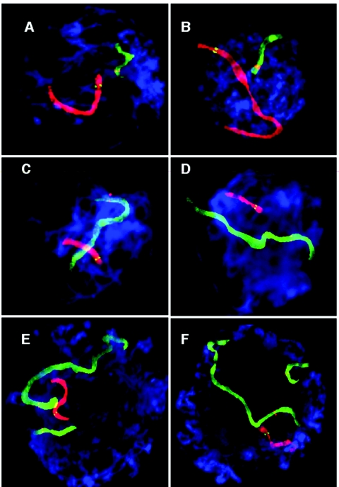
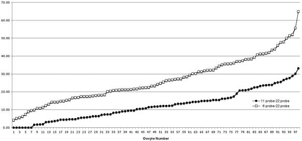
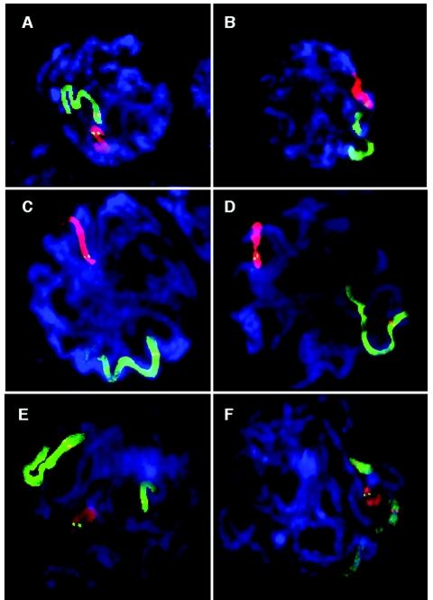
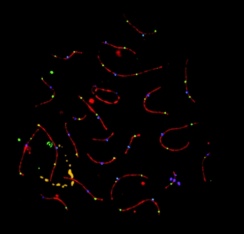
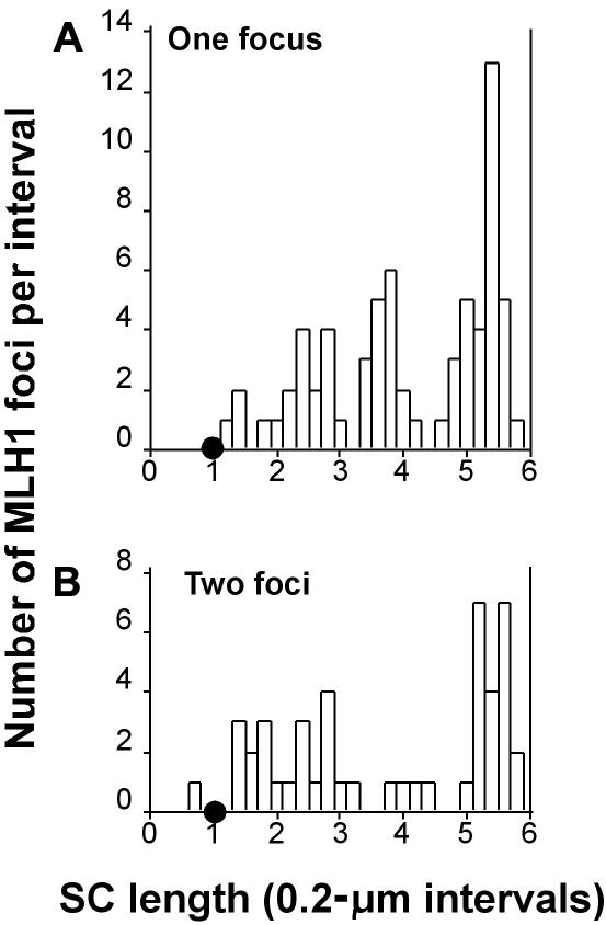
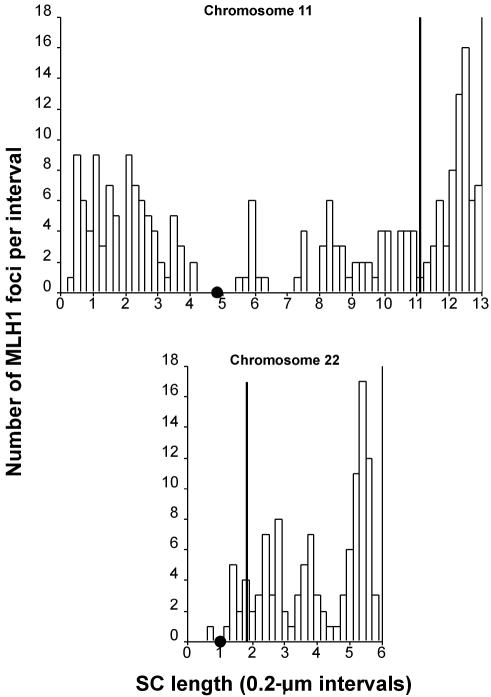
Although balanced translocations are among the most common human chromosomal aberrations, the constitutional t(11;22)(q23;q11) is the only known recurrent non-Robertsonian translocation. Evidence indicates that de novo formation of the t(11;22) occurs during meiosis. To test the hypothesis that spatial proximity of chromosomes 11 and 22 in meiotic prophase oocytes and spermatocytes plays a role in the rearrangement, the positions of the 11q23 and 22q11 translocation breakpoints were examined. Fluorescence in situ hybridization with use of DNA probes for these sites demonstrates that 11q23 is closer to 22q11 in meiosis than to a control at 6q26. Although chromosome 21p11, another control, often lies as close to 11q23 as does 22q11 during meiosis, chromosome 21 rarely rearranges with 11q23, and the DNA sequence of chromosome 21 appears to be less susceptible than 22q11 to double-strand breaks (DSBs). It has been suggested that the rearrangement recurs as a result of the palindromic AT-rich repeats at both 11q23 and 22q11, which extrude hairpin structures that are susceptible to DSBs. To determine whether the DSBs at these sites coincide with normal hotspots of meiotic recombination, immunocytochemical mapping of MLH1, a protein involved in crossing over, was employed. The results indicate that the translocation breakpoints do not coincide with recombination hotspots and therefore are unlikely to be the result of meiotic programmed DSBs, although MRE11 is likely to be involved. Previous analysis indicated that the DSBs appear to be repaired by a mechanism similar to nonhomologous end joining (NHEJ), although NHEJ is normally suppressed during meiosis. Taken together, these studies support the hypothesis that physical proximity between 11q23 and 22q11--but not typical meiotic recombinational activity in meiotic prophase--plays an important role in the generation of the constitutional t(11;22) rearrangement.


Figures

Figure  1.
Figure 1.
FISH-labeled human pachytene oocytes (3:1 preparation). Chromosomes were cohybridized with WCPs and locus-specific probes. For details of the probes and labeling scheme, see table 1. A and B, WCP11 is shown in red, with the 11q23 locus-specific probe shown in green; WCP22 is shown in green, with the 22q11 locus-specific probe in red (table 1 [experiments 1 and 2]). C and D, WCP6 is shown in green, with the 6q26 locus-specific probe shown in red; WCP22 is shown in red, with the 22q11 locus-specific probe shown in green (table 1 [experiments 3 and 4]). E and F, WCP11 is shown in green, with the 11q23 probe in red; WCP22 is shown in red, with the 22q11 probe shown in green; WCP21 is shown in green, with the 21q11 probe in red (table 1 [experiment 5]).
Figure  2.
Figure 2.
Comparison of distances between 11q23 and 22q11 and between 6q26 and 22q11. Probe-to-probe distance is shown in micrometers on the Y-axis. Distances between 11q23 and 22q11 locus-specific probes are shown as filled circles and between 6q26 and 22q11 as open squares.
Figure  3.
Figure 3.
FISH-labeled human pachytene spermatocytes (3:1 preparation). Chromosomes were cohybridized with WCPs and locus-specific probes. For details of the probes and labeling scheme, see table 1. A and B, WCP11 is shown in green, with the 11q23 locus-specific probe shown in red; WCP22 is shown in red, with the 22q11 probe shown in green (table 1 [experiment 6]). C and D, WCP6 is shown in green, with the 6q26 probe shown in red; WCP22 is shown in red, with the 22q11 probe shown in green (table 1 [experiment 7]). E and F, WCP11 is shown in green, with the 11q23 probe shown in red; WCP22 is shown in red, with the 22q11 probe shown in green; WCP21 is shown in green, with the 21q11 probe shown in red (table 1 [experiment 8]).
Figure  4.
Figure 4.
Immunostained and FISH-labeled human pachytene spermatocyte (microspread preparation). SCP3 is shown in red, MLH1 in green, and CREST in blue. In addition, the 11q23 probe is shown in gold, and the 22q11 probe is shown in magenta. The entire nucleus is stained with DAPI (cyan). MLH1 foci mark crossover sites.
Figure  5.
Figure 5.
Histograms of the distribution of MLH1 on chromosomal bivalent 11. The X-axis indicates the position (in μm) of MLH1 foci along the length of the bivalent. The position of the centromere is indicated by a small blackened circle; the p arm lies to the left, and the q arm lies to the right. The Y-axis indicates the number of MLH1 foci mapped to each interval. A, Distribution of MLH1 on bivalents with only one MLH1 focus. B, Distribution of MLH1 on bivalents with two foci. C, Distribution of MLH1 on bivalents with three foci. D, Distribution of MLH1 on the single bivalent with four foci.
Figure  6.
Figure 6.
Histograms of the distribution of MLH1 on chromosomal bivalent 22. The X-axis indicates the position (in μm) of MLH1 foci along the length of the bivalent. The position of the centromere is indicated by a small blackened circle; the p arm lies to the left, and the q arm lies to the right. The Y-axis indicates the number of MLH1 foci mapped to each interval. A, Distribution of MLH1 on bivalents with only one focus. B, Distribution of MLH1 on bivalents with two foci.
Figure  7.
Figure 7.
Histograms of the distribution of all MLH1 foci relative to the position of the probe for chromosomal bivalents 11 and 22. The X-axis indicates the position (in μm) of MLH1 foci (unblackened bars) and the probe (solid line) along the length of the bivalent. The chromosomes are positioned relative to one another on the X-axis on the basis of centromere position, which is indicated by a small blackened circle; the p arm lies to the left, and the q arm lies to the right. The Y-axis indicates the number of MLH1 foci mapped to each interval, as well as the location of the probe.

References

Web Resources

    1. GenBank, http://www.ncbi.nlm.nih.gov/Genbank/ (for BAC 442e11 [accession number AC007707], PATRR11 [accession number AF391129], PATRR17 [accession number AB195814], and PATRR22 [accession numbers AC087065 and AC074203])
    1. mfold, http://www.bioinfo.rpi.edu/applications/mfold/dna/form1.cgi
    1. MicroMeasure, http://www.colostate.edu/Depts/Biology/MicroMeasure
    1. Online Mendelian Inheritance in Man (OMIM), http://www.ncbi.nlm.nih.gov/Omim/ (for Emanuel syndrome)
    1. PALINDROME, http://bioweb.pasteur.fr/seqanal/interfaces/palindrome.html (for the EMBOSS palindrome recognition program)

References

    1. Zackai EH, Emanuel, BS (1980) Site-specific reciprocal translocation, t(11;22))q23;q11), in several unrelated families with 3:1 meiotic disjunction. Am J Med Genet 7:507–52110.1002/ajmg.1320070412 - DOI - PubMed
    1. Fraccaro M, Lindsten J, Ford C, Iselius L (1980) The 11q;22q translocation: a European collaborative analysis of 43 cases. Hum Genet 56:21–51 - PubMed
    1. Shaikh TH, Budarf M, Celle L, Zackai EH, Emanuel BS (1999) Clustered 11q23 and 22q11 breakpoints and 3:1 meiotic malsegragation in multiple unrelated t(11;22) families. Am J Hum Genet 65:1595–1607 - PMC - PubMed
    1. Lin TY, Viswanathan S, Wood C, Wilson PG, Wolf N, Fuller MT (1996) Coordinate developmental control of the meiotic cell cycle and spermatid defferentiation in Drosophila males. Development 122:1331–1341 - PubMed
    1. Kurahashi H, Shaikh TH, Hu P, Roe BA, Emanuel BS, Budarf M (2000) Regions of genomic instability on 22q11 and 11q23 as the etiology for the recurrent constitutional t(11;22). Hum Mol Genet 9:1665–167010.1093/hmg/9.11.1665 - DOI - PubMed

Publication types

LinkOut - more resources