. 2006 Sep;79(3):574-9.
doi: 10.1086/507568. Epub 2006 Jul 24.

Mutations in the gene KCNV2 encoding a voltage-gated potassium channel subunit cause "cone dystrophy with supernormal rod electroretinogram" in humans

Affiliations

Mutations in the gene KCNV2 encoding a voltage-gated potassium channel subunit cause "cone dystrophy with supernormal rod electroretinogram" in humans

Huimin Wu et al. Am J Hum Genet. 2006 Sep.

Abstract

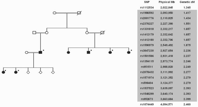
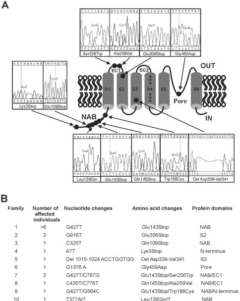
"Cone dystrophy with supernormal rod electroretinogram (ERG)" is an autosomal recessive disorder that causes lifelong visual loss combined with a supernormal ERG response to a bright flash of light. We have linked the disorder to a 0.98-cM (1.5-Mb) region on chromosome 9p24, flanked by rs1112534 and rs1074449, using homozygosity mapping in one large consanguineous pedigree. Analysis of one gene within this region, KCNV2, showed a homozygous nonsense mutation. Mutations were also found in 17 alleles of 10 other unrelated families with the same disorder. In situ hybridization demonstrated KCNV2 expression in human rod and cone photoreceptors. The precise function of KCNV2 in human photoreceptors remains to be determined, although this work suggests that mutations might perturb or abrogate I(KX), the potassium current within vertebrate photoreceptor inner segments, which has been shown to set their resting potential and voltage response.


Figures

Figure  1.
Figure  1.
Autofluorescence (A) and color image (B) of right and left retina of affected patient aged 59 years (family 4), showing areas of central atrophy of retinal pigment epithelium (RPE) and choroid. C, Color image of right and left retina of affected patient aged 24 years (family 1), showing subtle RPE depigmentation around fovea and crystals at the macula. D, Full-field ERGs (International Society for Clinical Electrophysiology of Vision [ISCEV]) in an unaffected subject (top row) and in a representative patient from family 9 (bottom row). In the patient, dark-adapted responses to the dimmest flash (0.002 candela [cd]-s/m2) are undetectable. Increasing stimulus intensity (“rod” 0.012 cd-s/m2) produces an abrupt increase in amplitude and a delayed rod ERG. At higher flash energies (“standard” 3.0 cd-s/m2 and “maximum” 11.5 cd-s/m2), the a-wave commences normally but develops a broadened trough before a high-amplitude, sharply rising b-wave that approaches the upper limit of normality (supernormal). Flicker and photopic single-flash ERGs were performed after 10 min of light adaptation. ISCEV-standard 30-Hz flicker ERGs show delay and marked reduction. The photopic single-flash ERG is delayed and subnormal, with a simplified waveform and delayed recovery after the beta-wave. Broken lines replace blink artifacts, frequently seen after the ERGs with strong flashes.
Figure  2.
Figure  2.
Linkage analysis. A, Pedigree of consanguineous family used for mapping with the GeneChip Human Mapping 10K SNP Array (Affymetrix). Individuals included in the analysis are marked by an asterisk. B, Contiguous homozygous SNPs at chromosome 9q24 (shaded), with flanking SNPs above and below.
Figure  3.
Figure  3.
Mutations in KCNV2 channel protein. A, Domain structure of KCNV2, with the approximate position of mutations (circles) linked to electropherograms showing sequence in patient DNA. B, KCNV2 disease-associated mutations. Affected individuals from families 1–6 were homozygotes for the variant indicated. Only a single missense mutation was found in family 10; the “missing” mutation is indicated by a quotation mark. Additionally, the proband of family 4 was heterozygous for L533V, and the proband of family 8 also had D147F in phase with Q145X.
Figure  4.
Figure  4.
Gel image showing PCR products of KCNV2, KCNB1, KCNC1, and KCNF1 amplified from human retinal cDNA. The fragments were amplified using primer pairs G, H, I, and J (table 1), were cloned into the pGEM-T-Easy vector, and were sequenced to confirm identity.
Figure  5.
Figure  5.
In situ hybridization of human retina probed with antisense and sense KCNV2 riboprobes. Sections 10 μm thick, from fixed and cryoprotected human retinal tissue, were probed with either antisense or sense digoxigenin (DIG)–labeled riboprobes generated from a 427-bp gene fragment, cloned into pGemT Easy vector, that spans the central portion of exon 1. Retinal sections were prepared for hybridization with the use of standard methods. Hybridization and washing was performed at 65°C. Signal was resolved using anti-DIG antibody at a 1:2,000 dilution, followed by color development. OS = Photoreceptor outer segment; IS = photoreceptor inner segment; ONL = outer nuclear layer.

References

Web Resources

    1. BLAST, http://www.ncbi.nlm.nih.gov/BLAST/
    1. ClustalW, ftp://ftp.ebi.ac.uk/pub/software/unix/clustalw/
    1. ECACC, http://www.ecacc.org.uk/ (for control DNAs from anonymous white donors)
    1. Ensembl, http://www.ensembl.org/
    1. International Society for Clinical Electrophysiology of Vision (ISCEV), http://www.iscev.org/

References

    1. Gouras P, Eggers HM, MacKay CJ (1983) Cone dystrophy, nyctalopia, and supernormal rod responses: a new retinal degeneration. Arch Ophthalmol 101:718–724 - PubMed
    1. Rosenberg T, Simonsen SE (1993) Retinal cone dysfunction of supernormal rod ERG type: five new cases. Acta Ophthalmol (Copenh) 71:246–255 - PubMed
    1. Sandberg MA, Miller S, Berson EL (1990) Rod electroretinograms in an elevated cyclic guanosine monophosphate-type human retinal degeneration: comparison with retinitis pigmentosa. Invest Ophthalmol Vis Sci 31:2283–2287 - PubMed
    1. Kato M, Kobayashi R, Watanabe I (1993) Cone dysfunction and supernormal scotopic electroretinogram with a high-intensity stimulus: a report of three cases. Doc Ophthalmol 84:71–81 10.1007/BF01203284 - DOI - PubMed
    1. Michaelides M, Holder GE, Webster AR, Hunt DM, Bird AC, Fitzke FW, Mollon JD, Moore AT (2005) A detailed phenotypic study of “cone dystrophy with supernormal rod ERG.” Br J Ophthalmol 89:332–339 - PMC - PubMed

Publication types

MeSH terms