. 2006;8(5):R141.
doi: 10.1186/ar2031.

Preventing autoimmune arthritis using antigen-specific immature dendritic cells: a novel tolerogenic vaccine

Affiliations

Preventing autoimmune arthritis using antigen-specific immature dendritic cells: a novel tolerogenic vaccine

Igor Popov et al. Arthritis Res Ther. 2006.

Abstract

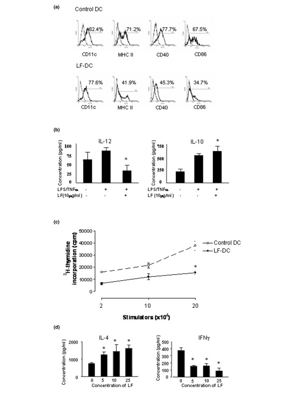
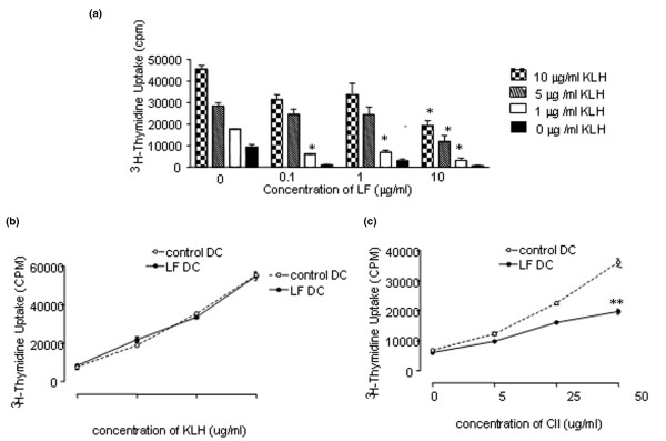
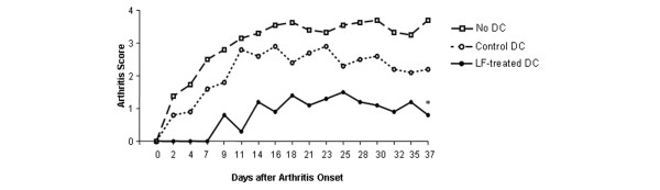
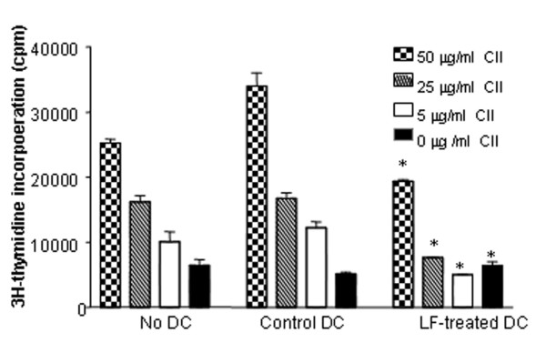
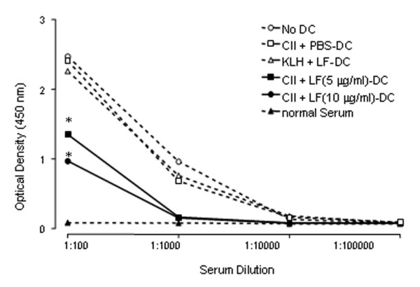
Conventional treatments for autoimmune diseases have relied heavily on nonspecific immune suppressants, which possess a variety of adverse effects without inhibiting the autoimmune process in a specific manner. In the present study we demonstrate the effectiveness of antigen-specific, maturation-resistant, tolerogenic dendritic cells (DC) in suppressing collagen-induced arthritis, a murine model of rheumatoid arthritis. Treatment of DC progenitors with the NF-kappaB inhibiting agent LF 15-0195 (LF) resulted in a population of tolerogenic DC that are characterized by low expression of MHC class II, CD40, and CD86 molecules, as well as by poor allostimulatory capacity in a mixed leukocyte reaction. Administering LF-treated DC pulsed with keyhole limpet hemocyanin antigen to naïve mice resulted hyporesponsiveness specific for this antigen. Furthermore, administration of LF-treated DC to mice with collagen-induced arthritis resulted in an improved clinical score, in an inhibited antigen-specific T-cell response, and in reduced antibody response to the collagen. The efficacy of LF-treated DC in preventing arthritis was substantiated by histological examination, which revealed a significant decrease in inflammatory cell infiltration in the joints. In conclusion, we demonstrate that in vitro-generated antigen-specific immature DC may have important potential as a tolerogenic vaccine for the treatment of autoimmune arthritis.


Figures

Figure 1
Figure 1
LF 15-0195 prevents maturation and function of dendritic cells. (a) Phenotypic analysis of LF-treated dendritic cells (DC). Bone-marrow-derived DC were cultured in the presence of granulocyte-macrophage colony-stimulating factor (10 ng/ml) and IL-4 (10 ng/ml) for 7 days. Control mature DC (upper panels) were activated using tumor necrosis factor alpha (TNFα)/lipopolysaccharide (LPS) in the last 24-hour culture. DC (lower panel) were treated by addition of LF (10 ng/ml) in the culture medium from day 4 onwards, and fresh medium was added every 24 hours. DC were stained with FITC-conjugated mAbs and analyzed by flow cytometry. Results represent one of three experiments (n = 4 per group/experiment). (b) LF regulates cytokine expression in DC. DC were treated with LF as in (a). The supernatants of DC culture were collected and used to measure IL-12 and IL-10 levels by ELISA as described in Materials and methods. *P < 0.05, comparing untreated control DC.(c) LF inhibits DC allostimulatory capacity in a mixed leukocyte reaction. DC were pretreated with LF and subsequently stimulated with 10 ng/ml TNFα/LPS as described in (a). DBA/1 control DC and LF-treated DC, at indicated concentrations, were used as stimulators, and BALB/c splenocytes (1 × 10⁵/well) were used as responders. Stimulators and responders were cocultured, and proliferation was assessed as described in Materials and methods. Data shown are representative of three independent experiments (n = 4 per group/experiment).P < 0.05, comparing untreated control DC.(d) LF-treated DC regulate T helper cell deviation. LF-treated DC and PBS-treated control DC (106) (DBA/1) were subsequently cultured with allogeneic (BALB/c) T cells (107) for 48 hours. Supernatants were collected from the cultures and interferon gamma (IFNγ; Th1) and IL-4 cytokine (Th2) levels were measured by ELISA. Results represent one of three experiments (n = 4 per group/experiment). P < 0.05, comparing untreated control DC.
Figure 2
Figure 2
LF 15-0195-treated dendritic cells inhibit antigen-specific T-cell responses. (a) LF-treated dendritic cells (DC) inhibit anti-keyhole limpet hemocyanin (KLH) T-cell responses. Day 4 bone-marrow-derived DC cultured in granulocyte-macrophage colony-stimulating factor (10 ng/ml) and IL-4 (10 ng/ml) were treated with different concentrations of LF (0.1, 1, and 10 μg/ml) or PBS alone. On day 7 of culture, 10 μg/ml KLH was added to the cells for 24 hours and then cells were activated with TNFα (10 ng/ml) and lipopolysaccharide (10 ng/ml). On day 9 of DC culture, 5 × 10⁵ cells/mouse were injected intraperitoneally into syngeneic BALB/c mice. After 10 days, the mice were sacrificed and T cells from lymph nodes were isolated. A KLH-specific recall response was determined by the proliferation, as described in Materials and methods. *P < 0.05 versus nontreated control DC. (b) and (c) LF-treated DC-induced immune suppression is antigen specific. DC were cultured, treated with LF, pulsed with type II collagen (CII) antigen, and immunized mice as described in (a). Two days prior to LF-treated DC or untreated control DC immunization, the mice were immunized with 10 μg KLH subcutaneously. Ten days after immunization, lymph node cells were harvested and proliferated in vitro in the presence of CII (b) and KLH (c), respectively, at the indicated concentrations. Results represent one of three experiments. *P < 0.05 versus nontreated control DC. cpm, counts per minute.
Figure 3
Figure 3
Type II collagen-pulsed LF 15-0195-treated dendritic cells inhibit clinical development of collagen-induced arthritis. Twelve days after intradermal challenge with type II collagen (CII) (200 μg/mouse in complete Freund's adjuvant), DBA/1 LacJ mice were injected intraperitoneally with LF-treated dendritic cells (DC) (5 μg/ml) and CII-pulsed DC (10 μg/ml) (5 × 10⁶ cells/mouse). Controls were either treated with non-LF-treated but CII-pulsed DC or remained untreated. All mice were boosted by an intraperitoneal injection with the same dose of CII in PBS 9 days later. The mice were observed for 37 days after arthritis onset. Each limb was graded on a scale from 0 to 4 and the average clinical score per affected paw was calculated. Each point denotes the score of six mice in each group. Results represent one of three experiments. *P < 0.05 versus the control DC-treated group.
Figure 4
Figure 4
T-cell hyporesponsiveness to type II collagen in collagen-induced arthritis-susceptible mice with LF-treated dendritic cells. Day 4 bone-marrow-derived dendritic cells (DC) cultured in granulocyte-macrophage colony-stimulating factor/IL-4 were treated with 5 μg/ml LF or PBS alone, and fresh medium was added every 24 hours. On day 7, both LF-treated DC and control PBS-treated DC were pulsed with type II collagen (CII) (10 μg/ml) for 24 hours. On day 8, CII-pulsed cell cultures were activated with TNFα/lipopolysaccharide for the next 24 hours, and 5 × 10⁶ cells/mouse were injected intraperitoneally into DBA/1 LacJ mice primed with CII (200 μg/mouse in complete Freund's adjuvant) 12 days earlier. Twenty-one days after priming, the mice were boosted intraperitoneally with the same dose of CII in PBS. At the end of clinical assessment of collagen-induced arthritis development, the mice were sacrificed and T cells from lymph nodes were isolated. A CII-specific response from different groups of animals was performed by proliferation, as described in Materials and methods. Lymphocytes were restimulated in vitro with different concentrations of CII (5, 25, and 50 μg/ml) or PBS alone and a 3H-labeled thymidine incorporation was measured. Results represent one of three experiments (n = 4 per group/experiment). *P < 0.05 versus the control DC-treated group. cpm, counts per minute.
Figure 5
Figure 5
Inhibition of CII-specific antibody production in arthritis mice with LF-treated dendritic cells. Blood was taken 40 days after arthritis onset and serum levels of anti-type II collagen (anti-CII) immunoglobulin were determined using sandwich ELISA. Results show average levels of antibody expressed as the optical density for experimental and control groups (n = 6 per group/experiment). P < 0.05 versus the control DC-treated group. KLH, keyhole limpet hemocyanin.
Figure 6
Figure 6
Histological joint sections from arthritic mice with CII-pulsed treated dendritic cells. H & E-stained sagittal sections of proximal interphalangeal joints from collagen-induced arthritis mice. (a) Control mouse shows severe edema, congestion, and monocyte infiltration; the bone surface became uneven. (b) The majority of joints from mice injected with LF 15-0195-treated dendritic cells have normal morphology with a smooth articulation cartilage surface, and an absence of inflammatory cell infiltrate and edema. Original magnification × 100.

Similar articles

Cited by

References

    1. Vosters O, Neve J, De Wit D, Willems F, Goldman M, Verhasselt V. Dendritic cells exposed to nacystelyn are refractory to maturation and promote the emergence of alloreactive regulatory t cells. Transplantation. 2003;75:383–389. doi: 10.1097/01.TP.0000043924.09647.61. - DOI - PubMed
    1. Mahnke K, Schmitt E, Bonifaz L, Enk AH, Jonuleit H. Immature, but not inactive: the tolerogenic function of immature dendritic cells. Immunol Cell Biol. 2002;80:477–483. doi: 10.1046/j.1440-1711.2002.01115.x. - DOI - PubMed
    1. Steinman RM, Hawiger D, Nussenzweig MC. Tolerogenic dendritic cells. Annu Rev Immunol. 2003;21:685–711. doi: 10.1146/annurev.immunol.21.120601.141040. - DOI - PubMed
    1. Roncarolo MG, Levings MK, Traversari C. Differentiation of T regulatory cells by immature dendritic cells. J Exp Med. 2001;193:F5–F9. doi: 10.1084/jem.193.2.F5. - DOI - PMC - PubMed
    1. de Heusch M, Oldenhove G, Urbain J, Thielemans K, Maliszewski C, Leo O, Moser M. Depending on their maturation state, splenic dendritic cells induce the differentiation of CD4(+) T lymphocytes into memory and/or effector cells in vivo. Eur J Immunol. 2004;34:1861–1869. doi: 10.1002/eji.200424878. - DOI - PubMed

Publication types

MeSH terms