

Adenoid basal lesions of the uterine cervix: evolving terminology and clinicopathological concepts
- PMID: 16911774
- PMCID: PMC1564042
- DOI: 10.1186/1746-1596-1-18
Adenoid basal lesions of the uterine cervix: evolving terminology and clinicopathological concepts
Abstract
The epithelial proliferations that are designated adenoid basal carcinoma (ABC) in the current classification from the World Health Organization represent <1% of all cervical malignancies. These lesions may be associated, and occasionally show morphologic transitions with, conventional cervical malignancies. The determination of the precise frequency with which these so-called ABCs show this association is hampered by the inherent selection bias in the reported cases. However, this frequency appears to be substantial (>15%). The biologic course of ABCs that are associated with separate malignancies is largely dependent on the clinicopathologic parameters of the associated malignancies. Morphologically pure lesions, in contrast, have largely been associated with favorable patient outcomes, as none of the 66 reported patients have experienced tumor recurrence, metastases or tumor-associated death, irrespective of the modality of treatment. Although the finding of genome integrated high-risk human papillomavirus (HPV) types and p53 alterations in adenoid basal lesions (ABL) argue in support of their neoplastic nature, we identified no lines evidence that suggest an inherent malignancy for morphologically pure lesions. The finding of morphologic transitions between ABLs and conventional malignancies and shared HPV types in these areas, suggest that ABLs have some malignant potential. However, the precise magnitude of this potential is not readily quantifiable and should not dictate the management of morphologically pure lesions that are entirely evaluable. ABLs continue to occupy a unique position in human oncology in which the term carcinoma (without an in-situ suffix) is applied to a tumor that has not been shown to recur, metastasize or cause death. We concur with a previous proposal that the term ABC should be discarded and replaced with Adenoid Basal Epithelioma (ABE). In our opinion, there is insufficient evidence at present time to expose patients with morphologically pure lesions to the ominous implications--social, psychological, medical, financial--of a "carcinoma" diagnosis. Morphologically impure lesions should not be designated ABC or ABE. Furthermore, given the uncertainties regarding the frequency with which ABE are associated with separate malignancies, we suggest that the ABE designation only be applied when the tumor in question is entirely evaluable e.g in a hysterectomy specimen or in an excisional biopsy with negative margins. Otherwise, the generic designation Adenoid Basal Tumor is preferable. This approach strikes an appropriate balance between the need to prevent over-treatment of pure lesions on one hand, and the need to ensure that the lesions are indeed pure on the other.
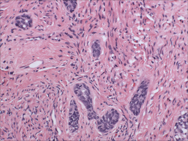
Figures


References
-
- Zamecnik M, Skrivanek A. Adenoid basal epithelioma of the uterine cervix in 21-year-old patient. Report of a case with histologic and immunohistochemical study. Cesk Patol. 2005;41:157–62. - PubMed
-
- Parwani AV, Smith Sehdev AE, Kurman RJ, Ronnett BM. Cervical adenoid basal tumors comprised of adenoid basal epithelioma associated with various types of invasive carcinoma: clinicopathologic features, human papillomavirus DNA detection, and P16 expression. Hum Pathol. 2005;36:82–90. doi: 10.1016/j.humpath.2004.08.015. - DOI - PubMed
-
- Khoury T, Lele S, Tan D. Pathologic quiz case: an asymptomatic 79-year-old woman with an abnormal Papanicolaou test. Adenoid basal carcinoma of the cervix. Arch Pathol Lab Med. 2004;128:485–6. - PubMed
LinkOut - more resources
Full Text Sources
Research Materials
Miscellaneous
