. 2006 Aug 15;20(16):2223-37.
doi: 10.1101/gad.380906.

A novel role for Xist RNA in the formation of a repressive nuclear compartment into which genes are recruited when silenced

Affiliations

A novel role for Xist RNA in the formation of a repressive nuclear compartment into which genes are recruited when silenced

Julie Chaumeil et al. Genes Dev. .

Abstract

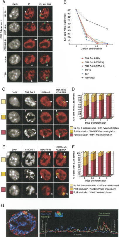
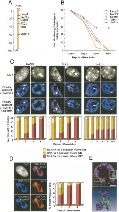
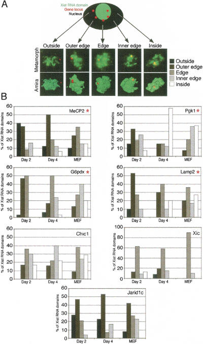
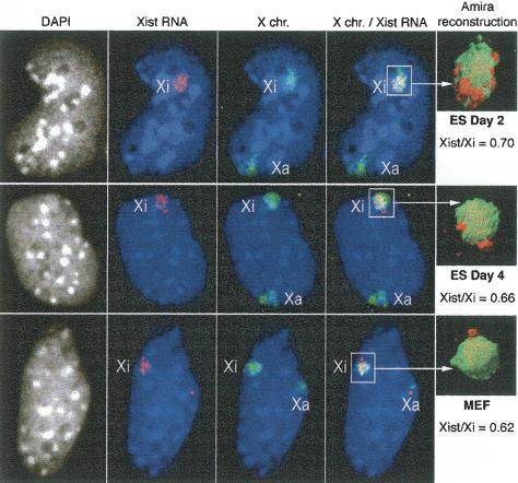
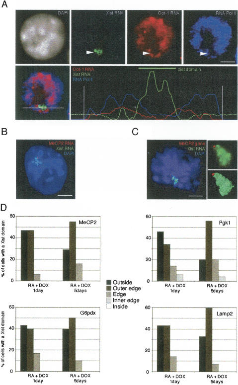
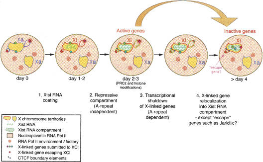
During early mammalian female development, one of the two X chromosomes becomes inactivated. Although X-chromosome coating by Xist RNA is essential for the initiation of X inactivation, little is known about how this signal is transformed into transcriptional silencing. Here we show that exclusion of RNA Polymerase II and transcription factors from the Xist RNA-coated X chromosome represents the earliest event following Xist RNA accumulation described so far in differentiating embryonic stem (ES) cells. Paradoxically, exclusion of the transcription machinery occurs before gene silencing is complete. However, examination of the three-dimensional organization of X-linked genes reveals that, when transcribed, they are always located at the periphery of, or outside, the Xist RNA domain, in contact with the transcription machinery. Upon silencing, genes shift to a more internal location, within the Xist RNA compartment devoid of transcription factors. Surprisingly, the appearance of this compartment is not dependent on the A-repeats of the Xist transcript, which are essential for gene silencing. However, the A-repeats are required for the relocation of genes into the Xist RNA silent domain. We propose that Xist RNA has multiple functions: A-repeat-independent creation of a transcriptionally silent nuclear compartment; and A-repeat-dependent induction of gene repression, which is associated with their translocation into this silent domain.


Figures

Figure 1.
Figure 1.
Exclusion of the transcription machinery from the Xist RNA domain during X inactivation of female ES cells undergoing differentiation. (A) Representative immunofluorescence (IF) using antibodies against several components of the transcription machinery (RNA Pol II, TAF10, and TBP; red) combined with Xist RNA FISH (green) on female ES cells after 2 d of differentiation. Three antibodies (H5, 8WG16, and CTD4H8) recognizing different phosphorylated forms of RNA Pol II were used. Arrowheads indicate the location of the Xist RNA domain. DNA is stained with DAPI (gray). Bar, 5 μm. (B) Kinetics of exclusion of the transcription machinery from the Xist RNA-coated X chromosome during female ES cell differentiation (day 1, n = 60; days 1.5 and 2, n = 100). (C,E) Dual IF for RNA Pol II (H5 Ab; gray) and histone H3K4me2 (C, red) or histone H3K27me3 (E, red) was combined with Xist RNA FISH (green) in female ES cells undergoing differentiation (day 1, top line; day 2, middle line; day 3, bottom line). The Xist RNA domain is shown with an arrowhead. DNA is stained with DAPI (gray). Bar, 5 μm. (D,F) Relative kinetics of the exclusion of the transcription machinery and the appearance of histone modifications during X inactivation on female ES cells undergoing differentiation (day 1, n = 60; days 1.5, 2, and 3, n = 100). (G) Representative example of 3D analysis, 3D reconstruction, and a scan line to demonstrate the relative exclusion of RNA Pol II (blue) and enrichment of H3K27me3 (red) with respect to the Xist RNA domain (green) in female ES cells differentiated for 3 d.
Figure 2.
Figure 2.
Exclusion of the transcription machinery occurs earlier than the transcriptional repression of three X-linked genes during X inactivation. (A) Locations of the Lamp2, MeCP2, G6pdx, Xist, Chic1, Pgk1, and Jarid1c genes on the X chromosome. (B) Kinetics of repression of Lamp2, MeCP2, G6pdx, Chic1, and Jarid1c during X inactivation (n > 50 at every time point). The percentage of cells with Xist RNA domains (except at day 0) showing biallelic expression of different X-linked genes is shown in ES cells differentiated for 0, 2, or 4 d, as well as in MEFs where X inactivation has been completed. (C) Representative IF of RNA Pol II (H5 Ab; blue) combined with Xist RNA FISH (green) and RNA FISH showing the primary transcript of three X-linked genes (MeCP2, Chic1, or Jarid1c; red) on female ES cells at day 2 (left column) and day 4 (right column) of differentiation. Arrowheads show primary transcripts on the inactive X chromosome, whereas asterisks indicate primary transcripts on the active X chromosome. DNA is stained with DAPI (gray). Bar, 5 μm. Relative kinetics of RNA Pol II exclusion and gene repression during X inactivation on female ES cell undergoing differentiation (day 1, n = 60; following days, n = 100). (D) Representative IF for RNA Pol II (H5 Ab; blue) combined with Cot-1 (red) and Xist (green) RNA FISH on female ES cells at day 2 of differentiation. Arrowheads show the exclusion of RNA Pol II and Cot-1 RNA on the Xist RNA domain. DNA is stained with DAPI (gray). Bar, 5 μm. Relative kinetics of RNA Pol II exclusion and Cot-1 repression during X inactivation on female ES cell undergoing differentiation (day 1, n = 60; days 2 and 3, n = 100). (E) Example of 3D analysis of RNA Pol II IF together with Chic1 and Xist RNA FISH in early differentiated ES cells, using Metamorph software (top) and after Amira reconstruction (bottom).
Figure 3.
Figure 3.
Location of three X-linked genes with respect to the Xist RNA domain during X inactivation. (A) 3D analysis of Xist RNA FISH (green) combined with DNA FISH to detect the location of X-linked genes (Lamp2, MeCP2, G6pdx, Pgk1, Chic1, Jarid1c, and the Xic locus; red) on female ES cells undergoing differentiation (days 2 and 4) and MEFs. Different profiles of X-linked gene location outside, at the outer edge, at the edge, at the inner edge, or inside the Xist RNA domain are shown following Amira reconstruction. (B) Distribution of the different locations of X-linked genes with respect to the Xist RNA domain before, during, and after X inactivation in differentiated ES cells at days 2 and 4, and in MEFs (n > 30). Genes showing a statistical difference in position distributions are indicated with red asterisks.
Figure 4.
Figure 4.
Comparative analysis of the relative volumes of Xist RNA domain and X-chromosome territory during X inactivation. Xist RNA FISH (red) combined with an X-chromosome paint DNA FISH (green) on female ES cells undergoing differentiation (days 2 and 4) and MEFs. 3D analysis using Amira software reconstruction (n = 30). DNA is stained with DAPI (gray or blue). Bar, 5 μm. Relative volumes were calculated using Amira software and are shown on the right of each panel.
Figure 5.
Figure 5.
Role of Xist RNA A-repeats using an inducible Xist gene. (A) RNA Pol II immunostaining (blue) combined with Cot-1 RNA FISH (red) and Xist RNA FISH (green) in 30–24–46 male ES cells after 2 d of differentiation and Doxicyclin-induction of XistΔA RNA transcription. DNA is stained with DAPI (gray). Bar, 5 μm. A line scan shows the relative intensities of the three signals across the nucleus. The large and the small peaks of Xist RNA likely correspond to the bulk of the X chromosome and the transcribed Xist locus itself, respectively. (B) Dual RNA FISH for MeCP2 RNA (red) and Xist RNA (green) following 1 d of differentiation and XistΔA expression. DNA is stained with DAPI (gray). Bar, 5 μm. (C) Representative 3D analysis of MeCP2 DNA FISH (red) combined with a Xist RNA FISH (green) using Metamorph software and Amira reconstruction, after 5 d of differentiation and XistΔA expression. DNA is stained with DAPI (gray). Bar, 5 μm. (D) Distribution of the different profiles of MeCP2, G6pdx, Lamp2, and Pgk1 locations with respect to the Xist RNA domain (based on the categories described in Fig. 3) after 1 or 5 d of differentiation and XistΔA expression (n = 30). No statistical differences in position distributions between days 1 and 5 are found.
Figure 6.
Figure 6.
Model for Xist RNA-mediated X-chromosome inactivation. In undifferentiated female ES cells, both X chromosomes are active (yellow). Only genes on the X chromosome undergoing inactivation (Xi) are depicted. (1) During early differentiation, Xist RNA starts to accumulate on the X chromosome chosen for inactivation. (2) The formation of this silent compartment is associated with the exclusion of the transcription machinery and the repression of internal repeat-rich regions. This step is independent of Xist A-repeats. (3) X-linked gene silencing is triggered via a Xist A-repeat-dependent mechanism, and histone modifications as well as PRC2/PRC1 accumulation (Xist A-repeat-independent) are observed. (4) Genes then become relocated into the Xist RNA compartment by default because they are no longer constrained by their requirement to be transcribed in an RNA Pol II-enriched environment (or potential transcription factory, see Osborne et al. 2004), and/or by an active mechanism of relocation, which may be dependent on the Xist A-repeats. This locus internalization may help maintain the silent state. Genes escaping inactivation, such as Jarid1c, have special elements (such as CTCF boundaries) that may prevent efficient internalization. Their location outside of the Xist repressive compartment could participate in, or facilitate, their reactivation.

References

    1. Avner P., Heard E. X-chromosome inactivation: Counting, choice and initiation. Nat. Rev. Genet. 2001;2:59–67. - PubMed
    1. Bantignies F., Grimaud C., Lavrov S., Gabut M., Cavalli G. Inheritance of Polycomb-dependent chromosomal interactions in Drosophila. Genes & Dev. 2003;17:2406–2420. - PMC - PubMed
    1. Boggs B.A., Connors B., Sobel R.E., Chinault A.C., Allis C.D. Reduced levels of histone H3 acetylation on the inactive X chromosome in human females. Chromosoma. 1996;105:303–309. - PubMed
    1. Boggs B.A., Cheung P., Heard E., Spector D.L., Chinault A.C., Allis C.D. Differentially methylated forms of his-tone H3 show unique association patterns with inactive human X chromosomes. Nat. Genet. 2002;30:73–76. - PubMed
    1. Brown C.J., Hendrich B.D., Rupert J.L., Lafreniere R.G., Xing Y., Lawrence J., Willard H.F. The human XIST gene: Analysis of a 17 kb inactive X-specific RNA that contains conserved repeats and is highly localized within the nucleus. Cell. 1992;71:527–542. - PubMed

Publication types

MeSH terms