. 2006 Sep;80(17):8390-401.
doi: 10.1128/JVI.00505-06.

Potential role for p53 in the permissive life cycle of human cytomegalovirus

Affiliations

Potential role for p53 in the permissive life cycle of human cytomegalovirus

N C Casavant et al. J Virol. 2006 Sep.

Abstract

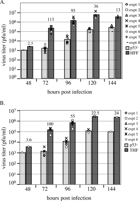
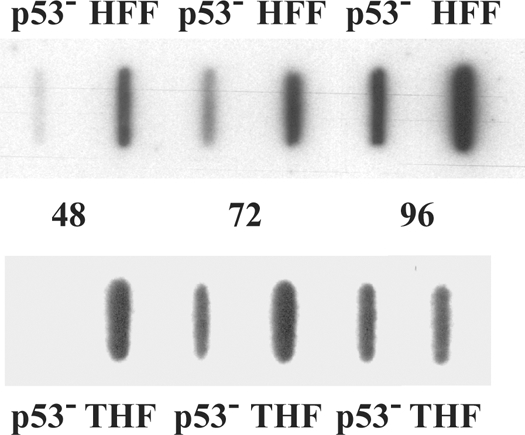
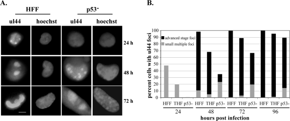
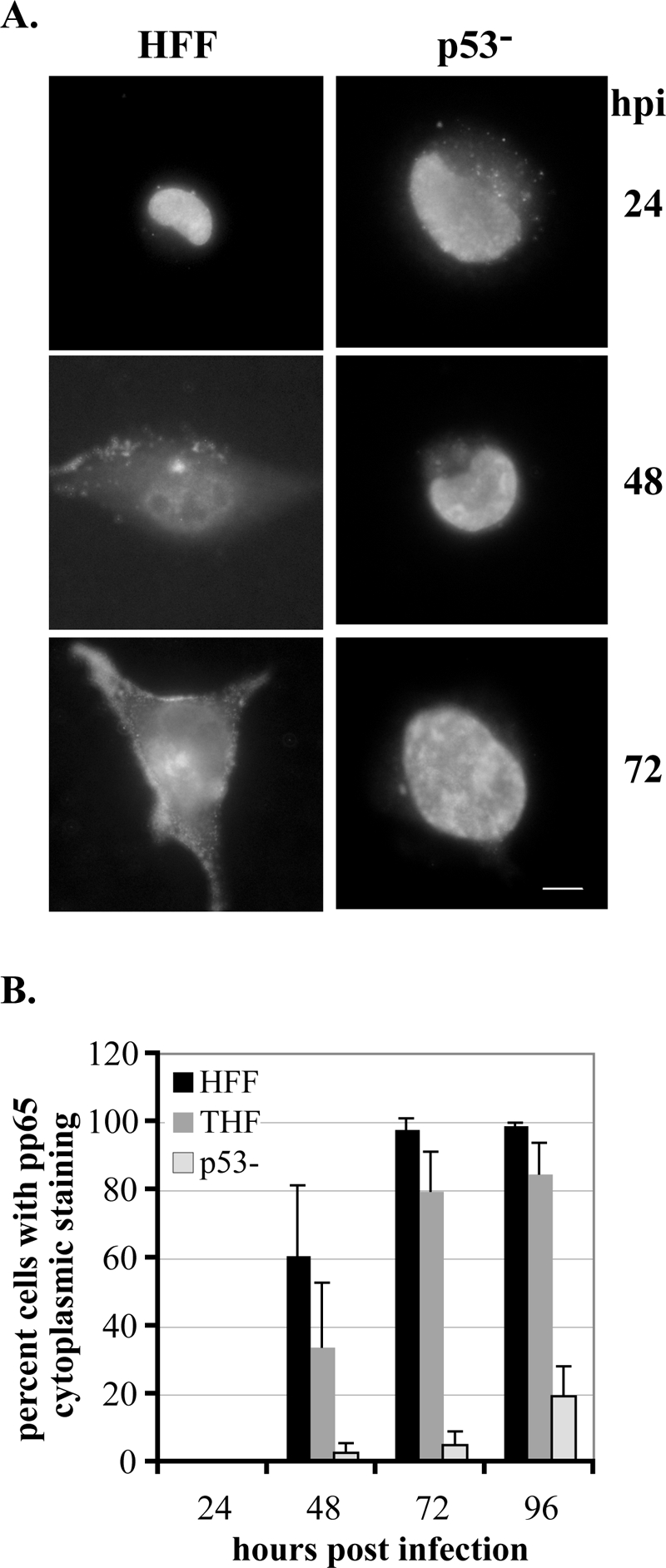
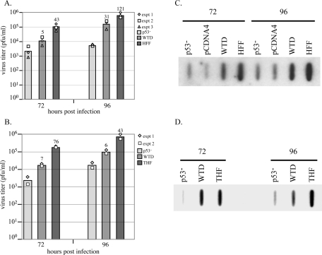
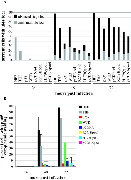
Infection of primary fibroblasts with human cytomegalovirus (HCMV) causes a rapid stabilization of the cellular protein p53. p53 is a major effector of the cellular damage response, and activation of this transcription factor can lead either to cell cycle arrest or to apoptosis. Viruses employ many tactics to avoid p53-mediated effects. One method HCMV uses to counteract p53 is sequestration into its viral replication centers. In order to determine whether or not HCMV benefits from this sequestration, we infected a p53(-/-) fibroblast line. We find that although these cells are permissive for viral infection, several parameters are substantially altered compared to wild-type (wt) fibroblasts. p53(-/-) cells show delayed and decreased accumulation of infectious viral particles compared to control fibroblasts, with the largest difference of 100-fold at 72 h post infection (p.i.) and peak titers decreased by approximately 10- to 20-fold at 144 h p.i. Viral DNA accumulation is also delayed and somewhat decreased in p53(-/-) cells; however, on average, levels of DNA are not more than fivefold lower than wt at any time p.i. and thus cannot account entirely for the observed differences in titers. In addition, there are delays in the expression of several key viral proteins, including the early replication protein UL44 and some of the late structural proteins, pp28 (UL99) and MCP (UL86). UL44 localization also indicates delayed formation and maturation of the replication centers throughout the course of infection. Localization of the major tegument protein pp65 (UL83) is also altered in these p53(-/-) cells. Partial reconstitution of the p53(-/-) cells with a wt copy of p53 returns all parameters toward wt, while reconstitution with mutant p53 does not. Taken together, our data suggest that wt p53 enhances the ability of HCMV to replicate and produce high concentrations of infectious virions in permissive cells.


Figures

FIG. 1.
FIG. 1.
p53−/− cells show delayed and decreased production of infectious virions. For each experiment performed, duplicate cultures of p53−/−, HFF, and THF cells were infected at an MOI of 5; at the times indicated, aliquots were removed from the supernatant and titered on fresh monolayers of HFF cells. Plaques were counted on days 7 and 9 p.i. (A) Experiments testing p53−/− versus HFF cells. (B) Experiments testing p53−/− versus THF cells. Each separate experiment is represented by a different symbol on the graphs. Bars represent the average titer as calculated from all experiments at that time point. Numbers above the control wt bars represent the differences (n = fold) between wt and p53−/− cell average titers for that time point. All time points were assessed in at least two experiments, except for the 48-h-p.i. time point in panels A and B and the 144-h-p.i. time point in panel B, which were performed only once.
FIG. 2.
FIG. 2.
p53−/− cells accumulate DNA more slowly than do wt cells. p53−/−, HFF, and THF cells were infected, and cells were harvested for total cellular DNA at the times indicated. Equivalent concentrations of total DNA were slotted in each well. Experiments were performed six (HFF) or three (THF) times. Representative slot blots demonstrating the differences between HFF (top) or THF (bottom) and p53−/− cell types for viral DNA accumulation over the course of infection are shown.
FIG. 3.
FIG. 3.
p53−/− cells exhibit substantial delays in the accumulation of several viral proteins. Cells were infected at an MOI of 5 and harvested at the indicated times p.i. Equivalent amounts of total cellular lysates were separated by SDS-polyacrylamide gel electrophoresis, transferred to a Protran membrane, and probed with the indicated Abs. Representative protein profiles are shown for IE, early, and late proteins. See Materials and Methods for Ab specifics. (A) Representative blots comparing p53−/− to HFF with 2 × 10⁵ cell equivalents. (B) Representative blots comparing p53−/− to THF with 1 × 10⁵ cell equivalents.
FIG. 4.
FIG. 4.
Replication center formation and development are delayed in p53−/− cells. Cells were seeded onto glass coverslips and infected, and coverslips were harvested at the indicated times p.i. Cells were stained for the presence of UL44 as described in Materials and Methods. Cells were scored for the presence of UL44 foci, and then focus-positive cells were scored for the size/stage of development of these foci as indicated in the text. (A) Representative staining for UL44 foci in HFF and p53−/− cells at the indicated times p.i. Images on the left of each grouping depict UL44 foci as small multiple foci, two large foci, or one single focus. Images on the right are Hoechst staining of the same nuclei. Scale bar, 5 μm. (B) Graphic representation of the results of scoring at least three experiments for each cell type for the presence and size of UL44 foci. At least 100 cells were scored for each cell type at each time point. Gray bars represent the percentages of cells with multiple small UL44 foci (see panel A, HFF, 24 h p.i.). Black bars represent the percentages of cells that have advanced-stage foci at that time point (see panel A, HFF, 48 or 72 h p.i.).
FIG. 5.
FIG. 5.
pp65 relocalization is impaired in p53−/− cells. Cells were seeded onto glass coverslips and infected, and coverslips were harvested at the indicated times p.i. Cells were stained for the presence of pp65 as described in Materials and Methods. Cells were scored for localization of pp65 within the cell as delineated in the text. (A) Representative staining for pp65 in HFF and p53−/− cells at the indicated times p.i. Images depict pp65 localization, with HFF images showing the three phases described in the text. Localization begins solely in the nucleus, gradually moves into the cytoplasm at 48 h p.i., and is almost completely in the cytoplasm by 72 h p.i. in these wt cells. For the purposes of our experiments, cells were scored as positive for pp65 in the cytoplasm if they progressed to the stage portrayed at 48 h p.i. in HFF cells. (B) Graphic representation of the results of scoring at least three experiments for each cell type for movement of pp65 into the cytoplasm. At least 100 cells were scored for each cell type at each time point in each experiment. Error bars represent 1 standard deviation.
FIG. 6.
FIG. 6.
Reintroduction of wt p53 into p53−/− cells restores production of infectious virions and accumulation of viral DNA toward wt levels. (A) The results of three separate titering experiments are shown for comparison of p53−/−, WTD, and HFF cells. Each experiment is represented by a different symbol, and bars represent the average titers for that time point. Differences (n = fold) of average titers compared to p53−/− titers are shown above these bars. (B) The results of two separate titer experiments between p53−/−, WTD, and THF cells are displayed. (C and D) All cells were infected and harvested for slot blot analysis as described in Materials and Methods. pCDNA4 is a representative clone that contains only the pPUR-pCDNA3 backbone construct.
FIG. 7.
FIG. 7.
Reintroduction of wt p53 but not mutant p53 into p53−/− cells restores UL44 focus formation and pp65 relocalization toward wt. (A) UL44 focus appearance and development were assayed via IF as described in the text and in the legend to Fig. 4. Percentages for HFF, THF, and p53−/− cells are the same as presented in Fig. 4. (B) pp65 movement into the cytoplasm was assessed via IF as described in the text and in the legend to Fig. 5. Percentages for HFF, THF, and p53−/− cells are the same as presented in Fig. 5. Assays were performed at least twice for each cell type. Error bars represent 1 standard deviation. pCDNA4 is a representative clone that contains only the pPUR-pCDNA3 backbone construct. R273Hpool and H179Qpool are pools of cells with mutant constructs reintroduced into the p53−/− background. pCDNApool is a pool of cells with the backbone construct reintroduced into the p53−/− cells.

References

    1. Adamo, J. E., J. Schroer, and T. Shenk. 2004. Human cytomegalovirus TRS1 protein is required for efficient assembly of DNA-containing capsids. J. Virol. 78:10221-10229. - PMC - PubMed
    1. Albrecht, T., M. P. Fons, I. Bologh, S. AbuBakar, C. Z. Deng, and D. Millinoff. 1991. Metabolic and cellular effects of human cytomegalovirus infection. Transplant. Proc. 23:48-55. - PubMed
    1. Blankenship, C. A., and T. Shenk. 2002. Mutant human cytomegalovirus lacking the immediate-early TRS1 coding region exhibits a late defect. J. Virol. 76:12290-12299. - PMC - PubMed
    1. Boldogh, I., S. AbuBakar, and T. Albrecht. 1990. Activation of proto-oncogenes: an immediate early event in human cytomegalovirus infection. Science 247:561-564. - PubMed
    1. Bonin, L. R., and J. K. McDougall. 1997. Human cytomegalovirus IE2 86-kilodalton protein binds p53 but does not abrogate G1 checkpoint function. J. Virol. 71:5861-5870. - PMC - PubMed

Publication types