. 2006 Sep;80(17):8493-502.
doi: 10.1128/JVI.00713-06.

Structural and functional basis for ADP-ribose and poly(ADP-ribose) binding by viral macro domains

Affiliations

Structural and functional basis for ADP-ribose and poly(ADP-ribose) binding by viral macro domains

Marie-Pierre Egloff et al. J Virol. 2006 Sep.

Abstract

Macro domains constitute a protein module family found associated with specific histones and proteins involved in chromatin metabolism. In addition, a small number of animal RNA viruses, such as corona- and toroviruses, alphaviruses, and hepatitis E virus, encode macro domains for which, however, structural and functional information is extremely limited. Here, we characterized the macro domains from hepatitis E virus, Semliki Forest virus, and severe acute respiratory syndrome coronavirus (SARS-CoV). The crystal structure of the SARS-CoV macro domain was determined at 1.8-Angstroms resolution in complex with ADP-ribose. Information derived from structural, mutational, and sequence analyses suggests a close phylogenetic and, most probably, functional relationship between viral and cellular macro domain homologs. The data revealed that viral macro domains have relatively poor ADP-ribose 1"-phosphohydrolase activities (which were previously proposed to be their biologically relevant function) but bind efficiently free and poly(ADP-ribose) polymerase 1-bound poly(ADP-ribose) in vitro. Collectively, these results suggest to further evaluate the role of viral macro domains in host response to viral infection.


Figures
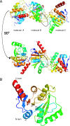
FIG. 1.
FIG. 1.
Ribbon representation of the SARS-CoV macro domain. (A) Two views rotated by 90° display the arrangement of the three molecules present in the asymmetric unit, each of them colored in a purple-to-red gradient (from N terminus to C terminus). In one molecule of the asymmetric unit only (molecule B), the ADP-ribose binding site is accessible to solvent and is surrounded by dotted lines. (B) Secondary structure elements are explicitly labeled.
FIG. 2.
FIG. 2.
ADP-ribose binding. (A) Representation of the σA-weighted Fo-Fc Fourier map, contoured at 3σ, as identified in the B molecule of the SARS-CoV macro domain during the first round of refinement. The final model of the ADP-ribose molecule is displayed in purple sticks in the purple electron density. The protein is represented in ribbons and colored according to secondary structure (α-helices in red, β-strands in yellow, and connecting loops in green). (B) Isothermal titration calorimetry profile for the binding of ADP-ribose to SARS-CoV macro domain.
FIG. 3.
FIG. 3.
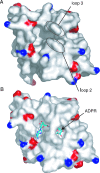
Conformational changes upon ADP-ribose binding. Panels A and B display the surface of the SARS-CoV macro domain colored according to electrostatic potential (positive and negative charges are indicated in blue and red, respectively), in its unliganded (A) and ADP-ribose bound (B) forms. Upon ADP-ribose binding, conformational changes implicating mainly loops 2 and 3 result in a small closing of the pocket around its ligand.
FIG. 4.
FIG. 4.
ADP-ribose 1"-phosphatase activity of viral macro domains. (A) Structure-based sequence alignment of the SARS-CoV macro domain sequence with macro domain sequences from Semliki Forest virus nsP3 (SFV), HCoV-OC43 nsp3, E. coli (protein COG2110), A. fulgidus (protein AF1521), Saccharomyces cerevisiae (protein YMX7_YEAST), and human (protein mH2A1.1). Strictly conserved residues are boxed in red, whereas residues for which the consensus is >70% are boxed in yellow. Secondary structure elements of the SARS-CoV macro domain are represented above the alignment. Yellow and orange thick arrows below the alignment indicate amino acids whose mutation decrease (yellow) or abolish (orange) ADP-ribose phosphatase activity. These are referred to in the text and in Fig. 5A and B. (B) TLC assay on the SARS-CoV macro domain activity. Markers were loaded in lanes 1 to 3, and the reaction mixture (described in Materials and Methods) was loaded in lane 4. Abbreviations: ADP-ribose (Appr), ADP-ribose 1"-phosphate (Appr1"-p), ADP-ribose 1",2"-cyclic phosphate (Appr>p). (C) TLC assay on the HEV macro domain activity. Lanes 1 to 5 contain reaction mixtures as indicated below the chromatogram. Abbreviations are the same as in panel B. Bovine serum albumin (BSA) was used as a negative control, and the yeast macro domain protein Poa1p (lane 5) was used as a positive control.
FIG. 5.
FIG. 5.
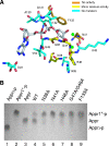
Structural determinants for the SARS-CoV macro domain ADP-ribose 1"-phosphatase activity. (A) The ADP-ribose molecule and the amino acid residues from the binding pocket are represented in sticks. Carbon atoms from the ADP-ribose molecule are shown in white, whereas carbon atoms from the surrounding residues are colored according to their role in ADP-ribose 1"-phosphatase activity: orange when substitution of the residue to alanine abolished activity and yellow when minor activity persists upon replacement by an alanine. For residues that were not replaced, carbon atoms are displayed in cyan. The two water molecules present in the ADP-ribose binding pocket are shown as green spheres. (B) TLC assay. Abbreviations are the same as in Fig. 4. In the first three lanes, Appr>p, Appr1"-p, and Appr were loaded as markers. In lanes 4 to 9, wild-type enzyme and several mutants (N38A, N41A, H46A, G47A/G48A, and F133A) were tested for their ADP-ribose 1"-phosphatase activity as described in Materials and Methods.
FIG. 6.
FIG. 6.
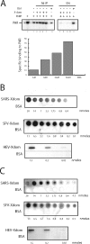
SARS-CoV, SFV, and HEV macro domains bind poly(ADP-Ribose) in vitro. (A) Ni precipitation of PAR bound to His-tagged SARS-CoV macro domain. BSA (50 μM, lanes 6 and 11) or increasing concentrations of the SARS-CoV macro domain (1 μΜ, lanes 2, 8, and 7; 5 μM, lanes 3, 9, and 8; 10 μM lanes 4, 10, and 9; 50 μM, lanes 5, 11, and 10) were incubated with radiolabeled PAR synthesized using hPARP-1 (lanes 2 to 11) and subsequently precipitated with Ni-beads (lanes 2 to 6). Unbound complexes present in the supernatant after Ni precipitation are shown in lanes 7 to 11. The amount of the radiolabeled PAR synthesized using hPARP-1 is shown as a control (lane c), and the amount of unspecific binding of PAR to Ni-beads is shown in lane 1. Migration position of PAR is shown on the left. After quantification, the ratio of the amount of PAR present after Ni precipitation over that present in the supernatant was calculated and used to determine the specific binding of PAR to the SARS-CoV macro domain. (B) 32P-labeled PAR-labeled PARP-1 was incubated with nitrocellulose membranes containing increasing concentrations of dot-blotted SARS-CoV, SFV, or HEV macro domains or BSA, which was used as a negative control. For each membrane, the same amount, indicated under the blot, of viral macro domain or control BSA was blotted. The membranes were analyzed by phosphoimaging. (C) Same experiment as in panel B except that membranes were incubated with 32P-labeled PAR purified after DNase and proteinase K treatment.
FIG. 7.
FIG. 7.
Model for PAR binding. Based on the position of ADP-ribose as observed in the crystal structure, modeling studies were performed to accommodate a di-ADP-ribose. One conformation for each of the best two clusters is represented in sticks (carbon atoms in pink and cyan, respectively). The SARS-CoV macro domain surface is colored as in Fig. 3.

References

    1. Aguiar, R. C., K. Takeyama, C. He, K. Kreinbrink, and M. A. Shipp. 2005. B-aggressive lymphoma family proteins have unique domains that modulate transcription and exhibit poly(ADP-ribose) polymerase activity. J. Biol. Chem. 280:33756-33765. - PubMed
    1. Allen, M. D., A. M. Buckle, S. C. Cordell, J. Lowe, and M. Bycroft. 2003. The crystal structure of AF1521 a protein from Archaeoglobus fulgidus with homology to the non-histone domain of macroH2A. J. Mol. Biol. 330:503-511. - PubMed
    1. Bick, M. J., J. W. Carroll, G. Gao, S. P. Goff, C. M. Rice, and M. R. MacDonald. 2003. Expression of the zinc-finger antiviral protein inhibits alphavirus replication. J. Virol. 77:11555-11562. - PMC - PubMed
    1. Brierley, I., P. Digard, and S. C. Inglis. 1989. Characterization of an efficient coronavirus ribosomal frameshifting signal: requirement for an RNA pseudoknot. Cell 57:537-547. - PMC - PubMed
    1. Cohen, S. X., R. J. Morris, F. J. Fernandez, M. Ben Jelloul, M. Kakaris, V. Parthasarathy, V. S. Lamzin, G. J. Kleywegt, and A. Perrakis. 2004. Towards complete validated models in the next generation of ARP/wARP. Acta Crystallogr. D Biol. Crystallogr. 60:2222-2229. - PubMed

Publication types

MeSH terms

LinkOut - more resources