. 2006 Sep;80(17):8566-81.
doi: 10.1128/JVI.00837-06.

Tertiary structural and functional analyses of a viroid RNA motif by isostericity matrix and mutagenesis reveal its essential role in replication

Affiliations

Tertiary structural and functional analyses of a viroid RNA motif by isostericity matrix and mutagenesis reveal its essential role in replication

Xuehua Zhong et al. J Virol. 2006 Sep.

Abstract

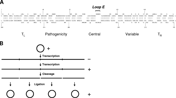
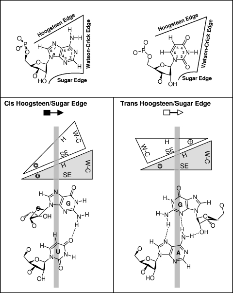
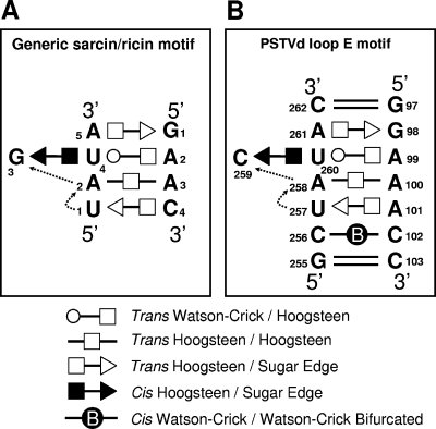
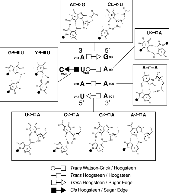
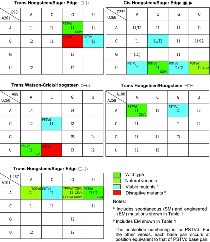
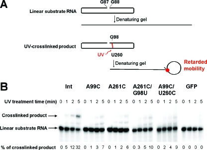
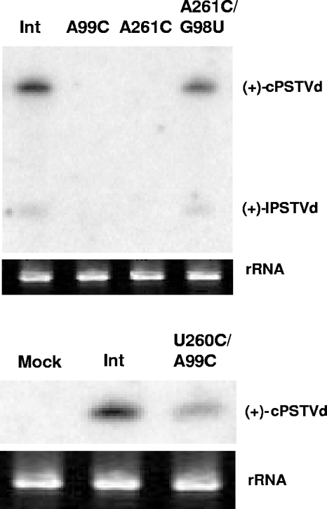
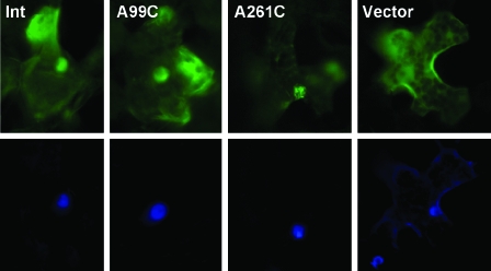
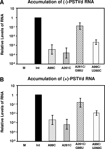
RNA-templated RNA replication is essential for viral or viroid infection, as well as for regulation of cellular gene expression. Specific RNA motifs likely regulate various aspects of this replication. Viroids of the Pospiviroidae family, as represented by the Potato spindle tuber viroid (PSTVd), replicate in the nucleus by utilizing DNA-dependent RNA polymerase II. We investigated the role of the loop E (sarcin/ricin) motif of the PSTVd genomic RNA in replication. A tertiary-structural model of this motif, inferred by comparative sequence analysis and comparison with nuclear magnetic resonance and X-ray crystal structures of loop E motifs in other RNAs, is presented in which core non-Watson-Crick base pairs are precisely specified. Isostericity matrix analysis of these base pairs showed that the model accounts for the reported natural sequence variations and viable experimental mutations in loop E motifs of PSTVd and other viroids. Furthermore, isostericity matrix analysis allowed us to design disruptive, as well as compensatory, mutations of PSTVd loop E. Functional analyses of such mutants by in vitro and in vivo experiments demonstrated that loop E structural integrity is crucial for replication, specifically during transcription. Our results suggest that the PSTVd loop E motif exists and functions in vivo and provide loss-of-function genetic evidence for the essential role of a viroid RNA three-dimensional motif in rolling-circle replication. The use of isostericity matrix analysis of non-Watson-Crick base pairing to rationalize mutagenesis of tertiary motifs and systematic in vitro and in vivo functional assays of mutants offers a novel, comprehensive approach to elucidate the tertiary-structure-function relationships for RNA motifs of general biological significance.


Figures

FIG. 1.
FIG. 1.
Secondary structure (A) and replication model (B) of PSTVd. (A) The rod-like secondary structure of PSTVd and the numbering of nucleotides are based on a report by Gross et al. (16). The five structural domains (26) include (i) the left terminal domain (TL, 1 to 46 and 315 to 359), (ii) the pathogenicity domain (47 to 73 and 286 to 314), (iii) the central domain (74 to 120 and 240 to 285), (iv) the variable domain (121 to 148 and 212 to 239), and (v) the right terminal domain (TR, 149 to 211). The loop E motif is in the central conserved region. (B) Asymmetric rolling-circle replication of PSTVd (5). The incoming circular monomeric plus-strand PSTVd serves as the initial template to synthesize linear, concatemeric minus-strand PSTVd. The latter functions as the replication intermediate to direct the synthesis of concatemeric plus-strand PSTVd, which is cleaved into unit length monomers and further ligated into circular forms.
FIG. 2.
FIG. 2.
Geometric classification of RNA base pairing. The upper panel shows that each nucleotide base has three edges (Watson-Crick, Hoogsteen, and sugar) that can potentially hydrogen bond with one of the three edges of another base. Thus, each base can be represented schematically by a triangle and can potentially pair with up to three other bases. The interacting bases can pair with a cis or trans relative orientation of their glycosidic bonds. This is illustrated in the lower panels for the cis and trans orientations of nucleotides pairing at the Hoogsteen edge of one base and the sugar edge of the second base. In these base pairs, the Watson-Crick edges of the interacting bases are available for further interactions—with other RNAs, proteins, or small molecules. The cross and circle in the triangle where the Hoogsteen and sugar edges meet indicate 5′→3′ and 3′→5′ orientations, respectively, of the sugar-phosphate backbones relative to the plane of the page. W-C, Watson-Crick edge; H, Hoogsteen edge; SE, sugar edge. (Adapted from reference with permission from the RNA Society.)
FIG. 3.
FIG. 3.
Tertiary-structural model of PSTVd loop E. (A) Paradigmatic sarcin/ricin motif based on X-ray crystal structures (adapted from reference with permission from Elsevier). (B) Inferred PSTVd loop E structural model. The dashed arrows indicate local changes in the strand orientation. All symbols that denote non-Watson-Crick base pairs and strand orientations are based on a report by Leontis and Westhof (34). Circles, squares, and triangles indicate the participation of Watson-Crick, Hoogsteen, and sugar edges, respectively. Open symbols indicate base pairs with a trans orientation of the glycosidic bonds, and closed symbols indicate base pairs with a cis orientation.
FIG. 4.
FIG. 4.
The five non-Watson-Crick base pairs that form the core of the PSTVd loop E motif. Examples of natural variants or experimentally designed mutations that can form isosteric base pairs are also illustrated. Y, pyrimidine (U or C); W, water molecule. Ribose is represented by a closed circle, except when it is involved in hydrogen bonding.
FIG. 5.
FIG. 5.
The functional status of viroid mutants and natural variants mapped onto the respective isostericity matrices for each base pair, according to the geometric family to which it belongs (30). I1 to I5 denote subfamilies within each of which the base pairs are isosteric (see Materials and Methods for the definition of isostericity and reference for details of the classification of subfamilies). Each blank and red box indicates that non-Watson-Crick base pairing between the two nucleotides in the family has not been reported in X-ray crystal structures or cannot be formed on the basis of isosteric predictions (30). Chrysanthemum stunt viroid (CSVd) has sequences identical to those of PSTVd in loop E. For sources of data and abbreviations of all other viroids, see Table 1.
FIG. 6.
FIG. 6.
Analysis of loop E formation by UV-cross-linking. Gel-purified substrate RNAs were preincubated under conditions that favor loop E formation and subjected to UV-cross-linking as described in Materials and Methods. (A) Diagram showing structural conformations of the substrate and cross-linked RNAs in their native state and in denaturing gels. G88 and G87 are the 5′ and 3′ ends, respectively, of the folded linear substrate RNA. The established cross-linking site (U260/G98) is marked in red. (B) Autoradiography of gel blots showing the presence or absence of UV-cross-linked products from the substrate RNAs as indicated. Efficiency of cross-linking is presented as the percentage of substrate RNAs that is converted into the cross-linked products. Note that UV treatment of the two compensatory mutants PSTVdIntA261C/G98U and PSTVdIntA99C/U260C, which are predicted by isostericity matrix analysis to restore loop E tertiary structure and showed restored replication, resulted in little cross-linked product formation. This indicates that G98/U260 base stacking, in addition to proper loop E tertiary structure, is required for specific UV cross-linking. The data from these two mutants thus provide evidence that UV cross-linking occurs specifically in loop E.
FIG. 7.
FIG. 7.
Northern blot assays showing accumulation of PSTVd RNAs in protoplasts of N. benthamiana. The rRNA served as a loading control. c-PSTVd and l-PSTVd indicate monomeric circular and monomeric linear PSTVd RNAs, respectively.
FIG. 8.
FIG. 8.
Nuclear import of fluorescently labeled in vitro transcripts of PSTVd. The transcripts of the wild type (Int) and the two mutants (A99C and A261C) appear in the nucleus within 30 min of injection into the cytoplasm (upper row). The vector transcripts do not show nuclear accumulation. DAPI staining (lower row) shows the position of the nucleus in each injected cell.
FIG. 9.
FIG. 9.
Circularization of loop E mutants in NbNE. Gel-purified and radiolabeled unit length linear plus-strand PSTVd transcripts were incubated in NbNE for 2 h and analyzed by urea-polyacrylamide gel electrophoresis. c-PSTVd and l-PSTVd indicate monomeric circular and monomeric linear PSTVd RNAs, respectively.
FIG. 10.
FIG. 10.
Determination of minus-strand PSTVd (A) and plus-strand PSTVd (B) RNA levels in infected N. benthamiana protoplasts by quantitative real-time RT-PCR. For details, see Materials and Methods. M, mock inoculation. To facilitate comparison, the accumulation level of the wild type (Int) was arbitrarily set to a value of 1 and the levels of the mutants are presented as relative values on a log scale.

Similar articles

Cited by

References

    1. Baulcombe, D. 2004. RNA silencing in plants. Nature 431:356-363. - PubMed
    1. Baumstark, T., and D. Riesner. 1995. Only one of four possible secondary structures of the central conserved region of potato spindle tuber viroid is a substrate for processing in a potato nuclear extract. Nucleic Acids Res. 23:4246-4254. - PMC - PubMed
    1. Baumstark, T., A. R. Schroder, and D. Riesner. 1997. Viroid processing: switch from cleavage to ligation is driven by a change from a tetraloop to a loop E conformation. EMBO J. 16:599-610. - PMC - PubMed
    1. Branch, A. D., B. J. Benenfeld, and H. D. Robertson. 1985. Ultraviolet light-induced crosslinking reveals a unique region of local tertiary structure in potato spindle tuber viroid and HeLa 5S RNA. Proc. Natl. Acad. Sci. USA 82:6590-6594. - PMC - PubMed
    1. Branch, A. D., and H. D. Robertson. 1984. A replication cycle for viroids and other small infectious RNA's. Science 223:450-455. - PubMed

Publication types

MeSH terms

LinkOut - more resources