. 2006 Sep;80(17):8763-77.
doi: 10.1128/JVI.00598-06.

Interaction between vaccinia virus extracellular virus envelope A33 and B5 glycoproteins

Affiliations

Interaction between vaccinia virus extracellular virus envelope A33 and B5 glycoproteins

Beatriz Perdiguero et al. J Virol. 2006 Sep.

Abstract

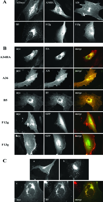
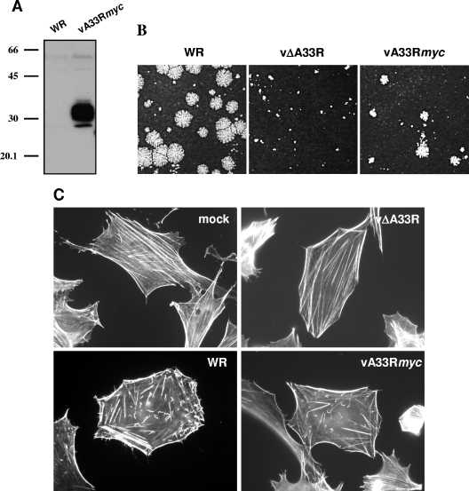
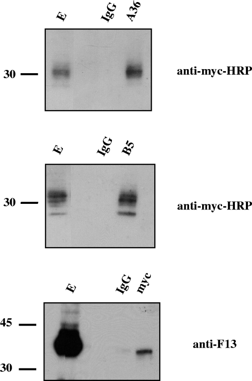
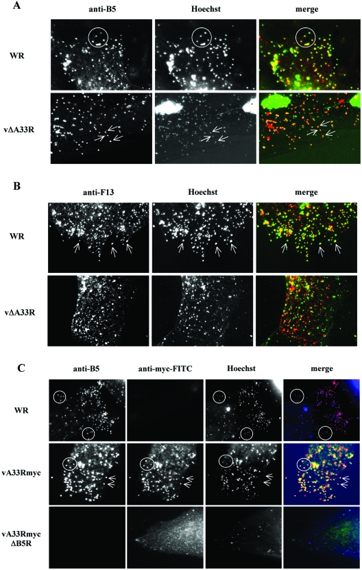
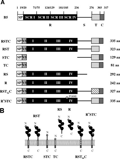
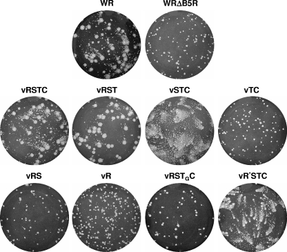
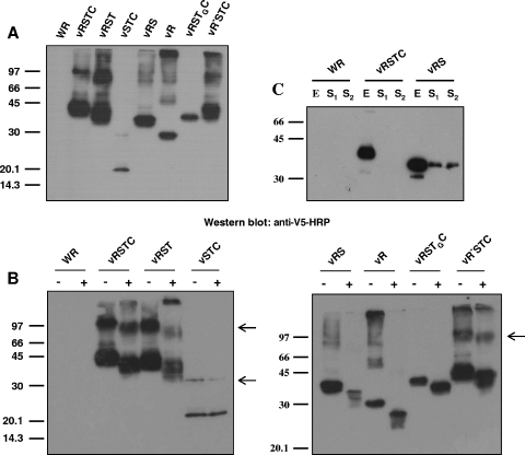
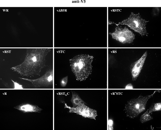
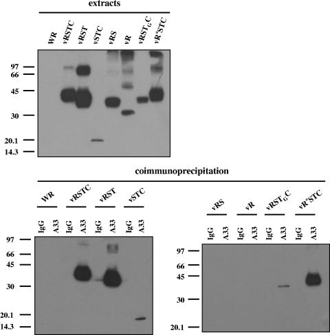
The extracellular form of vaccinia virus acquires its outer envelope by wrapping with cytoplasmic membranes that contain at least seven virus-encoded proteins, of which four are glycoproteins. We searched for interactions between the vaccinia virus A33 glycoprotein and proteins A34, A36, B5, F12, and F13. First, when myc epitope-tagged A33 was expressed in combination with other envelope proteins, A33 colocalized with B5 and A36, suggesting that direct A33-B5 and A33-A36 interactions occur in the absence of infection. A recombinant vaccinia virus (vA33Rmyc) was constructed by introduction of the myc-tagged A33 version (A33myc) into A33-deficient vaccinia virus. A33myc partially restored plaque formation and colocalized with enveloped virions in infected cells. Coimmunoprecipitation experiments with extracts of vA33Rmyc-infected cells confirmed the existence of a physical association of A33 with A36 and B5. Of these, the A33-B5 interaction is a novel finding, whereas the interaction between A33 and A36 has been previously characterized. A collection of vaccinia viruses expressing mutated versions of the B5 protein was used to investigate the domain(s) of B5 required for interaction with A33. Both the cytoplasmic domain and most of the extracellular domain, but not the transmembrane domain, of the B5 protein were dispensable for binding to A33. Mutations in the extracellular portions of B5 and A33 that enhance extracellular virus release did not affect the interaction between the two. In contrast, substituting the B5 transmembrane domain with that of the vesicular stomatitis virus G glycoprotein prevented the association with A33. Immunofluorescence experiments on virus mutants indicated that B5 is required for efficient targeting of A33 into enveloped virions. These results point to the transmembrane domain of B5 as the major determinant of the A33-B5 interaction and demonstrate that protein-protein interactions are crucial in determining the composition of the virus envelope.


Figures

FIG. 1.
FIG. 1.
Interactions of A33 protein with IEV envelope proteins in transfected cells. (A) Intracellular distribution of IEV envelope proteins individually expressed. Immunofluorescence staining was performed on BHK-21 cells transfected with plasmid pcDNA3.1/myc-HisB-A33R (A33myc), pcDNA 3.1/HA-HisB-A34R (A34HA), pcDNA3.1/A36R (A36), pSG5-B5R (B5), pQBI-F12L (F12g), or pcDNA3.1/p37g (F13g). At 24 h posttransfection, cells were fixed, permeabilized, and directly observed (F12g and F13g) or incubated with the corresponding specific antibodies to myc, HA, A36, or B5. Note the diffuse staining pattern of proteins F12 and F13 fused to GFP. (B) Coexpression of A33 with IEV envelope proteins. Immunofluorescence staining was carried out on BHK-21 cells transfected with pcDNA3.1/myc-HisB-A33R together with plasmids expressing the proteins indicated on the left. At 24 h posttransfection, cells were fixed, permeabilized, stained with specific antibodies (anti-myc, anti-HA-fluorescein, anti-myc-FITC, anti-A36, or anti-B5 followed by secondary rabbit anti-mouse IgG-TRITC), observed and photographed under a fluorescence microscope. The merged images result from the combination of the myc signal corresponding to A33myc and the signal corresponding to the second protein. (C) Distribution of A33 protein after transfection of a cell line stably expressing B5 protein. (a) BHK-21 cells transfected with plasmid pcDNA 3.1/myc-HisB-A33R were fixed at 24 h posttransfection, permeabilized, and incubated with anti-myc-FITC. (b) BHK-21 cells stably expressing B5 protein (BHK-B5) were fixed, permeabilized, and incubated with anti-B5 followed by anti-mouse-TRITC. (c) BHK-B5 cells transfected with plasmid pcDNA 3.1/myc-HisB-A33R were fixed at 24 h posttransfection, permeabilized, and incubated with anti-myc-FITC, anti-B5, and anti-mouse-TRITC. Note the colocalization of A33 and B5 signals in the merged image.
FIG. 2.
FIG. 2.
Characterization of a recombinant vaccinia virus expressing a myc-tagged version of the A33 protein. (A) Analysis of A33myc expression by Western blotting. Extracts of BSC-1 cells infected with the recombinant vaccinia virus expressing fusion protein A33myc (vA33Rmyc) or control virus (WR) were probed with anti-myc-HRP antibody. Positions of protein molecular mass markers (in kDa) are indicated. (B) Plaque formation by vA33Rmyc. BSC-1 cell monolayers infected with WR, parental vΔA33R, or vA33Rmyc were incubated for 2 days, stained with crystal violet, and photographed. (C) Induction of actin tails by vA33Rmyc. BHK-21 cells infected for 7 h were fixed, permeabilized, and incubated with 1 unit/ml Alexa594-phalloidin. Note the presence of actin tails in vA33Rmyc-infected cells.
FIG. 3.
FIG. 3.
Coimmunoprecipitation of A33 from infected cells. BSC-1 cells were infected with five PFU per cell of vA33Rmyc and lysed at 24 h postinfection. Immunoprecipitations were performed with either control immunoglobulin G (lanes IgG) or anti-myc, anti-A36, or anti-B5 antibodies (indicated at the top of each panel). Western blots were probed with the antibodies indicated on the right side of each panel. E, infected cell extract prior to immunoprecipitation. Immunoprecipitations were performed by use of digitonin-containing buffer, except for the case in which antibody to A36 was used, which was performed using NP-40 as detergent. Molecular-mass markers (in kDa) are indicated.
FIG. 4.
FIG. 4.
Distribution of IEV envelope proteins in A33- or B5-deficient viruses. BHK-21 cells were infected for 7 h, fixed, permeabilized, and stained with antibodies against IEV envelope protein A33, B5, or F13. DNA was visualized by Hoechst staining. (A) Colocalization of B5 and virus particles. Cells infected with WR or A33-deficient virus (vΔA33R) were stained with anti-B5 antibody. Merged images result from the combination of monochrome images pseudocolored to red (anti-B5 antibodies) and green (Hoechst). Note the colocalization of B5 protein with viral DNA in cells infected with WR (circles) and vΔA33R (arrows). (B) Colocalization of F13 and virus particles. Cells infected with WR or A33-deficient virus (vΔA33R) were stained with anti-F13 antibody. Merged images result from the combination of monochrome images pseudocolored to red (anti-F13 antibodies) and green (Hoechst). Note that the colocalization of F13 protein with viral DNA in cells infected with WR (arrows) is affected in vΔA33R-infected cells. (C) Colocalization of A33 and virus particles. Cells infected with WR, vA33Rmyc, or the corresponding B5R deletion mutant vA33RmycΔB5R (indicated on the left) were stained with anti-B5 and anti-myc antibodies. Merged images result from the combination of monochrome images pseudocolored to red (anti-B5), green (anti-myc), and blue (Hoechst). Note that, in the absence of B5 protein, no colocalization of A33myc with viral DNA is observed.
FIG. 5.
FIG. 5.
V5-tagged, mutated versions of the B5 protein. (A) Schematic representation of V5-tagged B5 versions used in this study. SP, signal peptide; V5, V5 epitope; R, four short consensus repeats (SCR I to IV); S, stalk; T, transmembrane domain; C, cytoplasmic tail; aa, amino acids. Versions were termed, as indicated on the left, according to the domains included in the construct. TG indicates the transmembrane domain of VSV G protein. R* indicates an R region including a P189S mutation located in SCR IV. (B) Predicted topology of mutated B5 versions in the outer membrane of the EEV particle. Domains above the lipid bilayer are in the lumen of the wrapping membranes or the extracellular space, whereas domains below the membrane are cytosolic. The N and C termini are indicated.
FIG. 6.
FIG. 6.
Plaque phenotypes of recombinant viruses expressing V5-tagged, mutated B5 versions. BSC-1 cell monolayers infected with WR, parental WRΔB5R, or the recombinant viruses indicated were incubated for 2 days, stained with crystal violet, and photographed.
FIG. 7.
FIG. 7.
Characterization of V5-tagged, mutated B5 versions. (A) Western blot analysis. BSC-1 cells were infected with the viruses indicated at the top of the panel at five PFU per cell for 24 h. Western blots were probed with anti-V5-HRP antibody. (B) Analysis of N glycosylation. BSC-1 cells infected with the viruses indicated in the presence (+) or absence (−) of 1 μg/ml tunicamycin were harvested at 24 h postinfection. Western blots were probed with antibody against the V5 epitope conjugated with horseradish peroxidase. Arrowheads point to B5 complexes, which are absent in extracts of cells infected with viruses expressing B5 versions RS, R, and RSTGC. Positions of protein molecular-mass markers are indicated (in kDa). (C) Secretion of B5 soluble version from vRS-infected cells. BSC-1 cells were infected with the viruses indicated at five PFU per cell, harvested at 24 h postinfection, and probed with anti-V5-HRP antibody. E, total cell lysate; S1, culture medium filtered through 0.1-μm filters (to eliminate vaccinia virus) and concentrated; S2, culture medium concentrated. Note the presence of the B5 version in the supernatant of vRS-infected cells.
FIG. 8.
FIG. 8.
Intracellular distribution of V5-tagged B5 versions in WR-infected cells. BHK-21 cells infected with the viruses indicated at five PFU per cell were fixed at 7 h postinfection, permeabilized, and labeled with antibody to the V5 epitope followed by secondary rabbit anti-mouse IgG-TRITC to detect the distributions of the different B5 versions.
FIG. 9.
FIG. 9.
Mapping of the A33-B5 interaction site by coimmunoprecipitation. BSC-1 cells were infected with the viruses indicated above the panels at five PFU per cell and harvested at 24 h postinfection. The top panel shows infected cell extracts prior to immunoprecipitation, probed with anti-V5 antibody conjugated with horseradish peroxidase. Immunoprecipitations were performed with either control immunoglobulin G (lanes IgG) or anti-A33 antibody (lanes A33), as indicated above the lower panels. Immunoprecipitated material was resolved by SDS-PAGE and subjected to Western blot analysis with anti-V5 antibody conjugated with horseradish peroxidase. Molecular-mass markers are indicated (in kDa).

Similar articles

Cited by

References

    1. Blasco, R., and B. Moss. 1991. Extracellular vaccinia virus formation and cell-to-cell virus transmission are prevented by deletion of the gene encoding the 37,000-dalton outer envelope protein. J. Virol. 65:5910-5920. - PMC - PubMed
    1. Blasco, R., and B. Moss. 1992. Role of cell-associated enveloped vaccinia virus in cell-to-cell virus spread. J. Virol. 66:4170-4179. - PMC - PubMed
    1. Blasco, R., and B. Moss. 1995. Selection of recombinant vaccinia viruses on the basis of plaque formation. Gene 158:157-162. - PubMed
    1. Blasco, R., J. R. Sisler, and B. Moss. 1993. Dissociation of progeny vaccinia virus from the cell membrane is regulated by a viral envelope glycoprotein: effect of a point mutation in the lectin homology domain of the A34R gene. J. Virol. 67:3319-3325. - PMC - PubMed
    1. Borrego, B., M. M. Lorenzo, and R. Blasco. 1999. Complementation of P37 (F13L gene) knock-out in vaccinia virus by a cell line expressing the gene constitutively. J. Gen. Virol. 80:425-432. - PubMed

Publication types

MeSH terms

LinkOut - more resources