. 2006 Sep;80(17):8778-86.
doi: 10.1128/JVI.00831-06.

Marek's disease virus encodes MicroRNAs that map to meq and the latency-associated transcript

Affiliations

Marek's disease virus encodes MicroRNAs that map to meq and the latency-associated transcript

Joan Burnside et al. J Virol. 2006 Sep.

Abstract

MicroRNAs (miRNAs) are a class of small (approximately 22-nucleotide) regulatory molecules that block translation or induce degradation of target mRNAs. These have been identified in a wide range of organisms, including viruses. In particular, the oncogenic gammaherpesviruses Kaposi's sarcoma herpesvirus and Epstein-Barr virus encode miRNAs that could potentially regulate either viral or host genes. To determine if Marek's disease virus (MDV), an oncogenic alphaherpesvirus of chickens, encodes miRNAs, we isolated small RNAs from MDV-infected chicken embryo fibroblasts (CEF) and used the 454 Life Sciences sequencing technology to obtain the sequences of 13,679 candidate host and viral small RNAs. Eight miRNAs were found, five of which flank the meq oncogene and three that map to the latency-associated transcript (LAT) region of the genome. The meq gene is unique to pathogenic serotypes of MDV and is transcriptionally active during latency and transformation, and the LAT region of the MDV genome is antisense to the immediate-early gene ICP4. Secondary structure analysis predicted that the regions flanking the miRNAs could form hairpin precursors. Northern blot analysis confirmed expression of all miRNAs in MDV-infected CEF, MDV-induced tumors, and MDV lymphoblastoid cell lines. We propose that the MDV miRNAs function to enable MDV pathogenesis and contribute to MDV-induced transformation of chicken T cells.


Figures

FIG. 1.
FIG. 1.
Secondary structure predictions of MDV pre-miRNAs. Folding and minimum free energy calculations were performed with mfold (80) without constraints. The mature cloned microRNAs are shown in bold, and the * strands are in italics (MDV-miR*).
FIG. 2.
FIG. 2.
Genomic location of MDV miRNAs. (A) Schematic and sequence of the MDV miRNAs flanking meq. (B) Schematic and sequence of the MDV miRNAs downstream of ICP4. Nucleotide and MDV gene numbering are according to Md5 (AF243438). In the schematics, small arrowheads indicate the locations of MDV miRNAs, and in the sequence, the individual miRNAs are shown in red and by an overline. A bent arrow depicts the transcriptional start site of the meq gene, and the TATAA and ATG for meq are boxed. Large orange arrows correspond to MDV transcripts, and the open reading frames are the hatched areas. The green arrow indicates the a-like sequence. Spliced introns are shown as solid lines linking exons. Additional spliced variants of the MSRs are described in reference .
FIG. 3.
FIG. 3.
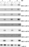
Northern blot of FlashPage-fractionated RNA (1 μg/lane) from uninfected CEFs (-) or CEFs infected with MDV (+) for 1, 3, or 5 days. RNA was electrophoresed on a 15% denaturing polyacrylamide gel, blotted, and hybridized to 32P-labeled oligos antisense to MDV miRNAs. Blots were stripped and hybridized to the miR-21 antisense probe to show the presence of microRNA in all lanes (a representative blot of this set is shown).
FIG. 4.
FIG. 4.
Northern blot analysis of FlashPage-fractionated RNA (1 μg/lane) from normal chicken spleen cells and MDV-induced tumors. Blots were prepared as described in the legend to Fig. 3.
FIG. 5.
FIG. 5.
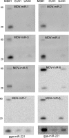
Northern blot analysis of PEG-purified RNA (1 μg/lane) from MSB1, CU91, and UA30 cells. Blots were prepared as described in the legend to Fig. 3, except that blots were hybridized to the miR-221 antisense probe as a loading control (only a representative blot is shown). Positions of DNA ladder markers are shown on the left.

References

    1. Afonso, C. L., E. R. Tulman, Z. Lu, L. Zsak, D. L. Rock, and G. F. Kutish. 2001. The genome of turkey herpesvirus. J. Virol. 75:971-978. - PMC - PubMed
    1. Akiyama, Y., S. Kato, and N. Iwa. 1973. Continuous cell culture from lymphoma of Marek's disease. Biken J. 16:177-179. - PubMed
    1. Altschul, S. F., T. L. Madden, A. A. Schäffer, J. Zhang, Z. Zhang, W. Miller, and D. J. Lipman. 1997. Gapped BLAST and PSI-BLAST: a new generation of protein database search programs. Nucleic Acids Res. 25:3389-3402. - PMC - PubMed
    1. Ambros, V., B. Bartel, D. P. Bartel, C. B. Burge, J. C. Carrington, X. Chen, G. Dreyfuss, S. R. Eddy, S. Griffiths-Jones, M. Marshall, M. Matzke, G. Ruvkun, and T. Tuschl. 2003. A uniform system for microRNA annotation. RNA 9:277-279. - PMC - PubMed
    1. Aravin, A. A., M. Lagos-Quintana, A. Yalcin, M. Zavolan, D. Marks, B. Snyder, T. Gaasterland, J. Meyer, and T. Tuschl. 2003. The small RNA profile during Drosophila melanogaster development. Dev. Cell 5:337-350. - PubMed

Publication types

MeSH terms