. 2006 Sep;133(18):3587-95.
doi: 10.1242/dev.02539. Epub 2006 Aug 16.

Mesodermal expression of Tbx1 is necessary and sufficient for pharyngeal arch and cardiac outflow tract development

Affiliations

Mesodermal expression of Tbx1 is necessary and sufficient for pharyngeal arch and cardiac outflow tract development

Zhen Zhang et al. Development. 2006 Sep.

Abstract

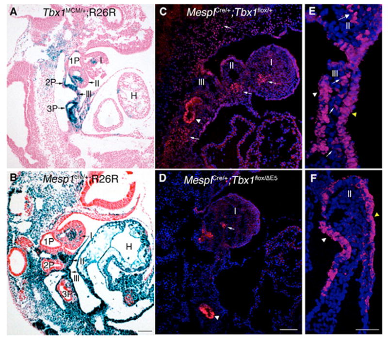
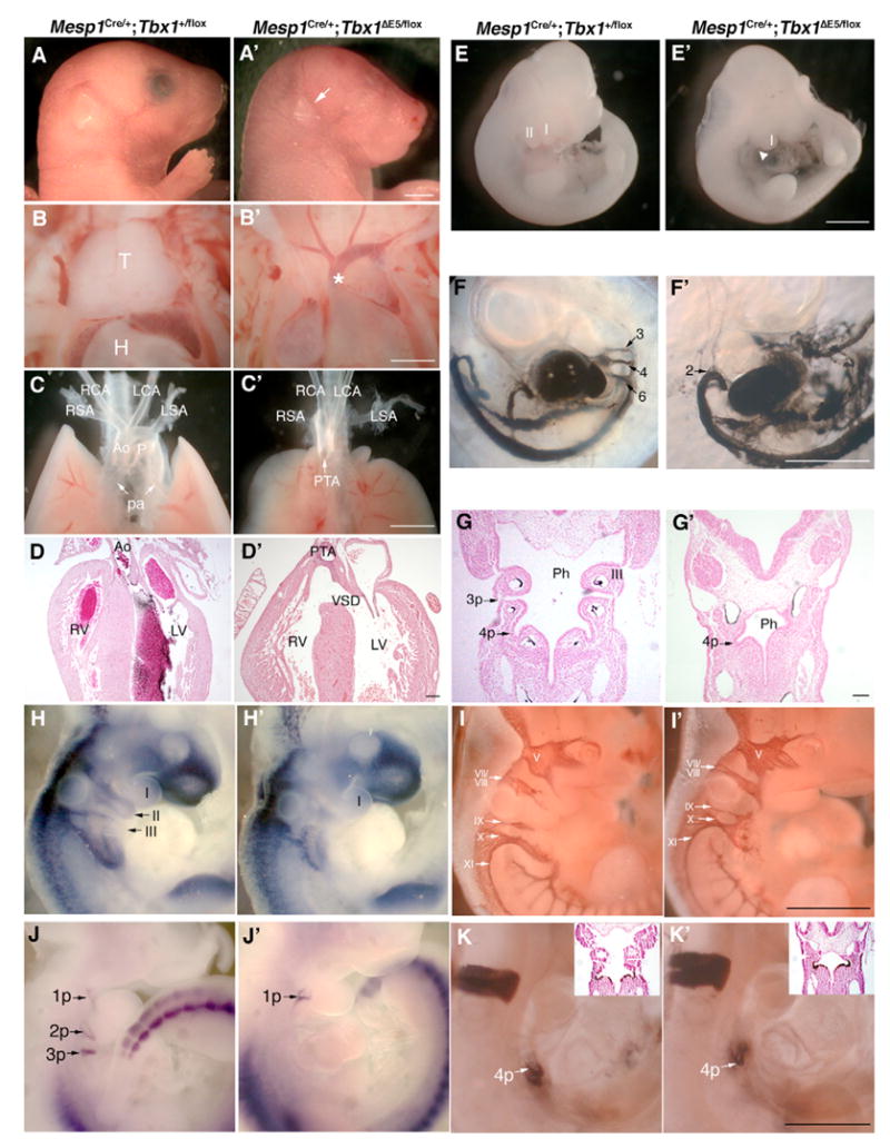
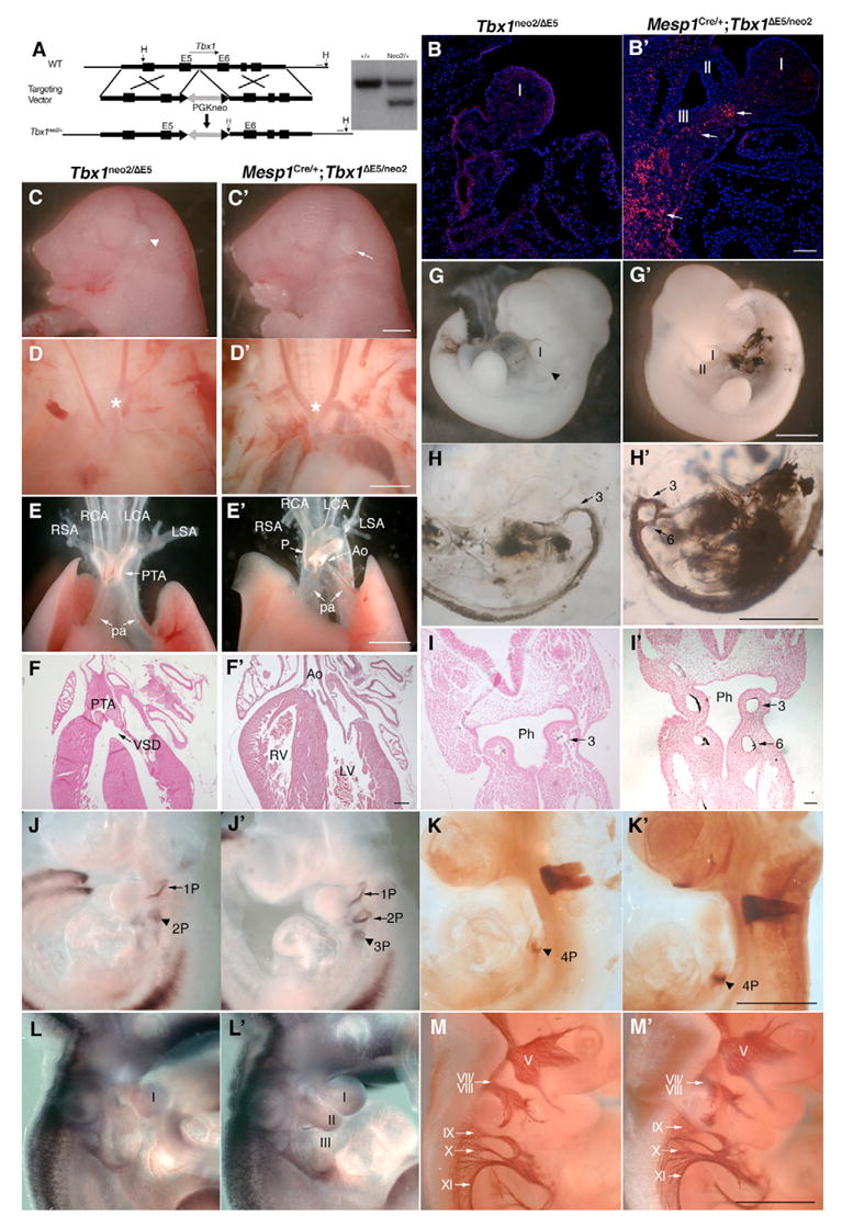
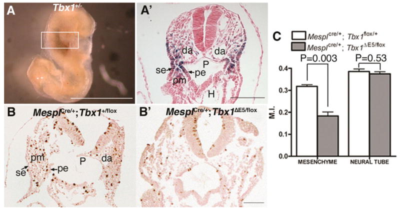
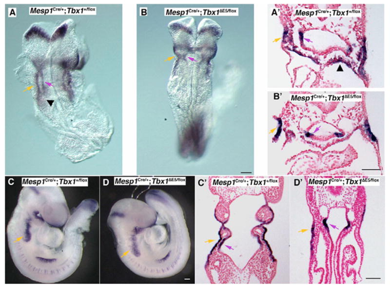
The development of the segmented pharyngeal apparatus involves complex interaction of tissues derived from all three germ layers. The role of mesoderm is the least studied, perhaps because of its apparent lack of anatomical boundaries and positionally restricted gene expression. Here, we report that the mesoderm-specific deletion of Tbx1, a T-box transcription factor, caused severe pharyngeal patterning and cardiovascular defects, while mesoderm-specific restoration of Tbx1 expression in a mutant background corrected most of those defects in the mouse. We show that some organs, e.g. the thymus, require Tbx1 expression in the mesoderm and in the epithelia. In addition, these experiments revealed that different pharyngeal arches require Tbx1 in different tissues. Finally, we show that Tbx1 in the mesoderm is required to sustain cell proliferation. Thus, the mesodermal transcription program is not only crucial for cardiovascular development, but is also key in the development and patterning of pharyngeal endoderm.


Figures

Fig. 1
Fig. 1. Mesodermal-specific deletion of Tbx1 by Mesp1Cre
(A–D) Sagittal sections of E9.5 embryos. (A) Distribution of Tbx1-traced cells visualized by crossing Tbx1mcm/+ mice with the reporter R26R, compared with the distribution of Mesp1Cre-traced cells (B). Recombination is absent in the endoderm in the latter experiment. (C,D) Immunofluorescent staining with an anti-Tbx1 antibody on control (C) and conditional mutant (D) E9.5 embryos. Tbx1 immunoreactivity is preserved in the endoderm (arrowheads) and, to a lesser extent, in the core of the 1st pharyngeal arch (arrow in D), but not in other mesodermal domains (arrows). (E,F) Coronal sections of E9 embryos. Immunofluorescent staining with an anti-Tbx1 antibody on control (E) and M-ko embryos (F). Mesodermal expression (arrows) is eliminated in M-ko embryos, but endodermal (white arrowhead) and ectodermal (yellow arrowhead) expression are maintained. I, II and III, 1st, 2nd and 3rd pharyngeal arches; 1P, 2P and 3P, 1st, 2nd and 3rd pharyngeal pouches. Scale bar: 100 μm in A–D; 50 μm in E,F.
Fig. 2
Fig. 2. Mesodermal ablation of Tbx1 causes severe pharyngeal and cardiovascular abnormalities
(A–K) Mesp1Cre/+;Tbx1+/flox control embryos compared with (A’–K’) Mesp1Cre/+;TbxΔE5/flox M-ko mutant embryos. Hypoplastic external ears (arrow A,A), absence of thymus (T and asterisk, B,B), persistent truncus arteriosus (PTA, C,C) originating from the right ventricle (D,D) and ventricular septal defect (VSD, D,D) in M-ko embryos at E18.5. (E,E) Hypoplasia of the 2nd pharyngeal arch (arrowhead) in an E10.5 M-ko embryo. (F,F) Cardiac ink injection in E10.5 embryos visualized the 3rd, 4th and 6th pharyngeal arch arteries in cotrols (F); M-ko embryos have only one pair of arteries connecting the aortic sac with the dorsal aorta (F). (G,G) Coronal sections of E10.5 embryos revealed severe hypoplasia of the pharynx (Ph) in M-ko mutants. (H,H) Whole-mount RNA in situ hybridization on E10 embryos using a Crabp1 probe as a neural crest marker. Conditional deletion mutants showed abnormal Crabp1 expressing pattern. (I,I) Whole-mount immunohistochemistry on E10 embryos with an anti-neurofilament M antibody revealed abnormalities of cranial nerve pathways in the mutant. (J,J) Whole-mount RNA in situ hybridization on E10 embryos with a Pax1 probe revealed a small 1st pouch and loss of labeling of pouches 2 and 3 in M-ko mutants. (K,K) Whole-mount immunohistochemistry on E10 embryos with an anti-Hoxb1 antibody revealed a normally specified, albeit slightly smaller, 4th pouch in M-ko mutants. The insets show coronal sections of the same embryos; the 4th pouches of M-ko embryos are not fully developed and not as close to the surface ectoderm as those of control embryos. T, thymus; H, heart; Ph, pharynx; Ao, aorta; P, pulmonary trunk; Pa, pulmonary artery; VSD, ventricular septum defect; RV/LV, right/left ventricle; RSA/LSA, right/left subclavian artery; RCA/LCA, right/left carotid artery; I, II and III, 1st, 2nd and 3rd pharyngeal arch; 3, 4 and 6, 3rd, 4th and 6th pharyngeal arch artery; 1p, 2p, 3p and 4p, 1st, 2nd, 3rd and 4th pharyngeal pouches; V, trigeminal nerve; VII/VIII, facial/acoustic nerve; IX, glossopharyngeal nerve; X, vagus nerve; XI, accessory nerve. Scale bars: 2 mm in A; 1 mm in B,C,E,F,I,K’; 100 μm in D and G.
Fig. 3
Fig. 3. Phenotypic analysis of compound mutants Tbx1neo2/− and conditional rescue mutants Mesp1Cre/+;Tbx1ΔE5/neo2
(A) Scheme of gene targeting strategy. H, HindIII. (B–M) Compound mutants Tbx1neo2/. (B’–M’) Conditional rescue mutants Mesp1Cre/+;Tbx1ΔE5/neo2. (B,B) Immunohistochemistry using an anti-Tbx1 antibody on sagittal sections of E9.5 embryos showed very low or undetectable signal in compound mutants (B) and reactivated expression in the mesoderm (arrows) of the rescued embryo (B). (C,C) External ears (arrowhead) are hypoplastic in compound mutants but are of normal size in rescued mutants (arrow) at E18.5. (D,D) Absence of thymus indicated by asterisk in both compound and rescued mutants at E18.5. (E,E,F,F) The cardiac outflow tract phenotype was normal in rescued embryos at E18.5. (G,G) The hypoplasia of the 2nd pharyngeal arch of compound mutants (arrowhead) was corrected in rescued embryos. (H,H) Ink injection into the heart of E10.5 embryos revealed normalization of the 6th pharyngeal arch arteries but not of the 4th pharyngeal arch arteries in rescued mutants. This is also evident from histological sections (I,I). (J,J) Whole-mount RNA in situ hybridization on E10 embryos with a PaxI probe revealed normalization of the 2nd pharyngeal pouch but only partial development of the 3rd pharyngeal pouch (arrowhead in J) in rescued embryos. (K,K) Whole-mount immunohistochemistry on E10 embryos with an anti-Hoxb1 antibody showed a more robust labeling of the 4th pouch (arrowhead) in rescued embryos. (L,L) Whole-mount RNA in situ hybridization on E10 embryos with a Crabp1 probe revealed only a marginal improvement of neural crest cell distribution in rescued embryos (compare with the normal pattern in Fig. 2H). Similarly, immunohistochemistry on E10 embryos using an anti neurofilament M antibody showed only modest improvement in rescued embryos, especially in the caudal region of the pharynx (M,M), compare with normal pattern in Fig. 2I). Ph, pharynx; Ao, aorta; P, pulmonary trunk; Pa, pulmonary artery; VSD, ventricular septum defect; RV/LV, right/left ventricle; RSA/LSA, right/left subclavian artery; RCA/LCA, right/left carotid artery; I and II, 1st and 2nd pharyngeal arch; 3 and 6: 3rd and 6th pharyngeal arch artery; 1p, 2p, 3p and 4p, 1st, 2nd, 3rd and 4th pharyngeal pouches; V, trigeminal nerve; VII/VIII, facial/acoustic nerve; IX, glossopharyngeal nerve; X, vagus nerve; XI, accessory nerve. Scale bars: 2mm in C; 1 mm in D,E,G,H,K,M; 100 μm in B,F,I.
Fig. 4
Fig. 4. Tbx1 regulates cell proliferation in mesenchyme cells
(A,A’) Tbx1 expression domain revealed by a Tbx1-lacZ knock-in allele (Tbx1+/−) at E8.5. (A) Transverse section of A. The area where cell proliferation was scored is indicated by the white box in A. (B) Transverse sections of E8.5 Mesp1Cre/+; Tbx1+/flox embryos as controls. (B’) Transverse sections of E8.5 Mesp1Cre/+; Tbx1ΔE5/flox embryos. (B,B) Immunohistochemistry staining with an anti-phospho-Histone H3 antibody to evaluate cell proliferation. (C) Mitotic index (M.I.) in pharyngeal mesenchyme at E8.5 embryos. Cells in neural tube were counted as an internal control. P values were calculated using Student’s t-test. da, dorsal aorta; se, surface ectoderm; pe, pharyngeal ectoderm; pm, pharyngeal mesenchyme; P, pharynx. Scale bars: 1 mm in A; 100 μm in A-B.
Fig. 5
Fig. 5. Mesodermal Tbx1 affects mesodermal but not epithelial Fgf8 expression
(A–D) Whole-mount RNA in situ hybridization with an Fgf8 probe. (A’,B’) Transverse sections of the E8.5 embryos shown in A and B, respectively. (C’,D’) Coronal sections of the E9 embryos shown in C and D, respectively. Orange arrows indicate Fgf8 expression in the ectoderm. Pink arrows indicate Fgf8 expression in endoderm. Black arrowheads indicate Fgf8 expression in splanchnic mesoderm/anterior heart field. Scale bar: 100 μm in B,D,D; 50 μm in B.

References

    1. Albrecht U, Eichele G, Helms JA, Lu HC. Visualization of gene expression patterns by in situ hybridization. In: Daston GP, editor. Molecular and Cellular Methods in Developmental Toxicology. New York: CRC Press; 1997. pp. 23–48.
    1. Arnold JS, Werling U, Braunstein EM, Liao J, Nowotschin S, Edelmann W, Hebert JM, Morrow BE. Inactivation of Tbx1 in the pharyngeal endoderm results in 22q11DS malformations. Development. 2006;133:977–987. - PubMed
    1. Ataliotis P, Ivins S, Mohun TJ, Scambler PJ. XTbx1 is a transcriptional activator involved in head and pharyngeal arch development in Xenopus laevis. Dev Dyn. 2005;232:979–991. - PubMed
    1. Chapman DL, Garvey N, Hancock S, Alexiou M, Agulnik SI, Gibson-Brown JJ, Cebra-Thomas J, Bollag RJ, Silver LM, Papaioannou VE. Expression of the T-box family genes, Tbx1-Tbx5, during early mouse development. Dev Dyn. 1996;206:379–390. - PubMed
    1. Chen A, Francis M, Ni L, Cremers CW, Kimberling WJ, Sato Y, Phelps PD, Bellman SC, Wagner MJ, Pembrey M, et al. Phenotypic manifestations of branchio-oto-renal syndrome. Am J Med Genet. 1995;58:365–370. - PubMed

Publication types

MeSH terms