. 2006 Sep;133(18):3549-62.
doi: 10.1242/dev.02512. Epub 2006 Aug 16.

Dynamic regulation of Drosophila nuclear receptor activity in vivo

Affiliations

Dynamic regulation of Drosophila nuclear receptor activity in vivo

Laura Palanker et al. Development. 2006 Sep.

Abstract

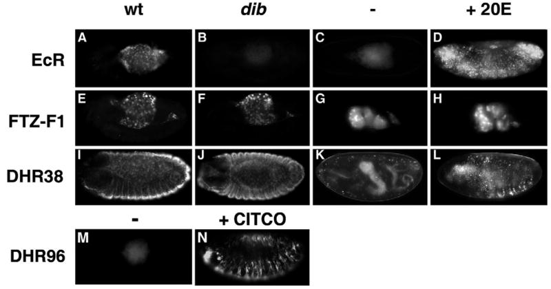
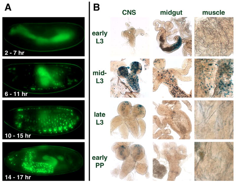
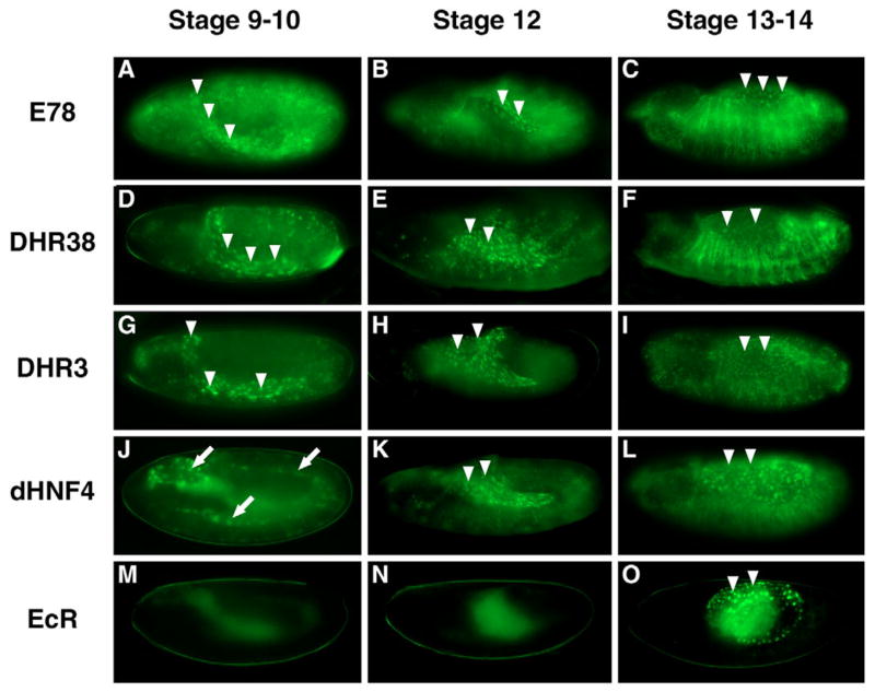
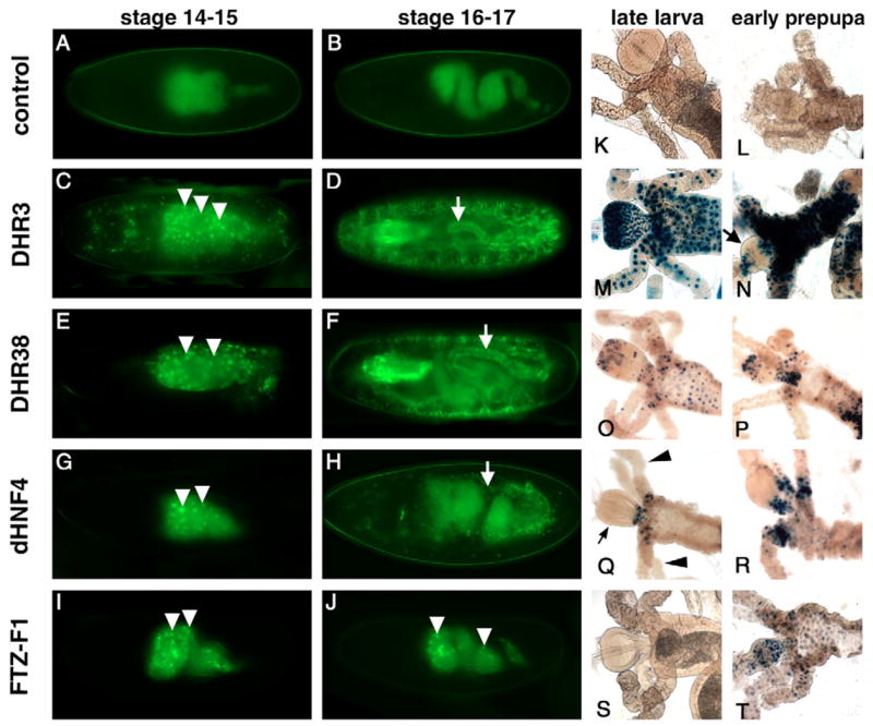
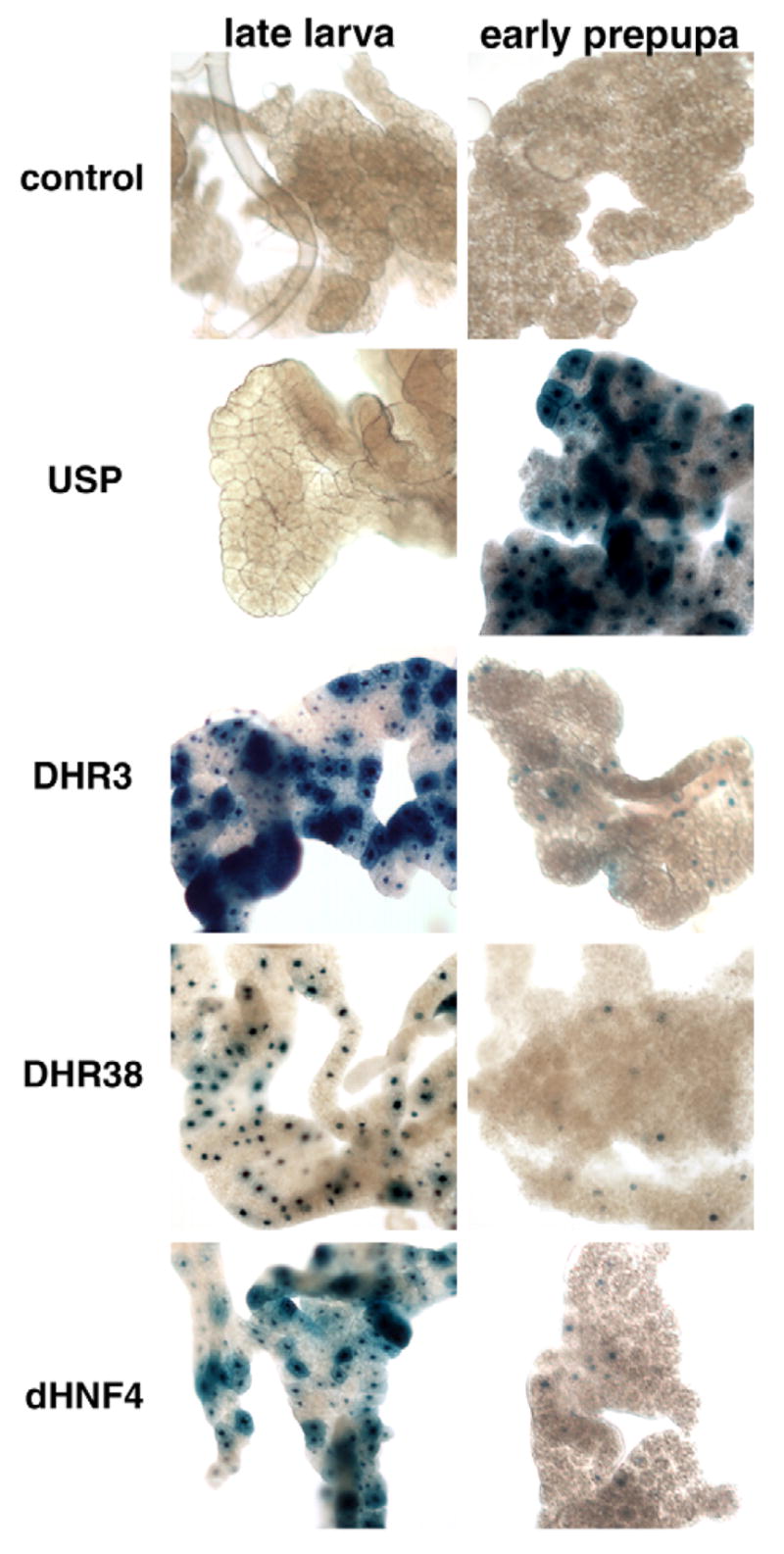
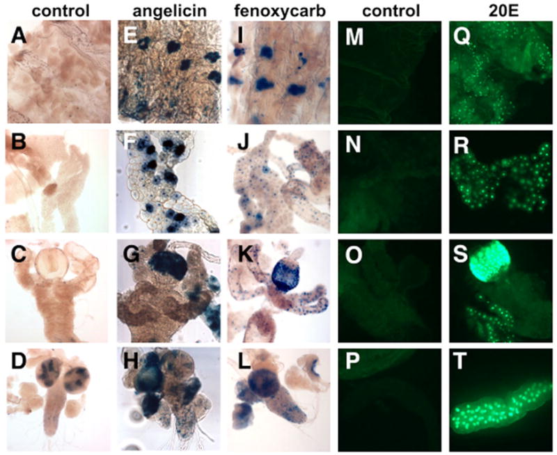
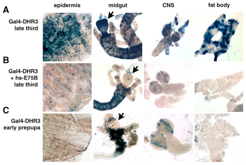
Nuclear receptors are a large family of transcription factors that play major roles in development, metamorphosis, metabolism and disease. To determine how, where and when nuclear receptors are regulated by small chemical ligands and/or protein partners, we have used a 'ligand sensor' system to visualize spatial activity patterns for each of the 18 Drosophila nuclear receptors in live developing animals. Transgenic lines were established that express the ligand binding domain of each nuclear receptor fused to the DNA-binding domain of yeast GAL4. When combined with a GAL4-responsive reporter gene, the fusion proteins show tissue- and stage-specific patterns of activation. We show that these responses accurately reflect the presence of endogenous and exogenously added hormone, and that they can be modulated by nuclear receptor partner proteins. The amnioserosa, yolk, midgut and fat body, which play major roles in lipid storage, metabolism and developmental timing, were identified as frequent sites of nuclear receptor activity. We also see dynamic changes in activation that are indicative of sweeping changes in ligand and/or co-factor production. The screening of a small compound library using this system identified the angular psoralen angelicin and the insect growth regulator fenoxycarb as activators of the Ultraspiracle (USP) ligand-binding domain. These results demonstrate the utility of this system for the functional dissection of nuclear receptor pathways and for the development of new receptor agonists and antagonists that can be used to modulate metabolism and disease and to develop more effective means of insect control.


Figures

Fig. 1
Fig. 1. The ligand sensor system
A schematic representation of the two transgenes that comprise the ligand sensor system is depicted. Upon heat treatment, the hsp70 promoter directs widespread expression of the GAL4 DNA-binding domain (DBD) fused to a nuclear receptor ligand-binding domain (LBD). This fusion protein is able to bind to a GAL4 UAS response element on a second transgene, activating reporter gene expression in cells that contain the necessary ligands and/or co-factors. Reporter genes that encode nuclear GFP or β-galactosidase are used to monitor GAL4-LBD ligand sensor activity in a cell-autonomous manner.
Fig. 2
Fig. 2. Ligand regulation of GAL4-LBD fusion protein activity
GAL4-LBD activation patterns are shown for three receptors in a wild-type background (wt; A,E,I) or disembodied mutant background (dib; B,F,J), in culture in either the absence (C,G,K) or presence of 5 μM 20-hydroxyecdysone (20E; D,H,L). GAL4-EcR is active in the amnioserosa of stage 14 wild-type embryos (A), but not in dib mutant embryos (B). Culturing in the presence of 20E induces ectopic activation in the epidermis (C,D). GAL4-FTZ-F1 is active in the yolk nuclei of embryos at stage 13 (E,F) and stage 16 (G,H) and is unaffected in a dib mutant background (F) or by the presence of exogenous 20E (H). GAL4-DHR38 is active in the epidermis and amnioserosa of stage 13 embryos (I,J) and is not affected in a dib mutant background (J, compare with K). The activity of GAL4-DHR38 in stage 17 cultured embryos is upregulated by exogenous 20E (L). The activity of GAL4-DHR96 in stage 13 embryos (M) is significantly increased by the addition of 5× 10⁻⁶ M CITCO (N).
Fig. 3
Fig. 3. Dynamic changes in the spatial and temporal patterns of ERR LBD activation
GAL4-ERR activation patterns are shown during embryogenesis (A) and third instar larval and prepupal stages (B). (A) ERR activation switches from myoblasts (6–11 hours AEL) and muscle (10–15 hours AEL) to predominantly CNS cells (14–17 hours AEL) in the late embryo. (B) In larvae, transient and widespread activation of GAL4-ERR occurs in the mid-third instar (mid-L3) in the muscle, CNS, midgut and fat body. Background bacterial β-galactosidase expression is seen in the larval midgut lumen of early third instar larvae (early L3). Background β-galactosidase expression is also present in the optic lobes of the CNS from larvae and early prepupae.
Fig. 4
Fig. 4. Distinct temporal patterns of GAL4-LBD activation in the amnioserosa
GAL4-LBD activation patterns are shown for five receptors: E78 (A–C), DHR38 (D–F), DHR3 (G–I), HNF4 (J–L) and EcR (M–O). The earliest activation in the amnioserosa is detected in stage 9–10 GAL4-E78 (A), GAL4-DHR38 (D), and GAL4-DHR3 (G) embryos. HNF4 embryos (J) show activation in the yolk nuclei at this stage (arrows). At stage 12, activation is detected in the amnioserosa of GAL4-E78 (B), GAL4-DHR38 (E), GAL4-DHR3 (H) and GAL4-HNF4 (K) embryos. At stage 13–14, activation in the amnioserosa is detected in all lines and becomes visible in GAL4-EcR embryos (O). The amnioserosa is indicated with arrowheads.
Fig. 5
Fig. 5. The yolk and midgut are hotspots for ligand sensor activation
GAL4-LBD activation patterns are depicted for the yolk and midgut during embryogenesis (AJ) and in the midgut at the onset of metamorphosis (KT). Representative embryos are shown for control (A,B), DHR3 (C,D), DHR38 (E,F), HNF4 (G,H), and FTZ-F1 (I,J) ligand sensors. The yolk is a major site of activation for GAL4-DHR3 (C), GAL4-DHR38 (E), GAL4-HNF4 (G) and GAL4-FTZ-F1 (I) embryos at stages 14–15 (arrowheads). Yolk activation remains prominent for GAL4-FTZ-F1 during stages 16–17 (J), but switches to the gut epithelium (arrows) for GAL4-DHR3 (D), GAL4-DHR38 (F) and GAL4-HNF4 (H). At later stages, GAL4-DHR3 displays strong and widespread activation in the proventriculus and midgut of late third instar larvae (M), and selectively reduced activation in the proventriculus after pupariation (arrow in N), while activation in the rest of the midgut is maintained. GAL4-DHR38 and GAL4-HNF4 display spatially restricted activation at the junction of the midgut, proventriculus (small arrow in Q) and gastric caeca (arrowheads in Q) (O–R). The FTZ-F1 ligand sensor is activated in the anterior midgut in a spatially and temporally specific fashion at puparium formation (S,T).
Fig. 6
Fig. 6. Dynamic changes in GAL4-LBD activation patterns in larval fat bodies at the onset of metamorphosis
As expected, GAL4-USP is activated in larval fat bodies by the 20E pulse at puparium formation. By contrast, the DHR3, DHR38 and HNF4 ligand sensors are active in the larval fat bodies of feeding third instar larvae and show reduced activation after pupariation.
Fig. 7
Fig. 7. GAL4-USP is activated by 20-hydroxyecdysone, angelicin and fenoxycarb in cultured larval organs
Organs dissected from hs-GAL4-USP; UAS-nlacZ (AL) or hs-GAL4-USP; UAS-nGFP (MT) mid-third instar larvae were cultured with either no hormone (control; A–D,M-P), 10μM angelicin (E–H), 100 μM fenoxycarb (I–L) or 5 μM 20E (Q–T). Activation is seen in the oenocytes (E,I), the fat body (F,J,R), the epidermis (Q), the proventriculus of the midgut (G,K,S), the larval salivary glands (T) and the CNS (H,L).
Fig. 8
Fig. 8. A repressive heterodimer partner can down-regulate GAL4-LBD activation
(A) GAL4-DHR3 is active in many tissues of a late third instar larva, including the epidermis, midgut, central nervous system (CNS) and fat body. (B) Ectopic expression of E75B results in a significantly reduced level of activation at this stage in development, recapitulating the pattern normally seen in early prepupae (C), in the presence of endogenous E75B expression. E75B-mediated downregulation of GAL4-DHR3 activity in the midgut is restricted to the proventriculus (compare arrows).

References

    1. Agoulnik IU, Tong XW, Fischer DC, Korner K, Atkinson NE, Edwards DP, Headon DR, Weigel NL, Kieback DG. A germline variation in the progesterone receptor gene increases transcriptional activity and may modify ovarian cancer risk. J Clin Endocrinol Metab. 2004;89:6340–6347. - PubMed
    1. Alcalay M, Zangrilli D, Pandolfi PP, Longo L, Mencarelli A, Giacomucci A, Rocchi M, Biondi A, Rambaldi A, Lo Coco F, et al. Translocation breakpoint of acute promyelocytic leukemia lies within the retinoic acid receptor alpha locus. Proc Natl Acad Sci USA. 1991;88:1977–1981. - PMC - PubMed
    1. Andres AJ, Thummel CS. Methods for quantitative analysis of transcription in larvae and prepupae. Methods Cell Biol. 1994;44:565–573. - PubMed
    1. Andres AJ, Fletcher JC, Karim FD, Thummel CS. Molecular analysis of the initiation of insect metamorphosis: a comparative study of Drosophila ecdysteroid-regulated transcription. Dev Biol. 1993;160:388–404. - PubMed
    1. Ashburner M. Patterns of puffing activity in the salivary gland chromosomes of Drosophila. VI Induction by ecdysone in salivary glands of D melanogaster cultured in vitro. Chromosoma. 1972;38:255–281. - PubMed

Publication types

MeSH terms