. 2006 Nov;17(11):4698-708.
doi: 10.1091/mbc.e06-06-0492. Epub 2006 Aug 16.

Invasion of host cells by Salmonella typhimurium requires focal adhesion kinase and p130Cas

Affiliations

Invasion of host cells by Salmonella typhimurium requires focal adhesion kinase and p130Cas

Jing Shi et al. Mol Biol Cell. 2006 Nov.

Abstract

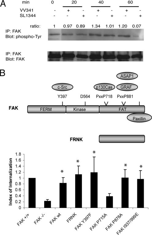
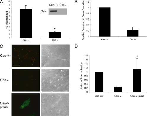
Salmonella typhimurium colonizes the intestinal epithelium by injecting an array of effector proteins into host cells that induces phagocytic uptake of attached bacteria. However, the host molecules targeted by these effectors remain poorly defined. Here, we demonstrate that S. typhimurium induces formation of focal adhesion-like complexes at sites of bacterial attachment and that both focal adhesion kinase (FAK) and the scaffolding protein p130Cas are required for Salmonella uptake. Entry of Salmonella into FAK(-/-) cells is dramatically impaired and can be restored to control levels by expression of wild-type FAK. Surprisingly, reconstitution of bacterial internalization requires neither the kinase domain of FAK nor activation of c-Src, but does require a C-terminal PXXP motif through which FAK interacts with Cas. Infection of Cas(-/-) cells is also impaired, and reconstitution of invasiveness requires the central Cas YXXP repeat domain. The invasion defect in Cas(-/-) cells can be suppressed by overexpression of FAK, suggesting a functional link between FAK and Cas in the regulation of Salmonella invasion. Together, these findings reveal a novel role for focal adhesion proteins in the invasion of host cells by Salmonella.


Figures

Figure 1.
Figure 1.
Focal adhesion-like complexes form at sites of Salmonella invasion. (A) MDCK cells were infected apically with S. typhimurium strain SL1344 for 20 min. For FAK, Cas, and paxillin staining, F-actin was labeled with rhodamine-conjugated phalloidin (red); endogenous FAK, Cas, or paxillin were detected with specific monoclonal antibodies, followed by a Cy2-conjugated secondary antibody (green); Salmonella were stained with a polyclonal antibody against bacterial LPS followed by a Cy5-conjugated secondary (blue). Merged images show colocalization of FAK, Cas, or paxillin with F-actin at sites of bacterial entry. For β1 integrin staining, F-actin was labeled with Alexa 647–conjugated phalloidin (blue); endogenous β1 integrins were stained with a polyclonal antibody E63E followed by a Cy2 secondary (green); Salmonella were labeled with a mouse anti-LPS IgA followed by a TR-conjugated secondary (red). Arrows indicate the bacterially induced membrane ruffles. Images were taken at 100× magnification on a Nikon C1 confocal microscope. Bars, 10 μm. (B) MDCK cells were infected apically with SL1344 or the invasion-defective mutant VV341 at a MOI = 100 for 30 min. Cas was immunoprecipitated with the polyclonal antibody Cas3B, and the precipitates were probed for associated FAK and paxillin. Blots were then stripped and reprobed for Cas.
Figure 2.
Figure 2.
FAK is required for Salmonella internalization. (A) FAK+/+ and FAK−/− MEFs were infected with wild-type S. typhimurium (SL1344) at a MOI = 30 for 1 h. Salmonella internalization was assayed by a standard gentamicin resistance assay as described in Materials and Methods. Expression levels of FAK in these cells were detected using the FAK mAb 4.47. *p < 0.001 (Student's t test). (B and C) FAK+/+, FAK−/− (mock transfected) and FAK−/− cells transfected with myc-FAK were infected with SL1344 at a MOI = 30 for 1 h before a two-color immunofluorescence invasion assay as described in Materials and Methods. (B) Transfected cells were labeled with an anti-myc antibody 9E10 followed by a Cy2-conjugated donkey anti-mouse secondary antibody. Phase-contrast images of the same field are shown on the right. Intracellular bacteria appear red and extracellular bacteria appear yellow. The bottom panel shows a FAK-reconstituted cell with internalized (red) and adhered bacteria (yellow). Bar, 50 μm. (C) Quantification of Salmonella internalization as measured using the two-color immunofluorescence invasion assay. *p < 0.01 compared with mock-transfected FAK−/− cells (Student's t test).
Figure 3.
Figure 3.
Rac1 activation is not impaired in FAK−/− MEFs. (A) FAK+/+ and FAK−/− cells were infected with wild-type S. typhimurium (SL1344) for 20 min before fixation. F-actin and bacteria were stained using rhodamine-conjugated phalloidin and rabbit anti-LPS, respectively. Frequency of focus formation was quantified as described in Materials and Methods. (B) FAK+/+ and FAK−/− cells were infected with SL1344 (lane 3 and 7) or the entry-deficient strain VV341 (lane 4 and 8), or maintained in HBSS+ (lane 1–2 and 5–6). GTP-bound Rac1 was isolated from cell lysates by affinity precipitation with GST-PBD (lane 2–4 and 6–8) as described in Materials and Methods. GST alone was used as negative control (lane 1 and 5). Pulldowns and a fraction of each lysate were simultaneously blotted for Rac1. The level of GTP-bound Rac is expressed relative to the basal level in uninfected cells (lane 2 and 6).
Figure 4.
Figure 4.
FAK kinase activity is not required for Salmonella entry. (A) MDCK cells were infected apically with wild-type S. typhimurium (SL1344) or the invasion-defective mutant VV341 (ΔHilA) at a MOI = 20 for 20, 40, or 60 min. Cells maintained in the absence of bacteria were used as controls. FAK was immunoprecipitated from the lysates using polyclonal antibody A-17. The precipitates were divided into two sets and probed with either anti-phospho-tyrosine antibody 4G10 or a mAb clone 4.47 against total FAK. (B) FAK+/+, FAK−/− (mock transfected), and FAK−/− cells transfected with wild-type FAK, FRNK, FAK Y397F, FAK P715A, FAK P878A, or FAK I937/999E mutant were subjected to the two-color immunofluorescence invasion assay. Bacterial internalization was normalized to control FAK+/+ cells. *p < 0.05 compared with mock-transfected FAK−/− cells (Student's t test).
Figure 5.
Figure 5.
p130Cas is necessary for Salmonella invasion. (A) Cas+/+ and Cas−/− MEFs were infected with wild-type S. typhimurium (SL1344) and subjected to the gentamicin resistant assay. *p < 0.001 (Student's t test). Expression levels of Cas in these cells were detected using a Cas mAb 8G4. (B) Frequency of focus formation in Cas+/+ and Cas−/− cells was determined as described Figure 3A. (C and D) Cas+/+, Cas−/− (mock transfected), and Cas−/− cells transfected with myc-Cas were subjected to the two-color immunofluorescence invasion assay. Transfected cells were labeled with 9E10 followed by a Cy2-conjugated donkey anti-mouse secondary antibody. (C) The bottom panel shows a Cas reconstituted cell with internalized (red) and adhered bacteria (yellow). Bar, 50 μm. (D) Quantification of Salmonella internalization using the two-color invasion assay. *p < 0.05 compared with mock-transfected Cas−/− cells (Student's t test).
Figure 6.
Figure 6.
Salmonella entry does not require increased Cas tyrosine phosphorylation, but does require the substrate-binding domain. (A) MDCK cells were infected with wild-type S. typhimurium (SL1344) or the invasion-defective mutant VV341 (ΔHilA) at a MOI = 20 for 20, 40, or 60 min. Cells maintained in the absence of bacteria during the period of infection were used as controls. Cas was immunoprecipitated from the lysates using a polyclonal antibody (Cas3B). The precipitates were split into two sets and probed with anti-phospho-tyrosine antibody 4G10 or monoclonal 8G4 against total Cas. (B) MDCK cells were treated with either DMSO (control) or 10 μM PP2 for 30 min before performing a gentamicin resistance invasion assay. (C) Cas+/+, Cas−/− (mock transfected), and Cas−/− cells transfected with full-length Cas, CasΔSH3, or CasΔYXXP mutant were subjected to the two-color immunofluorescence invasion assay. Bacterial internalization was normalized to invasion efficiency in Cas+/+ cells. *p < 0.05 compared with mock-transfected Cas−/− cells (Student's t test).
Figure 7.
Figure 7.
Aberrant phagocytic cups are induced in Cas−/− fibroblasts upon Salmonella infection. (A) Cas+/+ and Cas−/− cells were infected with wild-type S. typhimurium (SL1344) for 20 min before fixation, and F-actin was stained using FITC-conjugated phalloidin (green). Salmonella were labeled with polyclonal anti-LPS antibody followed by a TR-conjugated anti-rabbit IgG (red). Arrows indicate the bacterially induced phagocytic cups. Note the bright actin puncta within the enlarged phagocytic structure. Bar, 10 μm. (B) Quantification of ruffle size in Cas+/+ and Cas−/− cells; n = 40.
Figure 8.
Figure 8.
Paxillin is not necessary for Salmonella invasion. (A) Paxillin+/+, paxillin−/− (mock transfected), or paxillin−/− MEFs transfected with GFP-paxillin were infected with SL1344 at a MOI = 30 for 1 h and were subjected to the two-color immunofluorescence assay. (B) HeLa cells were transfected with scrambled siRNA or anti-paxillin siRNA 72 h before the gentamicin resistance invasion assay. The efficiency of paxillin knockdown is shown in the top panel. A corresponding actin immunoblot (bottom) is shown to demonstrate equal loading. *p < 0.001 compared with control cells transfected with scrambled siRNA (Student's t test).
Figure 9.
Figure 9.
Overexpression of FAK rescues Salmonella invasion in Cas−/− MEFs. (A) FAK−/− cells were either mock-transfected (FAK−/−) or transfected with an expression plasmid encoding p130Cas (FAK−/− pCas). FAK+/+cells were used as a positive control. Cells were infected with SL1344 at a MOI = 30 for 1 h, and bacterial invasion was quantified using the two-color immunofluorescence assay. (B) Cas−/− cells were either mock-transfected (Cas−/−) or transfected with an expression plasmid encoding FAK (Cas−/− pFAK). Cells were then infected using wild-type MEFs (Cas+/+) as a positive control and invasion efficiency quantified as in A. *p < 0.05 compared with mock-transfected Cas−/− cells (Student's t test).

Similar articles

Cited by

References

    1. Agerer F., Lux S., Michel A., Rohde M., Ohlsen K., Hauck C. R. Cellular invasion by Staphylococcus aureus reveals a functional link between focal adhesion kinase and cortactin in integrin-mediated internalisation. J. Cell Sci. 2005;118:2189–2200. - PubMed
    1. Alrutz M. A., Isberg R. R. Involvement of focal adhesion kinase in invasin-mediated uptake. Proc. Natl. Acad. Sci. USA. 1998;95:13658–13663. - PMC - PubMed
    1. Astier A., Avraham H., Manie S. N., Groopman J., Canty T., Avraham S., Freedman A. S. The related adhesion focal tyrosine kinase is tyrosine-phosphorylated after beta1-integrin stimulation in B cells and binds to p130cas. J. Biol. Chem. 1997;272:228–232. - PubMed
    1. Bourdet-Sicard R., Rudiger M., Jockusch B. M., Gounon P., Sansonetti P. J., Nhieu G. T. Binding of the Shigella protein IpaA to vinculin induces F-actin depolymerization. EMBO J. 1999;18:5853–5862. - PMC - PubMed
    1. Bouton A. H., Riggins R. B., Bruce-Staskal P. J. Functions of the adapter protein Cas: signal convergence and the determination of cellular responses. Oncogene. 2001;20:6448–6458. - PubMed

Publication types

MeSH terms