. 2006 Nov;17(11):4675-85.
doi: 10.1091/mbc.e06-05-0466. Epub 2006 Aug 16.

Gene targeting of Cdc42 and Cdc42GAP affirms the critical involvement of Cdc42 in filopodia induction, directed migration, and proliferation in primary mouse embryonic fibroblasts

Affiliations

Gene targeting of Cdc42 and Cdc42GAP affirms the critical involvement of Cdc42 in filopodia induction, directed migration, and proliferation in primary mouse embryonic fibroblasts

Linda Yang et al. Mol Biol Cell. 2006 Nov.

Abstract

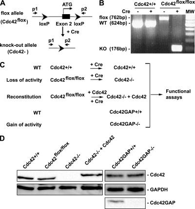
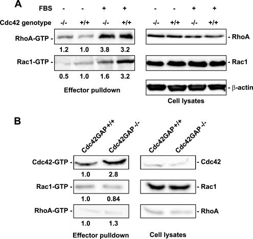
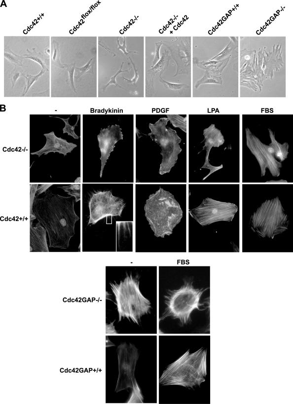
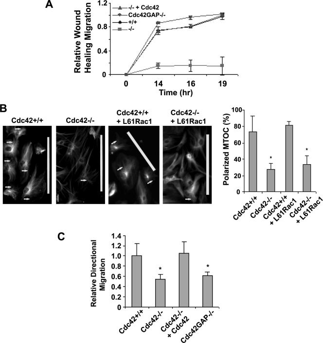
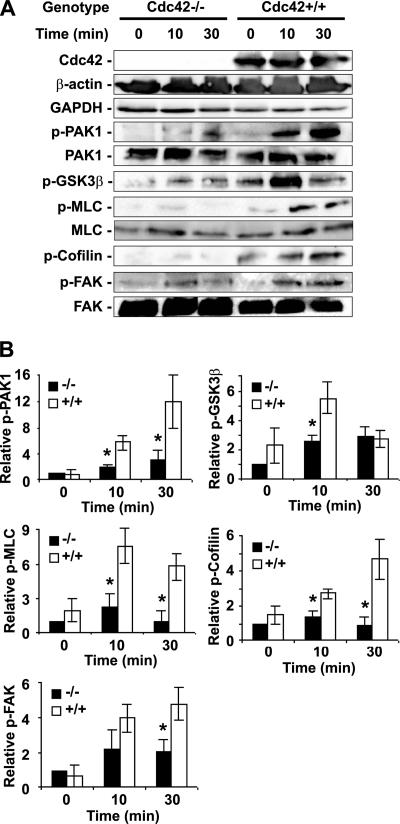
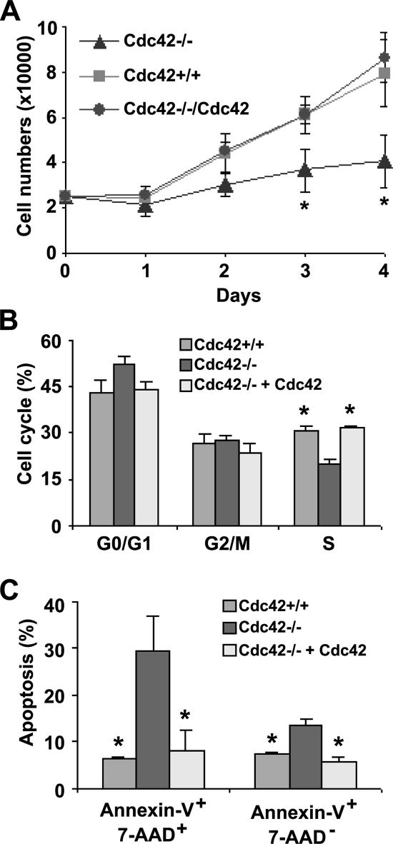
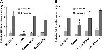
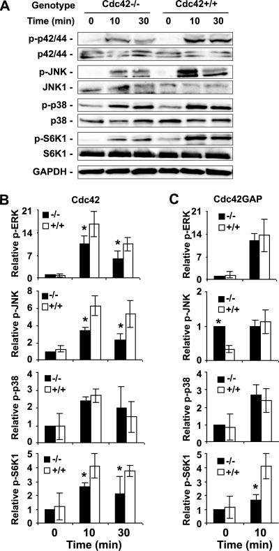
Recent studies in Cdc42 knockout mouse embryonic stem (ES) cells and ES-derived fibroblastoid cell lines raise concern on a body of literature derived by dominant mutant expression approach in a variety of cell lines implicating mammalian Cdc42 as a key regulator of filopodia induction, directional migration and cell cycle progression. To resolve the physiological function of mammalian Cdc42, we have characterized the Cdc42(-/-) and Cdc42GAP(-/-) primary mouse embryonic fibroblasts (MEFs) produced by gene targeting as the Cdc42 loss- or gain-of-activity cell model. The Cdc42(-/-) cells were defective in filopodia formation stimulated by bradykinin and in dorsal membrane ruffling stimulated by PDGF, whereas the Cdc42GAP(-/-) cells displayed spontaneous filopodia. The Cdc42 loss- or gain-of-activity cells were defective in adhesion to fibronectin, wound-healing, polarity establishment, and migration toward a serum gradient. These defects were associated with deficiencies of PAK1, GSK3beta, myosin light chain, and FAK phosphorylation. Furthermore, Cdc42(-/-) cells were defective in G1/S-phase transition and survival, correlating with deficient NF-kappaB transcription and defective JNK, p70 S6K, and ERK1/2 activation. These results demonstrate a different requirement of Cdc42 activity in primary MEFs from ES or ES-derived clonal fibroblastoid cells and suggest that Cdc42 plays cell-type-specific signaling roles.


Figures

Figure 1.
Figure 1.
Generation of Cdc42−/− and Cdc42GAP−/− primary MEFs by a loxP/Cre recombinase system or by a conventional gene targeting strategy respectively. (A) The loxP/Cre-mediated gene targeting strategy to generate Cdc42 gene–deleted (Cdc42) allele. (B) Genotyping results of primary MEFs obtained from cross-breeding of Cdc42flox/+ heterozygous mice with or without retrovirus-mediated Cre transduction. The 762-base pair band represents the floxed (flox) allele, the 624-base pair band represents the WT (+) allele, and the 176-base pair band represents the Cdc42 KO (−) allele in the cells. (C) Depiction of the primary MEF cell models of Cdc42 loss or gain of activity and the relevant controls. The Cdc42−/− cells were generated by retrovirus-mediated Cre transduction into the Cdc42flox/flox primary MEFs, whereas the Cdc42GAP−/− cells were produced by direct extraction of MEFs from the Cdc42GAP−/− mouse embryos. The relevant control cells, including the genetically matched Cdc42+/+ or Cdc42GAP+/+ MEFs derived from the same litters of Cdc42flox/flox or Cdc42GAP−/− embryos and the (HA)3-tagged, WT Cdc42 reconstituted (Cdc42−/− + Cdc42) cells, were produced in parallel. All cells were used for the studies at comparable low passages (passages 3–5). (D) Expression of Cdc42 in various primary MEFs, including the exogenous (HA)3-Cdc42 reconstituted cells, was analyzed by anti-Cdc42 Western blotting. Cdc42GAP deletion in the Cdc42GAP−/− MEFs was confirmed by anti-Cdc42GAP immunoblotting.
Figure 2.
Figure 2.
Loss or gain of Cdc42 activity and effects on Rac1 and RhoA activities in Cdc42 (A) or Cdc42GAP (B) knockout cells. The expression and the relative level of GTP-bound forms of Cdc42, Rac1, and RhoA in various primary MEFs were probed by the effector-domain (GST-PAK1 for Cdc42-GTP and Rac1-GTP; GST-Rhotekin for RhoA-GTP) pulldown assays followed by anti-Cdc42, -Rac1, or -RhoA Western blotting. The lysate inputs are shown on the right-hand side and the corresponding quantifications are indicated below each pulldown blot. The results are representative of three independent measurements.
Figure 3.
Figure 3.
Morphologies and actin cytoskeleton structures of Cdc42- or Cdc42GAP-deficient primary MEFs in response to stimuli. (A) The morphologies of Cdc42−/−, Cdc42GAP−/−, and control cells under normal tissue culture condition were examined by phase-contrast microscopy. (B) Various MEFs were grown on cover slides and stained with TRITC-phalloidin to reveal the F-actin structures. The Cdc42−/− and Cdc42+/+ cells were starved 16 h before stimulation with bradykinin (100 ng/ml), PDGF (10 ng/ml), LPA (20 ng/ml), or 10% FBS for 10 min. The Cdc42GAP−/− and matching Cdc42GAP+/+ cells were examined with or without 10% FBS stimulation.
Figure 4.
Figure 4.
Effect of Cdc42 loss or gain of activity on adhesion to fibronectin and focal complex assembly. The percentage of cells adhered to the fibronectin matrix after a 2-h incubation (A), and the focal adhesion complex visualized by anti-vinculin immunostaining (TRITC; B) are shown. *p < 0.05 between the Cdc42−/− or Cdc42GAP−/− and respective control WT cells.
Figure 5.
Figure 5.
Effect of loss or gain of Cdc42 activity on cell migration. (A) Wound-healing assays were carried out on a monolayer of confluent cells to determine cell movement toward the wound gap. The average migration distances to the center of the wound were quantified at the indicated times after the wound was introduced. (B) Immunostaining of α-tubulin revealed the MTOC orientation (arrows) toward the wound induction site (marked with white bars) 16 h after the wounds were introduced. The number of cells containing a wound-directed MTOC orientation was quantified under a microscope. (C) Transwell cell migration toward 10% FBS was measured to determine cell directional movement. The number of cells migrated across the membrane toward the serum gradient into the lower chamber of the Transwells was quantified 16 h after plating. The results are representative of three independent measurements. *p < 0.05 between the Cdc42−/−, Cdc42GAP−/−, or Cdc42−/−+L61Rac1 and the respective control cells.
Figure 6.
Figure 6.
Cdc42 activity regulates signaling responses to serum stimulation relevant to cell actin organization and migration. (A) Western blotting of p-GSK3β, p-PAK1, p-MLC, p-Cofilin, p-FAK, and relevant controls in the Cdc42−/− and Cdc42+/+ MEFs after 0, 10, or 30 min of 10% FBS stimulation after a 16-h serum withdrawal. (B) The relative Western blot levels were quantified by using a densitometer (mean ± SD from three independent experiments). All data were normalized to those of Cdc42−/− cells at time 0. *p < 0.05 between the Cdc42−/− and Cdc42+/+ cells.
Figure 7.
Figure 7.
Cdc42 activity is critical for primary MEF proliferation, cell cycle progression, and cell survival. (A) Cell proliferation potential of the respective MEFs under regular serum conditions was determined. MEF cells were plated in 12-well plates in triplicates in the presence of 10% FBS, and at the varying time points the cell numbers were quantified by Trypan blue exclusion analysis. (B) Various MEFs were labeled with PI/RNase, and the cell populations at various cell cycle phases were analyzed by FACS after 48-h serum starvation followed by a 24-h 10% FBS incubation. (C) The MEFs were labeled with Annexin-V/7AAD and subjected to FACS analysis to determine the percent of cell populations undergo apoptosis after 16-h serum starvation followed by 6-h stimulation by 10% FBS/DMEM. *p < 0.05 between the Cdc42−/− and Cdc42+/+ cells.
Figure 8.
Figure 8.
Effects of Cdc42 loss or gain of activity on NF-κB and SRF transcription activities. NF-κB (A) or SRF (B) transcriptional reporter plasmids were cotransfected into the respective MEF cells with a β-gal expressing reporter. After a 4–5-h incubation, cells were allowed to recover overnight in normal culture media. After 24 h with or without starvation, the luciferase activities were assayed of the cells. The luciferase reporter activities were normalized to those of the cotransfected β-gal activities and are expressed as fold of changes compared with those of WT control cells. *p < 0.05 between the Cdc42−/− and Cdc42+/+ cells under similar treatment.
Figure 9.
Figure 9.
Altered MAPK and p70S6K signaling activities in Cdc42−/− and Cdc42GAP−/− MEFs in response to serum stimulation. (A) Western blotting of p-ERK1/2, p-p38, p-JNK1/2 and p-S6K, and relevant controls as performed using MEF cell lysates after stimulation of the cells with 10% FBS for the indicated times after a 16-h serum withdrawal. (B and C) The relative Western blot levels were quantified by using a densitometer (mean ± SD from three independent experiments). *p < 0.05 between the knockout and WT cells.

References

    1. Bishop A. L., Hall A. Rho GTPases and their effector proteins. Biochem. J. 2000;348(Pt 2):241–255. - PMC - PubMed
    1. Chen F., et al. Cdc42 is required for PIP(2)-induced actin polymerization and early development but not for cell viability. Curr. Biol. 2000;10:758–765. - PubMed
    1. Chou M. M., Masuda-Robens J. M., Gupta M. L. Cdc42 promotes G1 progression through p70 S6 kinase-mediated induction of cyclin E expression. J. Biol. Chem. 2003;278:35241–35247. - PubMed
    1. Czuchra A., Wu X., Meyer H., van Hengel J., Schroeder T., Geffers R., Rottner K., Brakebusch C. Cdc42 is not essential for filopodium formation, directed migration, cell polarization, and mitosis in fibroblastoid cells. Mol. Biol. Cell. 2005;16:4473–4484. - PMC - PubMed
    1. Debidda M., Wang L., Zang H., Poli V., Zheng Y. A role of STAT3 in Rho GTPase-regulated cell migration and proliferation. J. Biol. Chem. 2005;280:17275–17285. - PubMed

Publication types

MeSH terms