. 2006 Nov;17(11):4645-55.
doi: 10.1091/mbc.e06-04-0282. Epub 2006 Aug 16.

Actin bodies in yeast quiescent cells: an immediately available actin reserve?

Affiliations

Actin bodies in yeast quiescent cells: an immediately available actin reserve?

Isabelle Sagot et al. Mol Biol Cell. 2006 Nov.

Abstract

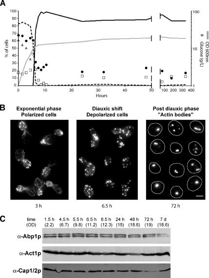
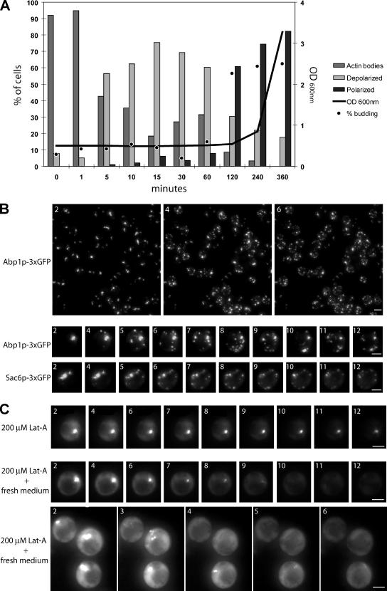
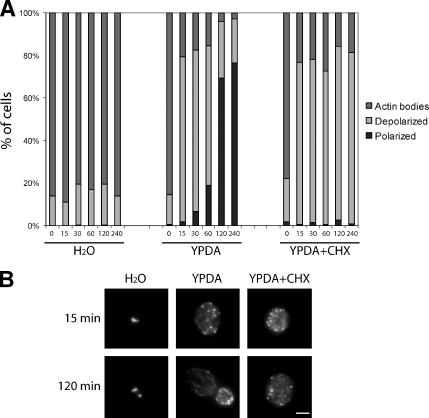
Most eukaryotic cells spend most of their life in a quiescent state, poised to respond to specific signals to proliferate. In Saccharomyces cerevisiae, entry into and exit from quiescence are dependent only on the availability of nutrients in the environment. The transition from quiescence to proliferation requires not only drastic metabolic changes but also a complete remodeling of various cellular structures. Here, we describe an actin cytoskeleton organization specific of the yeast quiescent state. When cells cease to divide, actin is reorganized into structures that we named "actin bodies." We show that actin bodies contain F-actin and several actin-binding proteins such as fimbrin and capping protein. Furthermore, by contrast to actin patches or cables, actin bodies are mostly immobile, and we could not detect any actin filament turnover. Finally, we show that upon cells refeeding, actin bodies rapidly disappear and actin cables and patches can be assembled in the absence of de novo protein synthesis. This led us to propose that actin bodies are a reserve of actin that can be immediately mobilized for actin cables and patches formation upon reentry into a proliferation cycle.


Figures

Figure 1.
Figure 1.
Actin cytoskeleton reorganization upon entry into quiescence. (A) Wild-type yeast cells were grown in YPDA at 30°C. Cell density was monitored by measuring OD600 nm (gray line). At various stage of growth, samples were taken, glucose concentration in the medium was measured (gray triangles), and cells were stained with Alexa-phalloidin. For each time point, cells with polarized actin cytoskeleton (black dashed curve), depolarized actin cytoskeleton (open squares), or actin bodies (black curve) were counted (n > 200 for each time point). The budding index (percentage of budding cells) is shown as black circles (n > 200 for each time point). (B) Left, examples of polarized cells in exponential phase of growth (time point 3 h in A). Middle, examples of depolarized cells upon diauxic shift (time point 6.5 h in A). Right, examples of quiescent cells bearing actin bodies (time point 72 h in A). Images are two-dimensional (2D) maximal projection of three-dimensional (3D) image stacks. Bar, 2 μm. (C) Western blot analysis of the steady-state levels of Abp1p, Act1p, and Cap1/2p upon entry into quiescence. Time points and OD correspond to time points in A.
Figure 2.
Figure 2.
Localization of actin bodies. (A) Localization of actin bodies within cells. Wild-type yeast cells expressing either Ilv3p-GFP (left) or Pho88p-GFP (right) staining the mitochondria or the endoplasmic reticulum, respectively, were grown for 7 d at 30°C in SD casa medium, fixed, and stained with DAPI and Alexa-phalloidin. In the merged bottom images, green is GFP-, blue is DAPI-, and red is Alexa-phalloidin–stained F-actin. Images are 2D maximal projection of 3D image stacks. Bar, 2 μm. (B) Immunogold localization of anti-actin antibodies linked to 10-nm gold particles on wild-type yeast cells grown for 3 d at 30°C in YPDA medium. Arrows point to clusters of gold particles. In the right top image, three clusters of gold particles within a cell are circled. Bar, 500 nm (top); 200 nm (bottom).
Figure 3.
Figure 3.
(A) Colocalization of actin bodies with various actin-binding proteins. Wild-type yeast cells expressing different ABPs fused to three tandem GFP copies at a native level were grown for 2 d at 30°C in SD casa medium. Cells were fixed and stained with Alexa-phalloidin. In the merged bottom panel, GFP is shown in green, and Alexa-phalloidin–stained F-actin is shown in red. Images are 2D maximal projection of 3D image stacks. (B) Actin bodies formation in two actin-bundling protein mutants: sac6Δ and scp1Δ. Wild-type (left), scp1Δ (middle), and sac6Δ (right) cells were grown for 3 d in YPDA medium, fixed, and stained with Alexa-phalloidin. Bar, 2 μm. (C) Actin-bundling mutants survival upon starvation and in stationary phase. Serial dilutions were spotted onto regular SD casa WAU medium or synthetic medium containing casa, WUA, 0.1% glucose, and erythrosine, a dye that stains dead cells pink (Bonneu et al., 1991). Viability of the strains after 7 d of growth at 30°C in YPDA medium is indicated. Viability was assessed as described in Materials and Methods. (D) Steady-state levels of Sac6p and Scp1p at different growth phases. Samples of wild-type cells were taken in exponential phase, after 3 d (3 d) or 7 d (7 d) of culture at 30°C in YPDA medium. (E) Localization of Sac6p and Scp1p in stationary phase. Yeast cells expressing either Sac6p-GFP or Scp1p-GFP were grown in SD casa medium at 30°C. Left, GFP fluorescence is shown in living exponentially growing cells. In the following panels, cells grown for 3 d at 30°C were fixed and stained with Alexa-phalloidin. In the merged panel, GFP is shown in green, and Alexa-phalloidin–stained F-actin is shown in red. Images are 2D maximal projection of 3D image stacks. Bar, 2 μm.
Figure 4.
Figure 4.
Actin bodies contain stable actin filaments. (A) Actin bodies are resistant to latrunculin A (A). Wild-type cells were grown at 30°C in YPDA medium. During exponential growth (top) or after 3 d of culture (bottom), cells were incubated 5 min or 2 h with either 200 μM Lat-A or dimethyl sulfoxide as a control. Cells were then fixed and stained with Alexa-phalloidin. (B) Fluorescence recovery after laser ablation of the Abp1p-3xGFP signal of an actin body in a cell with a single actin body. Wild-type cells expressing Abp1p-3xGFP were grown for 3 d at 30°C in SD casa medium. Images on the left are 2D maximal projection of 3D image stacks time-lapse series. Time is indicated in the top left corner. The small white circle indicates the bleached region. The graph on the right shows the kinetics of fluorescence course obtained for 10 different cells. The same kind of curves was obtained for 32 cells. (C) Fluorescence recovery after laser ablation of the Abp1p-3xGFP signal of an actin body in a cell displaying two actin bodies. Wild-type cells expressing Abp1p-3xGFP were grown for 3 d at 30°C in SD casa medium. Images on the left are 2D maximal projection of 3D image stacks time-lapse series. Time is indicated in the top left corner. The small white circle indicates the bleached region; the dotted line square indicates the unbleached actin body in the same cell. The graph on the right shows the kinetics of fluorescence course measured for the bleached (black dots) and the unbleached (open squares) regions. The same kind of curve was obtained for 12 different cells. (D) Fluorescence recovery after laser ablation of the Abp1p-3xGFP signal of one-half of an actin body. Wild-type cells expressing Abp1p-3xGFP were grown for 3 d at 30°C in SD casa medium. Images on the left are 2D maximal projection of 3D image stacks time-lapse series. Time is indicated in the top left corner. The small white circle indicates the bleached region; the dotted line square indicated the unbleached region of the same actin body. The graph on the right shows the kinetics of fluorescence course measured for the bleached (black dots) and the unbleached (open squares) regions. The same kind of curve was obtained for 14 different actin bodies. Bar, 2 μm.
Figure 5.
Figure 5.
Reorganization of the actin cytoskeleton upon stationary phase exit. (A) Wild-type cells were grown for 7 d at 30°C in YPDA medium. Cells were then washed and resuspended in fresh YPDA medium and grown at 30°C. Cell density was monitored by measuring OD600 nm (gray line). For each time points, cells were stained with Alexa-phalloidin. Cells with polarized actin cytoskeleton (dark gray bar), depolarized actin cytoskeleton (light gray bar), or actin bodies (gray bar) were counted (n > 200; SD <5%). The budding index (percentage of budding cells) is shown as black dots (n > 200; SD <5%). (B) Wild-type yeast cells expressing Abp1p-3xGFP (top) or Sac6–3xGFP were grown 2 d at 30°C in SD casa medium. After a brief centrifugation, the cell pellet was resuspended in fresh medium and cells were immediately imaged. Images are 2D maximal projection of 3D image stacks time-lapse series. Time in minutes after medium renewal is indicated in the left corner. Bar, 2 μm. (C) Wild-type yeast cells expressing Abp1p-3x GFP were grown for 3 d at 30°C in SD casa medium. The cell culture was concentrated and Lat-A was added at the final concentration of 200 μM. This concentrated culture was incubated for 30 min at 30°C. After this incubation, 20 μl of the cell suspension was mixed either to 20 μl of water containing 200 μM Lat-A (top) or to 20 μl of 2X fresh SD casa containing 200 μM Lat-A, and immediately imaged (two bottom panels). Images are 2D maximal projection of 3D image stacks time-lapse series. Time in minutes after addition of Lat-A–containing water or fresh medium is indicated in the left corner. Bar, 2 μm.
Figure 6.
Figure 6.
Actin cytoskeleton depolarization upon exit from quiescence can happen in the absence of protein synthesis. (A) Wild-type cells were grown for 7 d at 30°C in YPDA medium. Cells were then washed and resuspended in either water (left) or fresh YPDA medium (middle), and grown at 30°C. For cycloheximide (CHX) treatment, wild-type cells were grown for 7 d at 30°C in YPDA medium and then preincubated with 100 μg/ml cycloheximide by addition of the drug directly into the growth medium. After 1 h of pretreatment at 30°C, cells were washed and resuspended in fresh YPDA medium containing 100 μg/ml cycloheximide. Cells were then fixed and stained with Alexa-phalloidin. For each time point, cells with polarized actin cytoskeleton (dark gray bar), depolarized actin cytoskeleton (light gray bar), or actin bodies (gray bar) were counted (n > 200, SD <5%). (B) Images are 2D maximal projection of 3D image stacks of examples of cells observed for the experiment in A. Bar, 2 μm.

Similar articles

Cited by

References

    1. Adams A. E., Botstein D., Drubin D. G. Requirement of yeast fimbrin for actin organization and morphogenesis in vivo. Nature. 1991;354:404–408. - PubMed
    1. Amatruda J. F., Cooper J. A. Purification, characterization, and immunofluorescence localization of Saccharomyces cerevisiae capping protein. J. Cell Biol. 1992;117:1067–1076. - PMC - PubMed
    1. Asakura T. Isolation and characterization of a novel actin filament-binding protein from Saccharomyces cerevisiae. Oncogene. 1998;16:121–130. - PubMed
    1. Ayscough K. R., Stryker J., Pokala N., Sanders M., Crews P., Drubin D. G. High rates of actin filament turnover in budding yeast and roles for actin in establishment and maintenance of cell polarity revealed using the actin inhibitor latrunculin-A. J. Cell Biol. 1997;137:399–416. - PMC - PubMed
    1. Binley K. M., Radcliffe P. A., Trevethick J., Duffy K. A., Sudbery P. E. The yeast PRS3 gene is required for cell integrity, cell cycle arrest upon nutrient deprivation, ion homeostasis and the proper organization of the actin cytoskeleton. Yeast. 1999;15:1459–1469. - PubMed

Publication types

MeSH terms

LinkOut - more resources