. 2006 Aug 16;26(33):8570-7.
doi: 10.1523/JNEUROSCI.1478-06.2006.

Scaffolding protein INAD regulates deactivation of vision by promoting phosphorylation of transient receptor potential by eye protein kinase C in Drosophila

Affiliations

Scaffolding protein INAD regulates deactivation of vision by promoting phosphorylation of transient receptor potential by eye protein kinase C in Drosophila

Daniela C Popescu et al. J Neurosci. .

Abstract

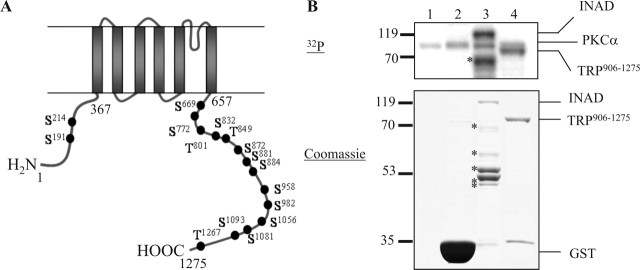
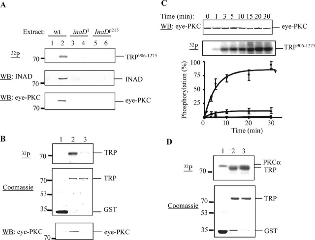
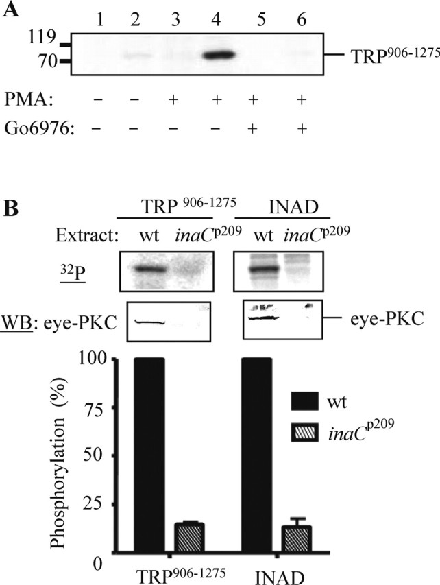
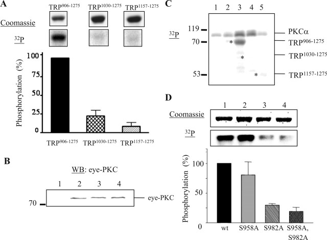
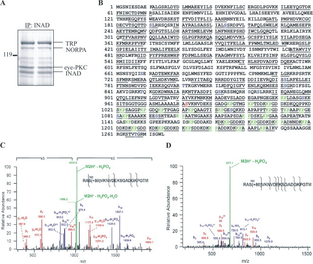
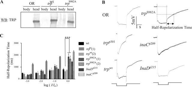
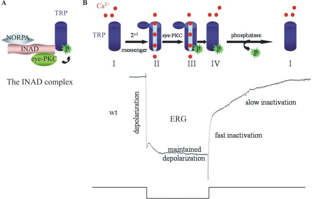
Drosophila visual signaling is one of the fastest G-protein-coupled transduction cascades, because effector and modulatory proteins are organized into a macromolecular complex ("transducisome"). Assembly of the complex is orchestrated by inactivation no afterpotential D (INAD), which colocalizes the transient receptor potential (TRP) Ca2+ channel, phospholipase Cbeta, and eye protein kinase C (eye-PKC), for more efficient signal transduction. Eye-PKC is critical for deactivation of vision. Moreover, deactivation is regulated by the interaction between INAD and TRP, because abrogation of this interaction in InaD(p215) results in slow deactivation similar to that of inaC(p209) lacking eye-PKC. To elucidate the mechanisms whereby eye-PKC modulates deactivation, here we demonstrate that eye-PKC, via tethering to INAD, phosphorylates TRP in vitro. We reveal that Ser982 of TRP is phosphorylated by eye-PKC in vitro and, importantly, in the fly eye, as shown by mass spectrometry. Furthermore, transgenic expression of modified TRP bearing an Ala substitution leads to slow deactivation of the visual response similar to that of InaD(p215). These results suggest that the INAD macromolecular complex plays an essential role in termination of the light response by promoting efficient phosphorylation at Ser982 of TRP for fast deactivation of the visual signaling.


Figures

Figure 1.
Figure 1.
The C-terminal tail of TRP is phosphorylated by recombinant PKCα. A, Distribution of the 16 putative PKC phosphorylation sites in TRP. B, Phosphorylation of TRP906–1275 and INAD by PKCα. Lane 1, No substrate; lane 2, GST; lane 3, INAD; lane 4, TRP906–1275. The asterisks indicate INAD degradation products. The protein standards (in kilodaltons) are denoted on the left.
Figure 2.
Figure 2.
TRP is phosphorylated in vitro by eye-PKC in a complex-dependent manner. A, Phosphorylation of TRP906–1275 using wild-type (wt), inaD1, and InaDp215 fly head extracts. Recovery of INAD (middle) and eye-PKC (bottom) was detected by Western blotting (WB). Lanes 1, 3, and 5, GST; lanes 2, 4, and 6, TRP906–1275. B, Reduced phosphorylation of a modified TRP906–1275 containing an Asp substitution at Val1266 by the complex-dependent kinase assay (top). The corresponding Coomassie-stained SDS-PAGE gel is shown in the middle. Recovery of eye-PKC was detected by Western blotting (bottom). Lane 1, GST; lane 2, TRP906–1275; lane 3, TRP906–1275, V1266D. C, Time course of the TRP phosphorylation using wild-type fly head extracts. Recovery of eye-PKC by wild-type TRP was detected by Western blotting (top). GST (▪); TRP906–1275 (▾); TRP906–1275, V1266D (▴). All quantifications were performed as described in Materials and Methods (n = 3). D, TRP906–1275, V1266D remains a substrate for recombinant PKCα (top). The corresponding Coomassie-stained SDS-PAGE gel is shown at the bottom. Lane 1, GST; lane 2, TRP906–1275; lane 3, TRP906–1275, V1266D.
Figure 3.
Figure 3.
TRP906–1275 is phosphorylated in vitro by eye-PKC. A, Phosphorylation of TRP906–1275 was eliminated by a specific conventional PKC inhibitor, Go6976. Lanes 1, 3, and 5, GST; lanes 2, 4, and 6, TRP906–1275. B, inaCp209 extracts failed to promote the complex-dependent phosphorylation of TRP and INAD. The incorporated radioactivity was normalized to protein levels obtained by densitometry (Odyssey analysis software), and the relative phosphorylation was presented as mean ± SEM (n = 3). WB, Western blot.
Figure 4.
Figure 4.
Mapping the PKC phosphorylation site in TRP906–1275. A, The complex-dependent phosphorylation of three TRP fusion proteins: TRP906–1275, TRP1030–1275, and TRP1157–1275. One relevant autoradiography and its corresponding Coomassie-stained proteins are shown. B, Eye-PKC is recovered by all three TRP fusion proteins. Lane 1, GST; lane 2, TRP906–1275; lane 3, TRP1030–1275; lane 4, TRP1157–1275. WB, Western blot. C, Phosphorylation of TRP fusion proteins by PKCα. Lane 1, No substrate; lane 2, GST; lane 3, TRP906–1275; lane 4, TRP1030–1275; lane 5, TRP1157–1275. The asterisks denote the location of each TRP fusion protein. D, TRP is phosphorylated in vitro at Ser982 by PKCα. Lane 1, TRP906–1275; lane 2, TRP906–1275, S958A; lane 3, TRP906–1275, S982A; lane 4, TRP906–1275, S958A, S982A. All quantifications were done as described previously (n = 3). All data are presented as mean ± SEM.
Figure 5.
Figure 5.
TRP is phosphorylated in vivo at Ser982 as revealed by LC-MS analysis. A, Colloidal Blue staining of a gel from which TRP was recovered after immunoprecipitation (IP). B, The sequence coverage of TRP was ∼70%, including 11 putative PKC sites. The solid lines denote tryptic and chymotryptic peptides identified by MS. Putative PKC phosphorylation sites are coded blue, Ser982 is coded red, and Lys-Pro repeats are coded green. C, Low-energy CID spectrum of the doubly charged phosphopeptide Arg980-Met999. The spectrum was zoomed in fivefold because of a very prominent neutral loss of ion. D, A low-energy CID spectrum of the triply charged phosphopeptide spanning Arg980-Met999. The insets show the sequence of the phosphopeptide; the black lines denote the identified cleavages. Fragment ions are labeled according to the accepted nomenclature. b-ions are coded blue, y-ions are coded red, and precursor ions are coded green. Spectra from the MS/MS/MS analysis of the neutral loss of phosphoric acid ions confirmed the sequence and the site of phosphorylation (data not shown).
Figure 6.
Figure 6.
Biochemical and electrophysiological characterization of transgenic flies lacking the phosphorylation site at Ser982. A, Western blotting (WB). The expression of TRP in the fly head or body was analyzed. B, ERG analysis. Shown are representative ERG recordings of wild-type (OR), trpp301, trpwt, trpS982A, inaCp209, and InaDp215 flies after stimulation of a 2 s pulse of the brightest light stimulus (log I/I0 = 0). C, A histogram that compares half-repolarization times (n = 5; mean ± SEM) at different light stimuli. The stimuli were 2 s white lights without any attenuation (log I/I0 = 0, where I represents stimulus intensity used and I0 represents maximum stimulus intensity available) or attenuated by 1 (log I/I0 = −1), 2 (log I/I0 = −2), or 3 (log I/I0 = −3) log units. The half-repolarization time is the time required to reach 50% of repolarization, as diagrammatically depicted for trpS982A flies in B. Two independent transgenic lines for both trpwt and trpS982A were used for quantification. ***p = 0.001.
Figure 7.
Figure 7.
A model of the TRP regulation by eye-PKC-mediated phosphorylation at Ser982. A, INAD facilitates phosphorylation of TRP at Ser982 by eye-PKC, by positioning eye-PKC in close proximity to TRP. B, Resting TRP channels (I) become open in response to light stimulation (II), leading to a massive influx of Ca2+ that results in depolarization of the photoreceptor cells. Meanwhile, increases in the concentrations of intracellular Ca2+ and DAG activate eye-PKC, which, in turn, phosphorylates TRP (III). When light stimulation is off, phosphorylated TRP rapidly becomes inactivated (IV). It is likely that eye-PKC-mediated phosphorylation at Ser982 of TRP facilitates the conformational changes associated with the closure of the channel. To return the TRP channels to the resting state, dephosphorylation with the participation of protein phosphatases may occur. Shown at the bottom is a representative ERG of a wild-type fly.

References

    1. Adamski FM, Zhu MY, Bahiraei F, Shieh BH (1998). Interaction of eye protein kinase C and INAD in Drosophila. Localization of binding domains and electrophysiological characterization of a loss of association in transgenic flies. J Biol Chem 273:17713–17719. - PubMed
    1. Ahmmed GU, Mehta D, Vogel S, Holinstat M, Paria BC, Tiruppathi C, Malik AB (2004). Protein kinase Cα phosphorylates the TRPC1 channel and regulates store-operated Ca2+ entry in endothelial cells. J Biol Chem 279:20941–20949. - PubMed
    1. Bloomquist BT, Shortridge RD, Schneuwly S, Perdew M, Montell C, Steller H, Rubin G, Pak WL (1988). Isolation of a putative phospholipase C gene of Drosophila, norpA, and its role in phototransduction. Cell 54:723–733. - PubMed
    1. Chevesich J, Kreuz AJ, Montell C (1997). Requirement for the PDZ domain protein, INAD, for localization of the TRP store-operated channel to a signaling complex. Neuron 18:95–105. - PubMed
    1. Chyb S, Raghu P, Hardie RC (1999). Polyunsaturated fatty acids activate the Drosophila light-sensitive channels TRP and TRPL. Nature 397:255–259. - PubMed

Publication types

MeSH terms

LinkOut - more resources