. 2006 Sep;26(17):6333-46.
doi: 10.1128/MCB.00207-06.

Exchange protein activated by cyclic AMP (Epac)-mediated induction of suppressor of cytokine signaling 3 (SOCS-3) in vascular endothelial cells

Affiliations

Exchange protein activated by cyclic AMP (Epac)-mediated induction of suppressor of cytokine signaling 3 (SOCS-3) in vascular endothelial cells

William A Sands et al. Mol Cell Biol. 2006 Sep.

Abstract

Here, we demonstrate that elevation of intracellular cyclic AMP (cAMP) in vascular endothelial cells (ECs) by either a direct activator of adenylyl cyclase or endogenous cAMP-mobilizing G protein-coupled receptors inhibited the tyrosine phosphorylation of STAT proteins by an interleukin 6 (IL-6) receptor trans-signaling complex (soluble IL-6Ralpha/IL-6). This was associated with the induction of suppressor of cytokine signaling 3 (SOCS-3), a bona fide inhibitor in vivo of gp130, the signal-transducing component of the IL-6 receptor complex. Attenuation of SOCS-3 induction in either ECs or SOCS-3-null murine embryonic fibroblasts abolished the inhibitory effect of cAMP, whereas inhibition of SHP-2, another negative regulator of gp130, was without effect. Interestingly, the inhibition of STAT phosphorylation and SOCS-3 induction did not require cAMP-dependent protein kinase activity but could be recapitulated upon selective activation of the alternative cAMP sensor Epac, a guanine nucleotide exchange factor for Rap1. Consistent with this hypothesis, small interfering RNA-mediated knockdown of Epac1 was sufficient to attenuate both cAMP-mediated SOCS-3 induction and inhibition of STAT phosphorylation, suggesting that Epac activation is both necessary and sufficient to observe these effects. Together, these data argue for the existence of a novel cAMP/Epac/Rap1/SOCS-3 pathway for limiting IL-6 receptor signaling in ECs and illuminate a new mechanism by which cAMP may mediate its potent anti-inflammatory effects.


Figures
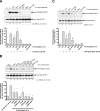
FIG. 1.
FIG. 1.
Effect of cAMP elevating agents on STAT3 phosphorylation by sIL-6Rα/IL-6 and leptin. (A) HUVECs were pretreated for 5 h with or without 10 μM Fsk and/or 10 μM Roli prior to the addition of vehicle or 25 ng/ml sIL-6Rα/5 ng/ml IL-6 for a further 30 min as indicated. Soluble cell extracts equalized for protein content were then fractionated by SDS-PAGE for immunoblotting with anti-Tyr705 phospho-STAT3 and total STAT3 antibodies. Quantitative analysis from three experiments is presented (*, P < 0.05 versus the response observed with sIL-6Rα/IL-6 alone). (B) HUVECs were pretreated for 5 h with 10 μM Roli and either 3 μM PGE2, 10 μM CGS21680, or 1 μM αMSH prior to the addition of vehicle or 2.5 ng/ml sIL-6Rα/0.5 ng/ml IL-6 for a further 30 min as indicated. Soluble cell extracts equalized for protein content were then fractionated by SDS-PAGE for immunoblotting with anti-Tyr705 phospho-STAT3 and total STAT3 antibodies. Quantitative analysis from three experiments is presented (*, P < 0.05 versus the response observed with sIL-6Rα/IL-6 alone). (C) HUVECs were pretreated for 5 h with or without Fsk+Roli prior to the addition of vehicle or 100 ng/ml leptin for a further 30 min as indicated. Soluble cell extracts equalized for protein content were then fractionated by SDS-PAGE for immunoblotting with anti-Tyr705 phospho-STAT3 and total STAT3 antibodies, as described for panel A. Quantitative analysis from three experiments is presented (*, P < 0.05 versus the response observed with leptin alone). Veh, vehicle.
FIG. 2.
FIG. 2.
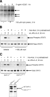
Effect of SHP-2 inhibition on cAMP-mediated inhibition of STAT3 phosphorylation by sIL-6Rα/IL-6. (A) HUVECs were incubated with or without the protein Tyr phosphatase inhibitor mpV (100 μM) for 5 h prior to treatment with or without 5 ng/ml vascular endothelial growth factor (VEGF) for 1 h as indicated. Soluble cell extracts equalized for protein content were then fractionated by SDS-PAGE for immunoblotting with anti-phospho-Tyr antibody. The three most prominent immunoreactive bands are indicated. preinc, preincubation. (B) HUVECs were incubated with or without the protein Tyr phosphatase inhibitor mpV (100 μM) for 30 min prior to treatment with or without Fsk+Roli for 5 h and vehicle or 25 ng/ml sIL-6Rα/5 ng/ml IL-6 for a further 30 min as indicated. Soluble cell extracts equalized for protein content were then fractionated by SDS-PAGE for immunoblotting with anti-Tyr705 phospho-STAT3, total STAT3, and SOCS-3 antibodies. (C) Upper panels, SHP-2+/+ and SHP-2Δ46-110 3T3 fibroblasts were pretreated with or without Fsk+Roli for 5 h prior to the addition of vehicle or sIL-6Rα/IL-6 for a further 30 min as shown in panel B. Soluble cell extracts equalized for protein content were then fractionated by SDS-PAGE for immunoblotting with anti-Tyr705 phospho-STAT3 and total STAT3 antibodies. Lower panel, lysates from SHP-2+/+ and SHP-2Δ46-110 3T3 fibroblasts were also immunoblotted with anti-SHP-2 antibody to identify wild-type (WT) and mutated SHP-2 Δ46-110, which migrates with enhanced mobility on SDS-PAGE due to the introduced deletion.
FIG. 3.
FIG. 3.
Effect of blocking SOCS-3 induction on cAMP-mediated inhibition of STAT3 phosphorylation by sIL-6Rα/IL-6. (A) HUVECs were incubated with or without the protein synthesis inhibitor emetine for 30 min prior to treatment with or without Fsk+Roli for 5 h and vehicle or 25 ng/ml sIL-6Rα and 5 ng/ml IL-6 for a further 30 min as indicated. Soluble cell extracts equalized for protein content were then fractionated by SDS-PAGE for immunoblotting with anti-Tyr705 phospho-STAT3, total STAT3, and SOCS-3 antibodies. (B) HUVECs were treated with SOCS-3 morpholino antisense oligonucleotide or a control morpholino for 48 h prior to treatment with or without 10 μM Fsk for 5 h as indicated and preparation of soluble cell extracts. Following equalization for protein content, samples were fractionated by SDS-PAGE for immunoblotting with anti-SOCS-3 and anti-GAPDH antibodies, the latter serving as a loading control. (C) HUVECs were treated with SOCS-3 morpholino antisense oligonucleotide or a control morpholino for 48 h prior to pretreatment with or without 10 μM Fsk for 5 h prior to the addition of vehicle or 25 ng/ml sIL-6Rα/5 ng/ml IL-6 for a further 30 min as indicated. Soluble cell extracts equalized for protein content were then fractionated by SDS-PAGE for immunoblotting with anti-Tyr705 phospho-STAT3 and total STAT3 antibodies. (D) SOCS-3+/+ and SOCS-3−/− MEFs were treated with Fsk+Roli for the indicated times prior to the preparation of soluble cell extracts. Following equalization for protein content, samples were fractionated by SDS-PAGE for immunoblotting with anti-SOCS-3 and anti-tubulin antibodies, the latter serving as a loading control. (E) SOCS-3+/+ and SOCS-3−/− MEFs were pretreated with or without Fsk+Roli for 5 h prior to the addition of vehicle or 25 ng/ml sIL-6Rα/5 ng/ml IL-6 for a further 30 min as indicated. Soluble cell extracts equalized for protein content were then fractionated by SDS-PAGE for immunoblotting with anti-Tyr705 phospho-STAT3 and total STAT3 antibodies.
FIG. 4.
FIG. 4.
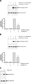
Effect of cAMP elevation on SOCS-3 induction and STAT3 phosphorylation by sIL-6Rα/IL-6 in HAECs. HAECs were pretreated for 5 h with or without 10 μM forskolin prior to the addition of vehicle or 25 ng/ml sIL-6Rα and 5 ng/ml IL-6 for a further 30 min as indicated. Soluble cell extracts equalized for protein content were then fractionated by SDS-PAGE for immunoblotting with either anti-Tyr701 phospho-STAT1 and total STAT1 antibodies (A) or anti-Tyr705 phospho-STAT3 and total STAT3 antibodies (B). Quantitative analysis from three experiments is presented (formula image, P < 0.05 versus the response observed with sIL-6Rα/IL-6 alone). (C) HAECs were pretreated for 5 h with 10 μM Fsk prior to the preparation of soluble cell extracts. Following equalization for protein content, samples were fractionated by SDS-PAGE for immunoblotting with anti-SOCS-3 and anti-GAPDH antibodies, with the latter serving as a loading control.
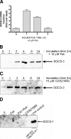
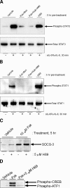
FIG. 5.
FIG. 5.
Characterization of SOCS-3 induction following cAMP elevation in HUVECs. (A) HUVECs were treated with Fsk for the indicated times prior to the preparation of RNA and analysis of SOCS-3 mRNA by quantitative real-time reverse transcription-PCR. Results from one of four experiments is presented. (B) HUVECs were treated with Fsk for the indicated times prior to the preparation of soluble cell extracts. Following equalization for protein content, samples were fractionated by SDS-PAGE for immunoblotting with anti-SOCS-3 antibody. (C) HUVECs were treated with the A2AAR-selective agonist CGS21680 (5 μM) for the indicated times prior to the preparation of soluble cell extracts. Following equalization for protein content, samples were fractionated by SDS-PAGE for immunoblotting with anti-SOCS-3 antibody. (D) HUVECs were treated with or without Fsk, Fsk+Roli, and CGS21680 for 5 h prior to the preparation of soluble cell extracts. Following equalization for protein content, samples were fractionated by SDS-PAGE for immunoblotting with anti-SOCS-1 antibody. Soluble extract (10 μg) prepared from HEK293 cells transiently expressing Flag epitope-tagged SOCS-1 (HEK293/Flag-SOCS-1) was run in parallel as a positive control for antibody immunoreactivity (+ve).
FIG. 6.
FIG. 6.
Effect of blocking PKA activity on cAMP-mediated SOCS-3 induction and inhibition of STAT3 phosphorylation by sIL-6Rα/IL-6. HUVECs were incubated with or without the PKA inhibitor H89 (5 μM) for 30 min prior to treatment with Fsk+Roli for 5 h and vehicle or 25 ng/ml sIL-6Rα/5 ng/ml IL-6 for a further 30 min as indicated. Soluble cell extracts equalized for protein content were then fractionated by SDS-PAGE for immunoblotting with anti-Tyr705 phospho-STAT3 and total STAT3 (A) or anti-Tyr701 phospho-STAT1 and total STAT1 antibodies (B). (C) HUVECs were incubated with or without the PKA inhibitor H89 (5 μM) for 30 min prior to treatment with 10 μM Fsk for 5 h as indicated. Soluble cell extracts equalized for protein content were then fractionated by SDS-PAGE for immunoblotting with anti-SOCS-3 antibody. (D) HUVECs were incubated with or without the PKA inhibitor H89 (5 μM) for 30 min prior to treatment with 10 μM Fsk for 30 min as indicated. Soluble cell extracts equalized for protein content were then fractionated by SDS-PAGE for immunoblotting with anti-Ser133 phospho-CREB antibody.
FIG. 7.
FIG. 7.
Contribution of Epac toward SOCS-3 induction and inhibition of STAT3 phosphorylation by sIL-6Rα/IL-6. (A) HUVECs were treated with the indicated concentrations of the Epac-selective activator 8-pCPT for 5 h prior to the preparation of soluble cell extracts. Following equalization for protein content, samples were fractionated by SDS-PAGE for immunoblotting with anti-SOCS-3 and antitubulin antibodies, with the latter serving as a loading control. Veh, vehicle. (B) HUVECs were treated with 50 μM 8-pCPT for the indicated times prior to the preparation of soluble cell extracts. Following equalization for protein content, samples were fractionated by SDS-PAGE for immunoblotting with anti-SOCS-3 antibody. (C) HUVECs were transiently transfected with empty vector or expression constructs encoding Leu61Cdc42 and Val12Rap1a as indicated. Forty-eight hours posttransfection, cells were treated with or without 6 μM MG132 for 6 h prior to the preparation of soluble cell extracts. Following equalization for protein content, samples were then fractionated by SDS-PAGE for immunoblotting with anti-SOCS-3 and anti-ERK1/2 antibodies, with the latter serving as a loading control. Quantitative analysis from three experiments is presented (formula image, P < 0.05 versus the response observed with vector plus MG132). (D) HUVECs were pretreated for 5 h with or without 50 μM 8-pCPT prior to the addition of vehicle, 2.5 ng/ml sIL-6Rα, and 0.5 ng/ml IL-6 for a further 30 min as indicated. Soluble cell extracts equalized for protein content were then fractionated by SDS-PAGE for immunoblotting with anti-Tyr705 phospho-STAT3 and total STAT3 antibodies. (E) HUVECs were transfected twice over 48 h with nontargeting control and Epac1-specific siRNAs prior to treatment with Fsk for 5 h as indicated. Soluble cell extracts equalized for protein content were then fractionated by SDS-PAGE for immunoblotting with anti-Epac1, SOCS-3, and GAPDH antibodies. (F) HUVECs were transfected with control and Epac1-specific siRNAs as described for panel E prior to pretreatment with or without Fsk+Roli for 5 h followed by exposure to 25 ng/ml sIL-6Rα and 5 ng/ml IL-6 for 30 min as indicated. Soluble cell extracts equalized for protein content were then fractionated by SDS-PAGE for immunoblotting with anti-Tyr705 phospho-STAT3 and total STAT3 antibodies.
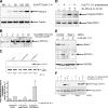
FIG. 8.
FIG. 8.
Effect of Epac activation on the accumulation of MCP-1. HUVECs were pretreated with either vehicle (Veh), Fsk+Roli, or 50 μM 8-pCPT for 5 h prior to treatment with 25 ng/ml sIL-6Rα and 5 ng/ml IL-6 for 24 h as indicated. Conditioned medium was then removed for measurement of MCP-1 levels. Data from three experiments are presented (formula image, P < 0.05 versus sIL-6Rα/IL-6-stimulated MCP-1 levels). Basal MCP-1 levels were 0.69 ± 0.42 ng/ml (n = 3).
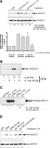
FIG. 9.
FIG. 9.
Identification of a signaling pathway controlling cAMP-mediated induction of SOCS-3. (A) HUVECs were pretreated for 30 min with or without 0.1 μM JAK inhibitor prior to the addition of either 25 ng/ml sIL-6Rα and 5 ng/ml IL-6 or Fsk+Roli for a further 3 or 5 h as indicated. Soluble cell extracts equalized for protein content were then fractionated by SDS-PAGE for immunoblotting with anti-SOCS-3 and anti-GAPDH antibodies, the latter serving as a loading control. Quantitative analysis from three experiments is presented (formula image, P < 0.05 versus sIL-6Rα/IL-6-stimulated SOCS-3 expression in the absence of JAK inhibitor). (B) HUVECs were pretreated for 30 min with or without MEK inhibitor U0126 (0.1 μM) prior to the addition of 5 μM Fsk for a further 5 h as indicated. Soluble cell extracts equalized for protein content were then fractionated by SDS-PAGE for immunoblotting with anti-SOCS-3 antibody. (C) HUVECs were pretreated with or without 0.1 μM U0126 prior to the addition of 5 μM Fsk or 1 μM PMA as indicated. Soluble cell extracts equalized for protein content were then fractionated by SDS-PAGE for immunoblotting with anti-Thr202/Tyr204 phospho-ERK1/2 antibody. (D) HUVECs were pretreated with or without 0.1 μM U0126 prior to the addition of 1 μM PMA or 25 ng/ml sIL-6Rα and 5 ng/ml IL-6 for 5 h as indicated. Soluble cell extracts equalized for protein content were then fractionated by SDS-PAGE for immunoblotting with anti-SOCS-3 and GAPDH antibodies. Veh, vehicle; pre-inc, preincubation.

References

    1. Aird, W. C. 2005. Spatial and temporal dynamics of the endothelium. J. Thromb. Haemost. 3:1392-1406. - PubMed
    1. Alexander, W. S., and D. J. Hilton. 2004. The role of suppressors of cytokine signaling (SOCS) proteins in regulation of the immune response. Annu. Rev. Immunol. 22:503-529. - PubMed
    1. Blanchard, F., W. Yanping, E. Kinzie, L. Duplomb, A. Godard, and H. Baumann. 2001. Oncostatin M regulates the synthesis and turnover of gp130, leukemia inhibitory factor receptor α, and oncostatin M receptor-β by distinct mechanisms. J. Biol. Chem. 276:47038-47045. - PubMed
    1. Blease, K., A. Burke-Gaffney, and P. G. Hellewell. 1998. Modulation of cell adhesion molecule expression and function on human lung microvascular endothelial cells by inhibition of phosphodiesterases 3 and 4. Br. J. Pharmacol. 124:229-237. - PMC - PubMed
    1. Bos, J. L. 2003. Epac: a new cAMP target and new avenues in cAMP research. Nat. Rev. Mol. Cell Biol. 4:733-738. - PubMed

Publication types

MeSH terms