. 2006 Sep;26(17):6381-94.
doi: 10.1128/MCB.00570-06.

Binding of SH2-B family members within a potential negative regulatory region maintains JAK2 in an active state

Affiliations

Binding of SH2-B family members within a potential negative regulatory region maintains JAK2 in an active state

Jason H Kurzer et al. Mol Cell Biol. 2006 Sep.

Abstract

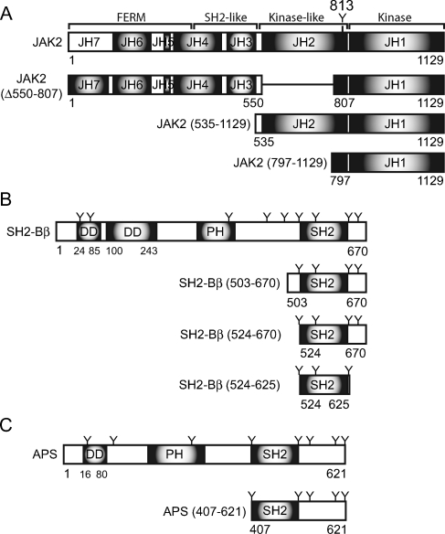
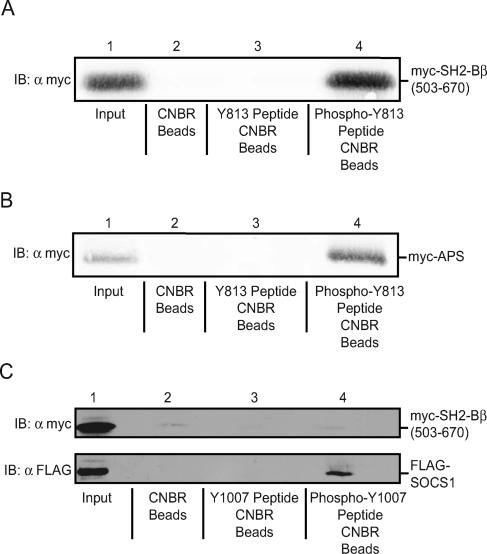
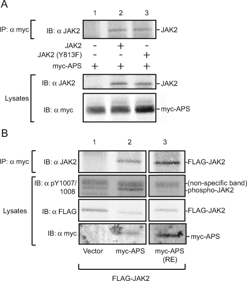
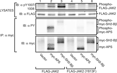
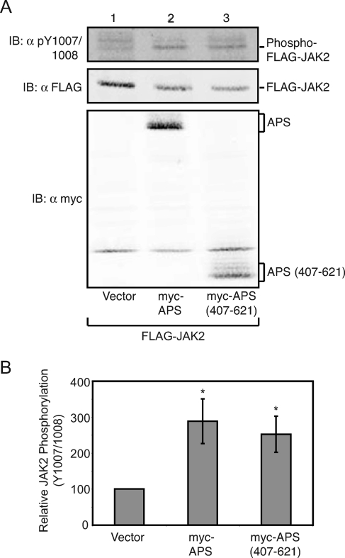
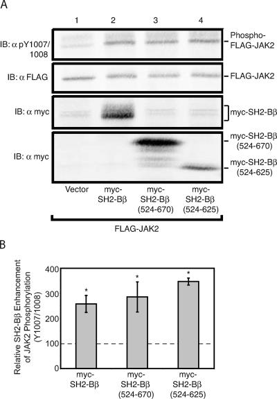
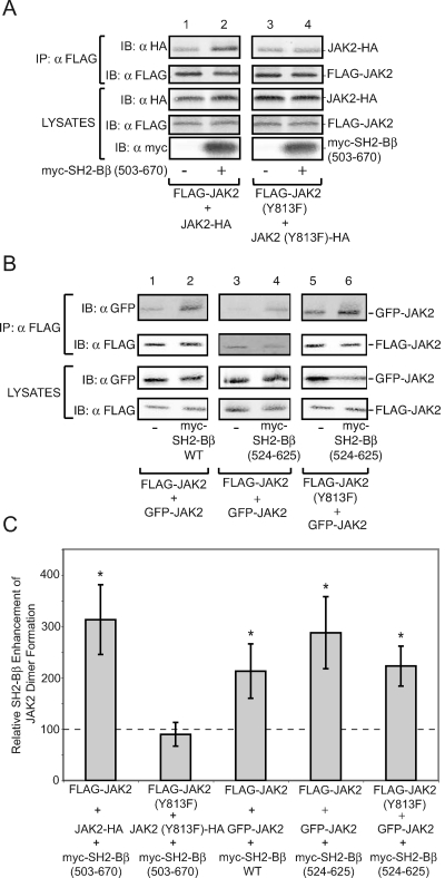
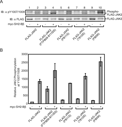
The tyrosine kinase Janus kinase 2 (JAK2) transduces signaling for the majority of known cytokine receptor family members and is constitutively activated in some cancers. Here we examine the mechanisms by which the adapter proteins SH2-Bbeta and APS regulate the activity of JAK2. We show that like SH2-Bbeta, APS binds JAK2 at multiple sites and that binding to phosphotyrosine 813 is essential for APS to increase active JAK2 and to be phosphorylated by JAK2. Binding of APS to a phosphotyrosine 813-independent site inhibits JAK2. Both APS and SH2-Bbeta increase JAK2 activity independent of their N-terminal dimerization domains. SH2-Bbeta-induced increases in JAK2 dimerization require only the SH2 domain and only one SH2-Bbeta to be bound to a JAK2 dimer. JAK2 mutations and truncations revealed that amino acids 809 to 811 in JAK2 are a critical component of a larger regulatory region within JAK2, most likely including amino acids within the JAK homology 1 (JH1) and JH2 domains and possibly the FERM domain. Together, our data suggest that SH2-Bbeta and APS do not activate JAK2 as a consequence of their own dimerization, recruitment of an activator of JAK2, or direct competition with a JAK2 inhibitor for binding to JAK2. Rather, they most likely induce or stabilize an active conformation of JAK2.


Figures

FIG. 1.
FIG. 1.
Schematic representation of JAK2, SH2-Bβ, and APS. (A) JAK2 is composed of seven JH domains. JAK2 (Δ550-807), JAK2 (535-1129), and JAK2 (797-1129) represent JAK2 mutants used in Fig. 10. (B) SH2-B is composed of an SH2 domain, a PH domain, and two identified dimerization domains (DD), as well as nine tyrosines (Y). SH2-Bβ (524-670) is used in Fig. 6, SH2-Bβ (524-625) is used in Fig. 6 and 7, and SH2-Bβ (503-670) is used in Fig. 7. (C) APS is composed of an SH2 domain, a PH domain, eight tyrosines (Y), and at least one identified dimerization domain. APS (407-621) is used in Fig. 5.
FIG. 2.
FIG. 2.
APS and SH2-Bβ bind phosphorylated tyrosine 813 of JAK2 but not unphosphorylated tyrosine 813. (A and B) Lysates of COS7 cells transfected with cDNA (5.0 μg) for myc-SH2-Bβ (503-670) (A) or myc-APS (B) were incubated with unconjugated beads (lanes 2), beads coupled to the Y813 peptide (lanes 3), or beads coupled to the phospho-Y813 peptide (lanes 4). Proteins in cell lysates (lanes 1) and proteins that bound to the beads (lanes 2 to 4) were separated by SDS-PAGE and immunoblotted with αmyc. (C) Lysates of COS7 cells transfected with cDNA (5.0 μg) encoding either FLAG-SOCS-1 or myc-SH2-Bβ (503-670) were incubated with unconjugated beads (lane 2), beads coupled to the Y1007 peptide (lane 3), or beads coupled to the phospho-Y1007 peptide (lane 4). Proteins in cell lysates (lane 1) and proteins that bound to the beads (lanes 2 to 4) were separated by SDS-PAGE and immunoblotted with αmyc (upper panel) or αFLAG (lower panel). IB, immunoblot.
FIG. 3.
FIG. 3.
APS binds phosphotyrosine 813 and other regions of JAK2. (A) COS7 cells were transfected with 2.0 μg cDNA for APS along with 1.0 μg of empty vector (lane 1) or with cDNA encoding FLAG-JAK2 (lane 2) or FLAG-JAK2 (Y813F) (lane 3). Proteins in cell lysates were immunoprecipitated with αmyc and immunoblotted with αJAK2 (top panel). Cell lysates were immunoblotted with αJAK2 (middle panel) or αmyc (bottom panel). (B) cDNA (1.0 μg) encoding FLAG-JAK2 was coexpressed in COS7 cells with 2.0 μg of empty vector (lane 1) or with cDNA encoding myc-APS (lane 2) or myc-APS (R437E) (lane 3). Cell lysates were immunoprecipitated with αmyc, and immunoprecipitated proteins were immunoblotted with αJAK2 (top panel). Cell lysates were immunoblotted with αpY1007/1008 (second panel), αFLAG (third panel), and αmyc (bottom panel). All three lanes were obtained from the same gel. IP, immunoprecipitation; IB, immunoblot.
FIG. 4.
FIG. 4.
Tyrosine 813 of JAK2 is essential for APS to increase active JAK2. COS7 cells were transfected with 1.0 μg cDNA encoding either FLAG-JAK2 (lanes 1 through 3) or FLAG-JAK2 (Y813F) (lanes 4 through 6), along with 2.0 μg cDNA encoding empty vector (lanes 1 and 4), myc-APS (lanes 2 and 5), or myc-SH2-Bβ (lanes 3 and 6). Proteins in cell lysates were immunoprecipitated (IP) with αmyc, and immunoprecipitated proteins and aliquots of cell lysates were separated by SDS-PAGE. Cell lysates were immunoblotted (IB) with αpY1007/1008 (top panel) or αFLAG (second panel). The immunoprecipitated proteins were immunoblotted with αPY (third panel) or αmyc (bottom panel).
FIG. 5.
FIG. 5.
Amino acids 407 to 621 are sufficient for APS to increase active JAK2. cDNA (1.0 μg) encoding FLAG-JAK2 was coexpressed with 2.0 μg of either empty prk5 myc vector (lane 1) or cDNA encoding myc-APS (lane 2) or myc-APS (407-621) (lane 3). Cell lysates were immunoblotted (IB) with αpY1007/1008 (top panel), αFLAG (FLAG-JAK2) (middle panel), or αmyc (bottom panel). (B) Replicates for the experiment shown in panel A were quantified, normalized to expression levels of JAK2, and expressed as percentages of JAK2 phosphorylation in the absence of APS (vector control). The data are expressed as means ± standard errors (SE) (n = 4). Conditions which yield statistically significant differences (P < 0.05) in JAK2 phosphorylation at tyrosine 1007/1008 compared to vector control are marked with asterisks.
FIG. 6.
FIG. 6.
The SH2 domain of SH2-B is sufficient to increase active JAK2. cDNA (1.0 μg) encoding FLAG-JAK2 was coexpressed with 2.0 μg cDNA encoding either empty prk5 myc vector (lane 1), myc-SH2-Bβ (lane 2), myc-SH2-Bβ (524-670) (lane 3), or myc-SH2-Bβ (524-625) (lane 4). (A) Cell lysates were immunoblotted (IB) with αFLAG (second panel) and reprobed with αpY1007/1008 (top panel) and αmyc (bottom two panels). (B) Replicates for the experiment shown in panel A were quantified, normalized to expression levels of JAK2, and expressed as percentages of JAK2 phosphorylation in the absence of SH2-Bβ (vector control). The data are expressed as means ± SE (n = 4, 3, and 3). Conditions which yield statistically significant differences (P < 0.05) in JAK2 phosphorylation of Y1007/1008 compared to the vector (no SH2-Bβ) control are marked with asterisks. Control levels are denoted by a dashed line.
FIG. 7.
FIG. 7.
SH2-Bβ enhances the dimerization of JAK2 in a manner independent of SH2-Bβ dimerization. (A) COS7 cells were transfected with 0.5 μg cDNA encoding FLAG-JAK2 and JAK2-HA (lanes 1 and 2) or FLAG-JAK2 (Y813F) and JAK2-HA (Y813F) (lanes 3 and 4) along with 2.0 μg cDNA encoding empty vector (lanes 1 and 3) or myc-SH2-Bβ (503-670) (lanes 2 and 4). Proteins in cell lysates were immunoprecipitated with αFLAG and immunoblotted with αHA (top panel) and αFLAG (second panel). Cell lysates were immunoblotted with αHA (third panel), αFLAG (fourth panel), and αmyc (bottom panel). (B) cDNA (1.5 μg) encoding GFP-JAK2 was transfected into COS7 cells with 0.5 μg cDNA encoding FLAG-JAK2 (lanes 1 through 4) or FLAG-JAK2 (Y813F) (lanes 5 and 6), along with 2.0 μg cDNA encoding empty vector (lanes 1, 3, and 5), myc-SH2-Bβ (lane 2), or myc-SH2-Bβ (524-625) (lanes 4 and 6). Proteins in cell lysates were immunoprecipitated with αFLAG and immunoblotted with αGFP (top panel) and αFLAG (second panel from top). Cell lysates were immunoblotted with αGFP (third panel from top) or αFLAG (bottom panel). Lanes 1, 2, 5, and 6 were obtained from the same gel. Lanes 3 and 4 were from a separate experiment and a separate gel. (C) Replicates for the experiments shown in panels A and B were quantified, and signal in the presence of SH2-Bβ (even-numbered lanes) was expressed as a percentage of the signal in the absence of SH2-Bβ (odd-numbered lanes). The data are expressed as means ± SE (n = 3). Statistically significant differences (P < 0.05) in JAK2 dimerization compared to conditions without SH2-Bβ are marked with asterisks. Control levels are denoted by a dashed line. IP, immunoprecipitation; IB, immunoblot.
FIG. 8.
FIG. 8.
Mutation of amino acids 809 to 811 of JAK2 enhances JAK2 activity. (A) cDNA (1.0 μg) encoding FLAG-JAK2 (lanes 1 and 2), FLAG-JAK2 (FTP809-811ISS) (lanes 3 and 4), FLAG-JAK2 (P811S) (lanes 5 and 6), FLAG-JAK2 (T810S) (lanes 7 and 8), or FLAG-JAK2 (F809I) (lanes 9 and 10) was cotransfected with 2.0 μg cDNA encoding prk5 myc (lanes 1, 3, 5, 7, and 9) or myc-SH2-Bβ (lanes 2, 4, 6, 8, and 10) into COS7 cells. Cell lysates were immunoblotted (IB) with αFLAG (bottom panel), and the membrane was reprobed with αpY1007/1008 (top panel). (B) Replicates for the experiment shown in panel A were quantified, normalized to expression levels of JAK2, and expressed as percentages of JAK2 phosphorylation in the absence of SH2-Bβ (vector control). The data are expressed as means ± ranges (n = 2).
FIG. 9.
FIG. 9.
Mutation of amino acids 781 to 783 in JAK3 does not alter JAK3 activity. (A) cDNA (1.0 μg) encoding JAK3 (lanes 1 and 2) or JAK3 (ISS781-783FTP) (lanes 3 and 4) was cotransfected with 2.0 μg cDNA encoding prk5 myc (lanes 1 and 3) or myc-SH2-Bβ (lanes 2 and 4). Cell lysates were immunoblotted (IB) with αmyc (bottom panel) or αJAK3 (middle panel). The membrane was reprobed with αPY (top panel). (B) Replicates for the experiment shown in panel A were quantified, normalized to expression levels of JAK3, and expressed as percentages of JAK3 phosphorylation in the absence of SH2-Bβ (vector control). The data are expressed as means ± SE (n = 3). No significant difference in phosphorylation levels was detected between the conditions.
FIG. 10.
FIG. 10.
Amino acids 1 to 806 of JAK2 are dispensable for SH2-Bβ to increase the amount of active JAK2. (A) cDNA (1.0 μg) encoding either FLAG-JAK2 (lanes 1 and 2), JAK2 (Δ550-807)-HA (lanes 3 and 4), JAK2 (535-1129)-HA (lanes 5 and 6), or FLAG-JAK2 (797-1129) (lanes 7 and 8) was transfected into COS7 cells along with 2.0 μg cDNA encoding either prk5 myc (lanes 1, 3, 5, and 7) or myc-SH2-Bβ (lanes 2, 4, 6, and 8). Cell lysates were immunoblotted with αFLAG (second panel, lanes 1, 2, 7, and 8) or with αHA (third panel, lanes 3 to 6). The nitrocellulose was reprobed with αpY1007/1008 (top panel). Cell lysates were immunoblotted (IB) with αmyc (bottom panel). (B) Replicates for the experiment shown in panel A were quantified, normalized to expression levels of each corresponding JAK2 mutant, and expressed as percentages of JAK2/JAK2 mutant phosphorylation in the absence of SH2-Bβ (vector control). The data are expressed as means ± SE [n = 4, 3, 4, and 3 for FLAG-JAK2, JAK2 (Δ550-807)-HA, JAK2 (535-1129)-HA, and FLAG-JAK2 (797-1129), respectively]. Control levels are denoted by a dashed line.

References

    1. Brown, R. J., J. J. Adams, R. A. Pelekanos, Y. Wan, W. J. McKinstry, K. Palethorpe, R. M. Seeber, T. A. Monks, K. A. Eidne, M. W. Parker, and M. J. Waters. 2005. Model for growth hormone receptor activation based on subunit rotation within a receptor dimer. Nat. Struct. Mol. Biol. 12:814-821. - PubMed
    1. Chen, C., and H. Okayama. 1987. High-efficiency transformation of mammalian cells by plasmid DNA. Mol. Cell. Biol. 7:2745-2752. - PMC - PubMed
    1. Darnell, J. E., Jr., I. M. Kerr, and G. R. Stark. 1994. Jak-STAT pathways and transcriptional activation in response to IFNs and other extracellular signaling proteins. Science 264:1415-1421. - PubMed
    1. Dhe-Paganon, S., E. D. Werner, M. Nishi, L. Hansen, Y. I. Chi, and S. E. Shoelson. 2004. A phenylalanine zipper mediates APS dimerization. Nat. Struct. Mol. Biol. 11:968-974. - PubMed
    1. Duan, C., H. Yang, M. F. White, and L. Rui. 2004. Disruption of SH2-B causes age-dependent insulin resistance and glucose intolerance. Mol. Cell. Biol. 24:743507443. - PMC - PubMed

Publication types

MeSH terms

LinkOut - more resources