. 2006 Sep;26(17):6469-86.
doi: 10.1128/MCB.00353-06.

Activation of receptor activator of NF-kappaB ligand gene expression by 1,25-dihydroxyvitamin D3 is mediated through multiple long-range enhancers

Affiliations

Activation of receptor activator of NF-kappaB ligand gene expression by 1,25-dihydroxyvitamin D3 is mediated through multiple long-range enhancers

Sungtae Kim et al. Mol Cell Biol. 2006 Sep.

Abstract

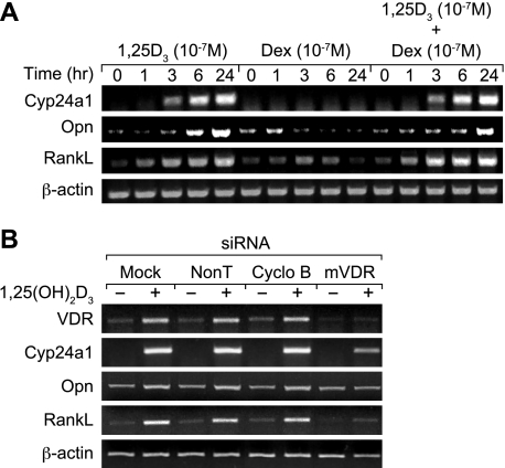
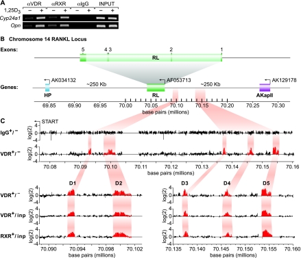
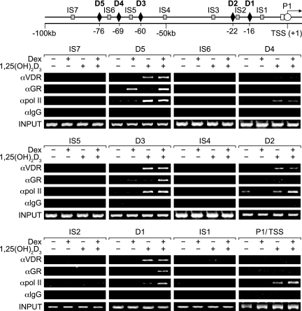
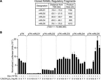
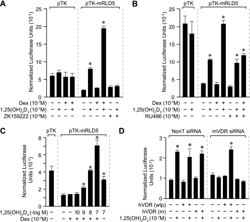
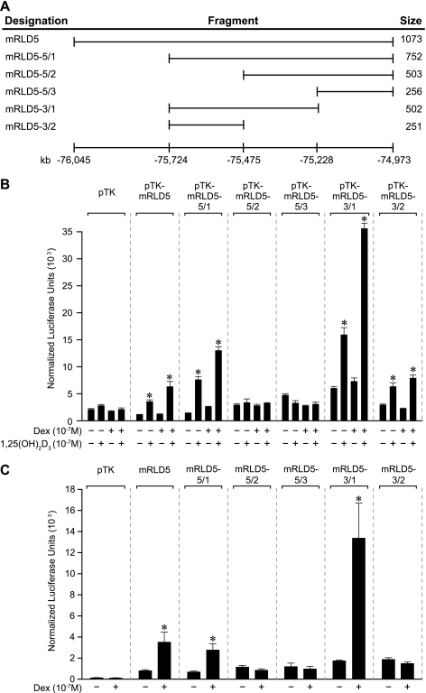
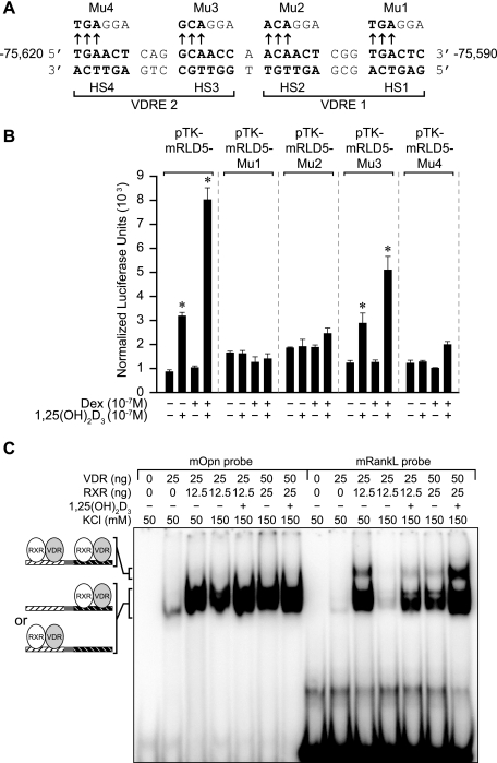
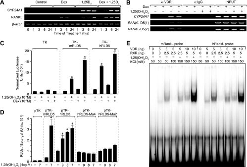
RANKL is a tumor necrosis factor (TNF)-like factor secreted by mesenchymal cells, osteoblast derivatives, and T cells that is essential for osteoclastogenesis. In osteoblasts, RANKL expression is regulated by two major calcemic hormones, 1,25-dihydroxyvitamin D(3) [1,25(OH)(2)D(3)] and parathyroid hormone (PTH), as well as by several inflammatory/osteoclastogenic cytokines; the molecular mechanisms for this regulation are unclear. To identify such mechanisms, we screened a DNA microarray which tiled across the entire mouse RankL gene locus at a 50-bp resolution using chromatin immunoprecipitation (ChIP)-derived DNA precipitated with antibodies to the vitamin D receptor (VDR) and the retinoid X receptor (RXR). Five sites of dimer interaction were observed on the RankL gene centered at 16, 22, 60, 69, and 76 kb upstream of the TSS. These regions contained binding sites for not only VDR and RXR, but also the glucocorticoid receptor (GR). The most distant of these regions, termed the distal control region (RL-DCR), conferred both VDR-dependent 1,25(OH)(2)D(3) and GR-dependent glucocorticoid (GC) responses. We mapped these activities to an unusual but functionally active vitamin D response element and to several potential GC response elements located over a more extensive region within the RL-DCR. An evolutionarily conserved region within the human RANKL gene contained a similar vitamin D response element and exhibited an equivalent behavior. Importantly, hormonal activation of the RankL gene was also associated with chromatin modification and RNA polymerase II recruitment. Our studies demonstrate that regulation of RankL gene expression by 1,25(OH)(2)D(3) is complex and mediated by at least five distal regions, one of which contains a specific element capable of mediating direct transcriptional activation.


Figures

FIG. 1.
FIG. 1.
Induction of mRankL mRNA by 1,25(OH)2D3 in ST2 cells is enhanced by DEX and mediated by the VDR. (A) Induction of RankL expression levels by 1,25(OH)2D3 and DEX in vitro. ST2 cells were treated for periods of up to 24 h with either 1,25(OH)2D3 (10−7 M), DEX (10−7 M), or both (at 10−7 M). Total RNA was isolated and subjected to reverse transcription-PCR (RT-PCR) analysis using primers specific to mouse Cyp24a1 (30 cycles), osteopontin (Opn) (15 cycles), RankL (30 cycles), or β-actin (20 cycles) as documented in Materials and Methods. The results are typical of multiple similar experiments. (B) Effects of mVDR siRNA on 1,25(OH)2D3-induced RankL expression levels in ST2 cells. ST2 cells were transfected with 20 nM nontargeting siRNA, cyclophilin B (Cyclo B) siRNA, or mVDR siRNA. After 48 h, the cells were treated for an additional 6 h with either vehicle or 1,25(OH)2D3 (10−7 M). Total RNA was isolated and subjected to standard RT-PCR analysis using the primers identified in panel A above. The numbers of cycles for amplification of each transcript are as follows: VDR, 20 cycles; Cyp24a1, 25 cycles; Opn, 15 cycles, and β-actin, 15 cycles. These results are typical of several independent experiments.
FIG. 2.
FIG. 2.
ChIP/chip analysis reveals five VDR/RXR-interacting regions at significant distances upstream of the mRankL gene TSS. (A) ST2 cells were treated with either vehicle or 1,25(OH)2D3 (10−7 M) for 6 h and then subjected to ChIP analysis using antibodies to VDR, RXR(pan), or control IgG (αVDR, αRXR, and αIgG, respectively). Immunoprecipitated DNA was isolated and then amplified using ChIP primer sets to either Cyp24a1 or Opn as indicated in Materials and Methods and Table 1. Input DNA was obtained prior to precipitation. (B) Schematic diagram of the mouse RankL gene with its five exons and its position relative to adjacent downstream (AK034132) and upstream (AK129178) genes on chromosome 14. The reverse arrow indicates the direction of transcription on the reverse strand. The nucleotide base pairs indicate nucleotide location on chromosome 14 (December 2004 assembly). (C, upper panel) Individual data tracks representing the enrichment ratio of Cy5-to-Cy3 hybridization intensity (log2) for IgG ± hormone or VDR ± hormone. The nucleotide base pairs on the x axis indicate the position on chromosome 14. (C, lower panel) An expanded view of the Cy5/Cy3 signal enrichment ratios for VDR ± hormone, VDR ± versus input, and RXR plus hormone versus input. The highlighted areas indicate the peaks of interest which are designated mRLD1 to mRLD5.
FIG. 3.
FIG. 3.
ChIP analysis of the upstream region of the mRankL gene reveals complex 1,25(OH)2D3- and DEX-stimulated transactivator DNA binding and RNA pol II recruitment. ST2 cells were treated with either vehicle, 1,25(OH)2D3 (10−7 M), DEX (10−7 M), or 1,25(OH)2D3 and DEX (10−7 M) for 6 h and then subjected to ChIP analysis using antibodies to VDR, GR, RNA pol II, or control IgG (αVDR, αGR, αpol II, and αIgG, respectively). The immunoprecipitated DNA was isolated and then amplified using the primer sets whose positions are illustrated in the top panel and whose sequences are documented in Table 1. Amplification utilized 31 cycles for mRLD1 to mRLD5 and mRLIS1 to mRLIS7 and 34 cycles for the TSS. PCR analyses were all performed within the linear range of amplification. These results are typical of several similar studies.
FIG. 4.
FIG. 4.
The mRLD5 region of the mRankL gene mediates transcriptional induction by 1,25(OH)2D3 and DEX. (A) Features of the cloned mRLD1-to-mRLD5 regions of the mRankL gene, including the sizes of the fragments cloned for evaluation, the distances from the RankL TSS, and the boundaries of the fragments on chromosome 14 (February 2006 assembly). (B) Basal and hormone-inducible activities of mRLD1 to mRLD5 in ST2 cells. ST2 cells were transfected with pCH110-βgal (50 ng), pcDNA-hVDR (50 ng), and either the pTK control vector (250 ng) or pTK-mRLD1, pTK-mRLD2, pTK-mRLD3, pTK-mRLD4, or pTK-mRLD5. Cells were treated with either vehicle, 1,25(OH)2D3 (10−7 M), DEX (10−7 M), or DEX (10−7 M) plus increasing concentrations of 1,25(OH)2D3 (10−10 to 10−7 M) and evaluated after 24 h for both luciferase and β-galactosidase activities as described in Materials and Methods. Each point represents the normalized relative light unit average ± standard error of the mean for a triplicate set of transfections. These data are representative of three or more similar experiments.
FIG. 5.
FIG. 5.
Hormone-inducible activity of the mRLD5 region is mediated by the VDR and GR. (A) The VDR antagonist ZK159222 blocks activation of mRLD5 by 1,25(OH)2D3. ST2 cells were transfected with pCH110-βgal (50 ng), pcDNA-hVDR (50 ng), and either the pTK control vector (250 ng) or pTK-mRLD5. Cells were treated with either vehicle, 1,25(OH)2D3 (10−7 M), DEX (10−7 M), DEX (10−7 M) plus 1,25(OH)2D3 (10−7 M), ZK159222 (10−6 M), or 1,25(OH)2D3 (10−7 M) plus ZK159222 (10−6 M) and evaluated after 24 h for both luciferase and β-galactosidase activities as described in Materials and Methods. (B) The GR antagonist blocks activation of mRLD5 by DEX. ST2 cells were treated as in panel A above with either vehicle, 1,25(OH)2D3 (10−7 M), DEX (10−7 M), DEX (10−7 M) plus 1,25(OH)2D3 (10−7 M), RU486 (10−6 M), RU486 (10−6 M) plus 1,25(OH)2D3 (10−7 M), or RU486 (10−6 M) plus 1,25(OH)2D3 (10−7 M) plus DEX (10−7 M) and evaluated after 24 h for both luciferase and β-galactosidase activities as described in Materials and Methods. (C) 1,25(OH)2D3 activates mRLD5 via endogenous VDR. ST2 cells were transfected with pCH110-βgal (50 ng) and either the pTK control vector (250 ng) or pTK-mRLD5 and then treated with either vehicle, DEX (10−7 M), DEX (10−7 M) plus 1,25(OH)2D3 (10−10 to 10−7 M), or 1,25(OH)2D3 (10−7 M) alone. Luciferase and β-galactosidase activities were assessed 24 h later as described in Materials and Methods. (D) The ability of VDR siRNA to block activation of mRLD5-mediated transcription by 1,25(OH)2D3 is rescued through the addition of a functional VDR expression vector. ST2 cells were cotransfected with 10 ng pCH110-βgal, pTK-mRLD5, pcDNA-hVDR(wtp) (wild type), or pcDNA-hVDR(m) (mutant) and 50 nM of either nontargeted siRNA or mVDR siRNA as described in Materials and Methods. Transfected cells were cultured for 48 h and then treated for an additional 24 h with either vehicle or 1,25(OH)2D3 (10−7 M) and processed as in panel B. Each point represents the normalized relative light unit average ± standard error of the mean for a triplicate set of transfections. All studies were repeated with similar results.
FIG. 6.
FIG. 6.
Deletion analysis of the mRLD5 region reveals the approximate location of transcriptional response to both 1,25(OH)2D3 and DEX. (A) Schematic of the mRankL deletion fragments of the mRLD5 region that were used to map 1,25(OH)2D3 and DEX responses. The numbering at the bottom represents the distance from the RankL TSS (February 2006 assembly). (B) Transcriptional activities of the mRLD5 subfragments in response to 1,25(OH)2D3 and DEX. ST2 cells were transfected with pCH110-βgal (50 ng), pcDNA-hVDR (50 ng), and either the pTK control vector (250 ng) or the pTK-mRLD5 deletion constructs indicated. Cells were treated with either vehicle, 1,25(OH)2D3 (10−7 M), DEX (10−7 M), or DEX (10−7 M) plus 1,25(OH)2D3 (10−7 M) and evaluated after 24 h for both luciferase and β-galactosidase activities as described in Materials and Methods. (C) Transcriptional activities of the mRLD5 subfragments in response to DEX. ST2 cells were transfected with pCH110-βgal (50 ng), pRSV-hGR (50 ng), and either the pTK control vector (250 ng) or the pTK-mRLD5 deletion constructs indicated. Cells were treated with either vehicle or DEX (10−7 M) and evaluated after 24 h as in panel B. These results were confirmed via at least three separate experiments.
FIG. 7.
FIG. 7.
Mapping the mRL-VDRE. (A) DNA sequence of a putative mRL-VDRE revealed by in silico analysis (http://mordor.cgb.ki.se/cgi-bin/CONSITE/consite). The nucleotide numbering represents the boundaries of the mRL-VDRE relative to the RankL TSS (February 2006 assembly). Nucleotide bases above the arrows represent the triplet alterations introduced by site-directed mutagenesis into each of the half-sites (HS1 to HS4) of the two VDREs (VDRE1 and VDRE2) that constitute the mRL-VDRE. Mutants are designated Mu1 to Mu4. (B) Transcriptional activity of wild-type pTK-mRLD5 or pTK-mRLD5 containing mutation 1 (Mu1), Mu2, Mu3, or Mu4. ST2 cells were transfected with pCH110-βgal (50 ng), pcDNA-hVDR (50 ng), and either the pTK control vector (250 ng), pTK-mRLD5, or pTK-mRLD5 containing Mu1 to Mu4 as illustrated. Cells were treated with either vehicle, 1,25(OH)2D3 (10−7 M), DEX (10−7 M), or the combination and evaluated after 24 h for both luciferase and β-galactosidase activities. These results were repeated with similar findings. (C) Purified VDR and RXR bind to the mRL-VDRE as a pair of heterodimers. Labeled duplex DNA probes from the mouse osteopontin VDRE (mOpn DR3) or the sequence corresponding to the mRL-VDRE depicted in panel A above were incubated with the indicated amounts of purified VDR and RXRα in 50 mM or 150 mM KCl without (−) or with (+) 1,25(OH)2D3 (5 × 10⁻⁹ M) as indicated. Complexes were resolved on 6% nondenaturing polyacrylamide gels, dried, and visualized by autoradiography. Complexes 1 and 2 are indicated. These results were confirmed through at least three separate EMSA analyses.
FIG. 8.
FIG. 8.
The mRL-VDRE confers significant VDR-dependent, 1,25(OH)2D3 response to the heterologous TK promoter. (A) Transcriptional activity of the mRL-VDRE or its two-component VDREs in ST2 cells. ST2 cells were transfected with pCH110-βgal (50 ng), pcDNA-hVDR (50 ng), and either the pTK control vector (250 ng), pTK-mRL-VDRE, pTK-mRL-VDRE1, or pTK-mRL-VDRE2. Cells were treated with either vehicle or increasing concentrations of 1,25(OH)2D3 (10−10 to 10−7 M) and evaluated after 24 h for both luciferase and β-galactosidase activities. (B) Effect of half-site mutations within the mRL-VDRE on response to 1,25(OH)2D3. Duplex mRL-VDRE oligonucleotides containing either the wild-type sequence or individual triplet mutations as depicted in Fig. 7A were cloned into the TK vector and transfected into ST2 cells, and their activity was evaluated in response to 1,25(OH)2D3 (10−7 M) after 24 h. Due to baseline differences, induction (fold) is reported. (C) Ability of mVDR siRNA to block activation of mRL-VDRE-mediated transcription by 1,25(OH)2D3 is rescued through the addition of a functional VDR expression vector. ST2 cells were cotransfected with 10 ng pCH110-βgal, pTK-mRL-VDRE, pcDNA-hVDR(wtp) (wild type), or pcDNA hVDR(m) (mutant) and 50 nM either nontargeted siRNA or mVDR siRNA as described in the legend to Fig. 5. Transfected cells were cultured for 48 h and then treated for an additional 24 h with either vehicle or 1,25(OH)2D3 (10−7 M) and processed as in panel B. Each point represents the normalized relative light unit average ± standard error of the mean for a triplicate set of transfections.
FIG. 9.
FIG. 9.
1,25(OH)2D3 induces histone acetylation in the upstream regions of the mRankL gene. ST2 cells were treated with 1,25(OH)2D3 (10−7 M) for periods of up to 6 h and then subjected to ChIP analysis using antibodies to VDR, tetra-acetylated histone 4, or IgG (αVDR, αAcH4, and αIgG, respectively). Precipitated DNA was isolated and evaluated by PCR using Cyp24a1 primers (see Materials and Methods) or RankL primers whose locations are depicted in Fig. 3 (upper panel) and whose sequences are delineated in Table 1. Amplifications were carried out as in Fig. 3. Similar results were obtained in at least four separate experiments.
FIG. 10.
FIG. 10.
The highly evolutionarily conserved hRLD5 region mediates the transcriptional activity of 1,25(OH)2D3 in the human RANKL gene. (A) RANKL mRNA is induced by 1,25(OH)2D3 and enhanced by DEX in MG63 cells. MG63 cells were treated for periods up to 24 h with either vehicle, DEX (10−7 M), 1,25(OH)2D3 (10−7 M), or both (at 10−7 M). Total RNA was isolated and subjected to reverse transcription-PCR (RT-PCR) analysis using primers specific to human CYP24A1 (27 cycles), RANKL (30 cycles), or β-actin (20 cycles) as documented in Materials and Methods. The results are typical of multiple similar experiments. (B) 1,25(OH)2D3 induces VDR binding to the hRLD5 region of the human RANKL gene. MG63 cells were treated with either vehicle, DEX (10−7 M), 1,25(OH)2D3 (10−7 M), or 1,25(OH)2D3 and DEX (10−7 M) and then subjected to ChIP analysis using antibodies to VDR or control IgG (αVDR and αIgG, respectively). The immunoprecipitated DNA was isolated and then amplified using the primer sets documented in Table 1. PCR was performed for 31 cycles. Two separate primer sets were used for validation. (C) The hRLD5 region of the hRANKL gene mediates transcriptional induction by 1,25(OH)2D3 and DEX. MG63 cells were transfected with pCH110-βgal (50 ng), pcDNA-hVDR (50 ng), and either the pTK control vector (250 ng), pTK-mRLD5, or pTK-hRLD5. Cells were treated with either vehicle, 1,25(OH)2D3 (10−7 M), DEX (10−7 M), or DEX (10−7 M) plus 1,25(OH)2D3 (10−7 M) and evaluated after 24 h for both luciferase and β-galactosidase activities. Each point represents the normalized relative light unit (RLU) average ± standard error of the mean for a triplicate set of transfections. (D) Transcriptional activities of wild-type pTK-hRLD5 or mutant pTK-hRLD5 containing triplet changes in the hRL-VDRE. MG63 cells were transfected with pCH110-βgal (50 ng), pcDNA-hVDR (50 ng), and either the pTK control vector (250 ng), pTK-mRLD5, pTK-hRLD5, or pTK-hRLD5 containing triplet mutations in half-site 2 (Mu2) or 4 (Mu4). Cells were treated with either vehicle or increasing concentrations of 1,25(OH)2D3 (10−9 to 10−7 M) and evaluated after 24 h for both luciferase and β-galactosidase activities. Each point represents the normalized RLU average ± standard error of the mean for a triplicate set of transfections. (E) VDR and RXR bind directly to the hRL-VDRE in a salt- and hormone-dependent fashion. Labeled duplex DNA probes comprising the mRL-VDRE or the hRL-VDRE were incubated with the indicated amounts of purified VDR and RXRα in 50 mM (−) or 150 mM (+) KCl without or with 1,25(OH)2D3 (10−9 M) as indicated. Complexes were resolved on 6% nondenaturing polyacrylamide gels, dried, and visualized using autoradiography. The results of these separate studies were reproduced at least three times.

References

    1. Bettoun, D. J., T. P. Burris, K. A. Houck, D. W. Buck II, K. R. Stayrook, B. Khalifa, J. Lu, W. W. Chin, and S. Nagpal. 2003. Retinoid X receptor is a nonsilent major contributor to vitamin D receptor-mediated transcriptional activation. Mol. Endocrinol. 17:2320-2328. [Epub ahead of print.] - PubMed
    1. Carroll, J. S., X. S. Liu, A. S. Brodsky, W. Li, C. A. Meyer, A. J. Szary, J. Eeckhoute, W. Shao, E. V. Hestermann, T. R. Geistlinger, E. A. Fox, P. A. Silver, and M. Brown. 2005. Chromosome-wide mapping of estrogen receptor binding reveals long-range regulation requiring the forkhead protein FoxA1. Cell 122:33-43. - PubMed
    1. Cawley, S., S. Bekiranov, H. H. Ng, P. Kapranov, E. A. Sekinger, D. Kampa, A. Piccolboni, V. Sementchenko, J. Cheng, A. J. Williams, R. Wheeler, B. Wong, J. Drenkow, M. Yamanaka, S. Patel, S. Brubaker, H. Tammana, G. Helt, K. Struhl, and T. R. Gingeras. 2004. Unbiased mapping of transcription factor binding sites along human chromosomes 21 and 22 points to widespread regulation of noncoding RNAs. Cell 116:499-509. - PubMed
    1. Cheng, J., P. Kapranov, J. Drenkow, S. Dike, S. Brubaker, S. Patel, J. Long, D. Stern, H. Tammana, G. Helt, V. Sementchenko, A. Piccolboni, S. Bekiranov, D. K. Bailey, M. Ganesh, S. Ghosh, I. Bell, D. S. Gerhard, and T. R. Gingeras. 2005. Transcriptional maps of 10 human chromosomes at 5-nucleotide resolution. Science 308:1149-1154. [Epub ahead of print.] - PubMed
    1. Cosgrove, M. S., J. D. Boeke, and C. Wolberger. 2004. Regulated nucleosome mobility and the histone code. Nat. Struct. Mol. Biol. 11:1037-1043. - PubMed

Publication types

MeSH terms

LinkOut - more resources