. 2006 Sep;26(17):6487-501.
doi: 10.1128/MCB.00117-06.

Actin-induced hyperactivation of the Ras signaling pathway leads to apoptosis in Saccharomyces cerevisiae

Affiliations

Actin-induced hyperactivation of the Ras signaling pathway leads to apoptosis in Saccharomyces cerevisiae

C W Gourlay et al. Mol Cell Biol. 2006 Sep.

Abstract

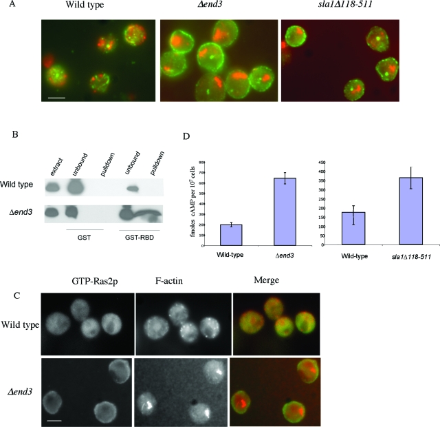
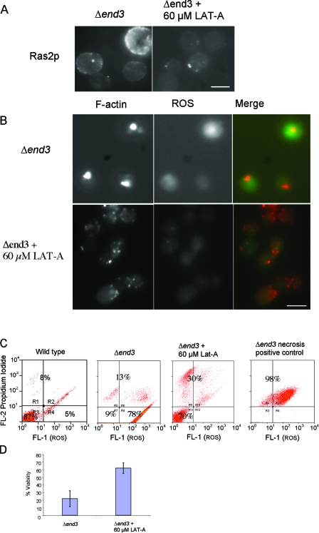
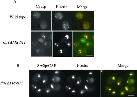
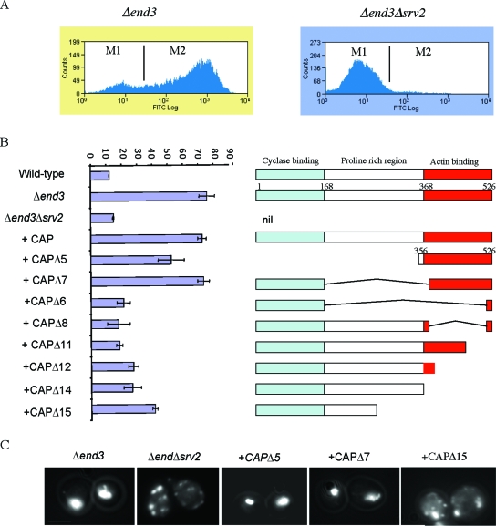
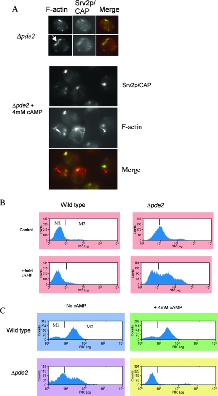
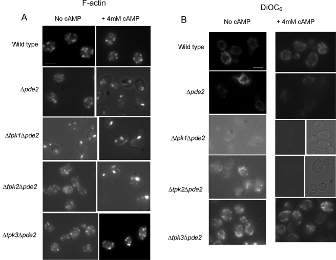
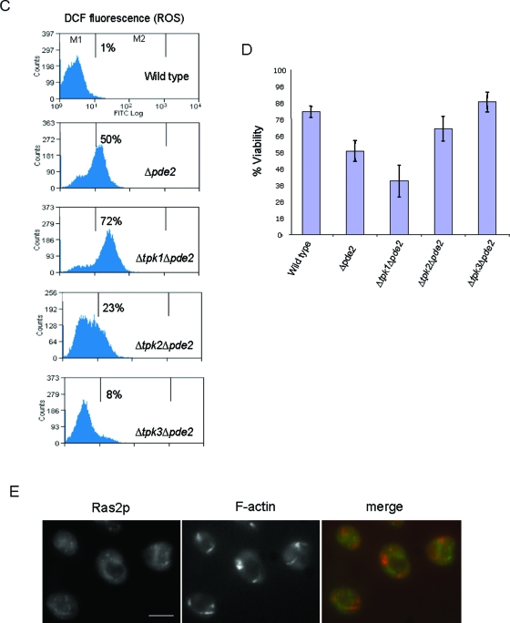
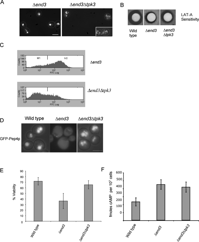
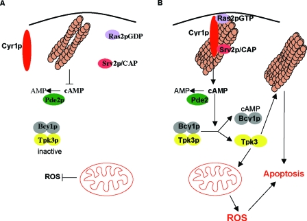
Recent research has revealed a conserved role for the actin cytoskeleton in the regulation of aging and apoptosis among eukaryotes. Here we show that the stabilization of the actin cytoskeleton caused by deletion of Sla1p or End3p leads to hyperactivation of the Ras signaling pathway. The consequent rise in cyclic AMP (cAMP) levels leads to the loss of mitochondrial membrane potential, accumulation of reactive oxygen species (ROS), and cell death. We have established a mechanistic link between Ras signaling and actin by demonstrating that ROS production in actin-stabilized cells is dependent on the G-actin binding region of the cyclase-associated protein Srv2p/CAP. Furthermore, the artificial elevation of cAMP directly mimics the apoptotic phenotypes displayed by actin-stabilized cells. The effect of cAMP elevation in inducing actin-mediated apoptosis functions primarily through the Tpk3p subunit of protein kinase A. This pathway represents the first defined link between environmental sensing, actin remodeling, and apoptosis in Saccharomyces cerevisiae.


Figures

FIG. 1.
FIG. 1.
Ras/cAMP pathway is hyperactivated in actin-stabilized cells. (A) Immunofluorescence was carried out to show colocalization of F-actin (red) with Ras2p (green) in wild-type, Δend3, and sla1Δ118-511 stationary-phase cells, as described in Materials and Methods. (B) To determine the state of Ras2p activity, GTP-bound Ras2p was detected in extracts prepared from wild-type and Δend3 stationary-phase cells by a pull-down method using purified GST-RafRBD, as described in Materials and Methods. (C) The activity of Ras2p was also investigated in vivo using immunofluorescence to show colocalization of F-actin (red) with active Ras2p (green). This was achieved by overlaying fixed wild-type and Δend3 stationary-phase cells with purified GST-RafRBD (see Materials and Methods) and utilizing anti-GST antibodies to detect RafRBD localization. Bar = 10 μm. (D) An assay to determine the cAMP levels in wild-type, Δend3, and sla1Δ118-511 stationary-phase cells was carried out in triplicate using a nonradioactive immunoassay (see Materials and Methods). Error bars represent standard deviations.
FIG. 2.
FIG. 2.
Actin stabilization directly activates Ras signaling and apoptosis. (A) The effect of the addition of 60 μM LAT-A to growth medium at the time of inoculation on F-actin organization and ROS accumulation in Δend3 cells was observed. The localization of Ras2p was also investigated in Δend3 cells grown to stationary phase with and without 60 μM LAT-A by using immunofluorescence. (B) F-actin was visualized by rhodamine-phalloidine staining and the presence of ROS was assessed by staining with H2DCF-DA in Δend3 cells, which were either untreated or treated with 60 μM LAT-A and allowed to grow to early stationary phase. Merged images of F-actin and ROS are also presented. Bar = 10 μm. (C) The effect of LAT-A treatment on cell death was investigated using flow cytometry to measure propidium iodide uptake and ROS accumulation in treated and untreated Δend3 cells. As a positive control to demarcate the necrotic cell fraction, Δend3 cells which had been heat treated at 80°C for 2 min were also stained. (D) A viability study was carried out with cells to assess the effects of the addition of 60 μM LAT-A to Δend3 cell cultures grown to stationary phase.
FIG. 3.
FIG. 3.
Components of the cAMP generation machinery are sequestered in actin aggregates. Immunofluorescence was used to show colocalization of components of the cAMP generation module in wild-type and sla1Δ118-511 stationary-phase cells. F-actin (red) was colocalized with adenylate cyclase, Cyr1p (green) (A), and Srv2p/CAP (green) (B). Bar = 10 μm.
FIG. 4.
FIG. 4.
Srv2p/CAP mediates actin aggregation and ROS formation in actin-stabilized cells. (A) ROS accumulation was assayed using H2DCF-DA in Δend3 and Δend3 Δsrv2 stationary-phase cells by flow cytometry (see Materials and Methods). The resultant data were plotted on histograms and divided into M1 and M2 regions, reflecting small and large ROS populations, respectively. (B) Similarly, ROS accumulation was assessed in triplicate in wild-type, Δend3, Δend3 Δsrv2, and Δend3Δsrv2 cells expressing either full-length Srv2/CAP or various truncations of Srv2/CAP (CAPΔ5-15). The percentages of cells appearing in the M2 region (large number of ROS cells) are presented. Error bars represent the standard errors obtained from triplicate experiments. (C) The F-actin architecture was visualized in Δend3 cells and in Δend3 Δsrv2 cells expressing various truncations of Srv2p/CAP by fluorescence microscopy using rhodamine-phalloidine as described previously. Bar = 10 μm.
FIG. 5.
FIG. 5.
cAMP elevation results in actin aggregation and apoptosis. (A) Immunofluorescence microscopy was used to show colocalization of F-actin (red) and Srv2p/CAP (green) in cells lacking Pde2p, which had been grown to stationary phase under normal growth conditions or in the presence of 4 mM cAMP. Bar = 10 μm. Under the same conditions, ROS accumulation was assayed using H2DCF-DA (B) and MMP was measured using DiOC6 (C) by flow cytometry. Histograms of single representative experiments are shown.
FIG.6.
FIG.6.
cAMP elevation signals through the Tpk3p subunit of PKA to induce actin aggregation and apoptosis. Rhodamine-phalloidine staining was used to visualize F-actin (A) and MMP was assessed using DiOC6 staining (B) by fluorescence microscopy of wild-type, Δpde2, Δtpk1 Δpde2, Δtpk2 Δpde2, and Δtpk3 Δpde2 cells which had been grown to stationary phase in the presence or absence of 4 mM cAMP. (C) ROS accumulation was also assayed, using H2DCF-DA, by flow cytometry of wild-type, Δtpk1 Δpde2, Δtpk2 Δpde2, and Δtpk3 Δpde2 cells grown to stationary phase in the presence of 4 mM cAMP. Histograms of single representative experiments are shown. (D) We also assessed the effects of growing wild-type, Δpde2, Δtpk1 Δpde2, Δtpk2 Δpde2, and Δtpk3 Δpde2 cells to stationary phase in the presence of 4 mM cAMP on culture viability. Error bars represent standard deviations from triplicate experiments. (E) To investigate whether an elevation of the cAMP level resulted in the activation of Ras2p, the localization of Ras2p was assessed in Δpde2 cells which had been grown to stationary phase in the presence of 4 mM cAMP. F-actin architecture was also assessed in these cells by rhodamine-phalloidine staining, and a merged image is also presented. Bar = 10 μm.
FIG.6.
FIG.6.
cAMP elevation signals through the Tpk3p subunit of PKA to induce actin aggregation and apoptosis. Rhodamine-phalloidine staining was used to visualize F-actin (A) and MMP was assessed using DiOC6 staining (B) by fluorescence microscopy of wild-type, Δpde2, Δtpk1 Δpde2, Δtpk2 Δpde2, and Δtpk3 Δpde2 cells which had been grown to stationary phase in the presence or absence of 4 mM cAMP. (C) ROS accumulation was also assayed, using H2DCF-DA, by flow cytometry of wild-type, Δtpk1 Δpde2, Δtpk2 Δpde2, and Δtpk3 Δpde2 cells grown to stationary phase in the presence of 4 mM cAMP. Histograms of single representative experiments are shown. (D) We also assessed the effects of growing wild-type, Δpde2, Δtpk1 Δpde2, Δtpk2 Δpde2, and Δtpk3 Δpde2 cells to stationary phase in the presence of 4 mM cAMP on culture viability. Error bars represent standard deviations from triplicate experiments. (E) To investigate whether an elevation of the cAMP level resulted in the activation of Ras2p, the localization of Ras2p was assessed in Δpde2 cells which had been grown to stationary phase in the presence of 4 mM cAMP. F-actin architecture was also assessed in these cells by rhodamine-phalloidine staining, and a merged image is also presented. Bar = 10 μm.
FIG. 7.
FIG. 7.
TPK3 is required for actin aggregation and ROS accumulation in Δend3 cells. F-actin staining was visualized using rhodamine-phalloidin staining (A), and ROS accumulation was assayed using H2DCF-DA for flow cytometry (C) of stationary-phase Δend3 and Δend Δtpk3 cells. (B) To further assess the effect of the loss of Tpk3p activity on actin dynamics in Δend3 cells, a LAT-A halo sensitivity assay was carried out as described in Materials and Methods. The role of Tpk3p in apoptosis was investigated by observing the localization of GFP-Pep4 (D) and by conducting a viability assay (E) with wild-type, Δend3, and Δend Δtpk3 cells. (F) To determine whether the loss of Tpk3p function led to reduced Ras/cAMP signaling activity, the levels of cAMP were determined in wild-type, Δend3, and Δend Δtpk3 cells. Bar = 10 μm.
FIG. 8.
FIG. 8.
Actin stabilization triggers constitutive Ras/cAMP/PKA signaling and apoptosis in stationary-phase S. cerevisiae cells. In wild-type cells, components of the Ras pathway are not found in association with F-actin during stationary phase. A reduction in Ras activity leads to a fall in cAMP, which results in PKA inactivation and the downregulation of signaling (A). (B) In actin-stabilized cells, components of the cAMP generation machinery, adenylyl cyclase (Cyr1p) and Srv2p, are found in association with large aggregates of actin during stationary phase and Ras2p is found to be constitutively active. This hyperactive Ras signaling state gives rise to the elevation of cAMP and the activation of PKA. This cascade of events triggers the elevation of ROS derived from the mitochondria and apoptosis. The PKA subunit Tpk3p is primarily responsible for the induction of apoptosis in response to actin-mediated hyperactivation of the Ras signaling pathway. It is likely that Tpk3p acts directly on both F-actin regulatory proteins and alters the enzymatic content of mitochondria to elicit this response. The combination of aggregated actin and constitutive Ras signaling may act together to establish conditions which lead to apoptotic cell death.

Similar articles

Cited by

References

    1. Ayscough, K. R., J. J. Eby, T. Lila, H. Dewar, K. G. Kozminski, and D. G. Drubin. 1999. Sla1p is a functionally modular component of the yeast cortical actin cytoskeleton required for correct localization of both Rho1p-GTPase and Sla2p, a protein with talin homology. Mol. Biol. Cell 10:1061-1075. - PMC - PubMed
    1. Balaban, R. S., S. Nemoto, and T. Finkel. 2005. Mitochondria, oxidants, and aging. Cell 120:483-495. - PubMed
    1. Benedetti, H., S. Raths, F. Crausaz, and H. Riezman. 1994. The END3 gene encodes a protein that is required for the internalization step of endocytosis and for actin cytoskeleton organization in yeast. Mol. Biol. Cell 5:1023-1037. - PMC - PubMed
    1. Chevtzoff, C., J. Vallortigara, N. Averet, M. Rigoulet, and A. Devin. 2005. The yeast cAMP protein kinase Tpk3p is involved in the regulation of mitochondrial enzymatic content during growth. Biochim. Biophys. Acta 1706:117-125. - PubMed
    1. Choi, J., H. D. Rees, S. T. Weintraub, A. I. Levey, L. S. Chin, and L. Li. 2005. Oxidative modifications and aggregation of Cu, Zn-superoxide dismutase associated with Alzheimer and Parkinson diseases. J. Biol. Chem. 280:11648-11655. - PubMed

Publication types

MeSH terms