. 2006 Sep;26(17):6609-22.
doi: 10.1128/MCB.00295-06.

Mice with a targeted mutation of patched2 are viable but develop alopecia and epidermal hyperplasia

Affiliations

Mice with a targeted mutation of patched2 are viable but develop alopecia and epidermal hyperplasia

Erica Nieuwenhuis et al. Mol Cell Biol. 2006 Sep.

Abstract

Hedgehog (Hh) signaling plays pivotal roles in tissue patterning and development in Drosophila melanogaster and vertebrates. The Patched1 (Ptc1) gene, encoding the Hh receptor, is mutated in nevoid basal cell carcinoma syndrome, a human genetic disorder associated with developmental abnormalities and increased incidences of basal cell carcinoma (BCC) and medulloblastoma (MB). Ptc1 mutations also occur in sporadic forms of BCC and MB. Mutational studies with mice have verified that Ptc1 is a tumor suppressor. We previously identified a second mammalian Patched gene, Ptc2, and demonstrated its distinct expression pattern during embryogenesis, suggesting a unique role in development. Most notably, Ptc2 is expressed in an overlapping pattern with Shh in the epidermal compartment of developing hair follicles and is highly expressed in the developing limb bud, cerebellum, and testis. Here, we describe the generation and phenotypic analysis of Ptc2(tm1/tm1) mice. Our molecular analysis suggests that Ptc2(tm1) likely represents a hypomorphic allele. Despite the dynamic expression of Ptc2 during embryogenesis, Ptc2(tm1/tm1) mice are viable, fertile, and apparently normal. Interestingly, adult Ptc2(tm1/tm1) male animals develop skin lesions consisting of alopecia, ulceration, and epidermal hyperplasia. While functional compensation by Ptc1 might account for the lack of a strong mutant phenotype in Ptc2-deficient mice, our results suggest that normal Ptc2 function is required for adult skin homeostasis.


Figures

FIG. 1.
FIG. 1.
Disruption of Ptc2 by gene targeting. (A) Targeting strategy illustrating the genomic organization, a restriction map of the wild-type (Wt) locus, the targeting vector, and the targeted allele (Mt). Ptc2 is located in chromosome 4 and consists of 22 predicted exons. The black bar indicates the position of the probe used for Southern hybridization. Arrows indicate positions of genotyping primers. X, XbaI; Xh, XhoI; B, BamHI; E, EcoRI. (B) Genotyping of progeny by PCR analysis. PCR amplification generated wild-type (150-bp) and mutant (350-bp) bands. (C) Northern blot analysis. The major transcript (a) encoded by Ptc2 is absent in Ptc2tm1/tm1 testis RNA. Four transcripts (b to e), ranging from 2.4 to 7.5 kb, are present in Ptc2tm1/tm1 testis RNA. (D) RT-PCR analysis of Ptc2 expression in skin, cerebellum, and testis of wild-type and mutant mice. The diagram indicates the locations of primer pairs Ptc2-A (325 bp), Ptc2-B (600 bp), and Ptc2-C (350 bp) relative to the insertion (arrowhead). Similar results were obtained for all transgenic tissues analyzed. Ptc2-A and Ptc2-C amplified fragments of the expected sizes in wild-type and Ptc2 mutant samples. Ptc2-B amplified expected transcript 1 in the wild type only, transcripts 2 and 3 in the mutant only, and transcript 4 present in the wild type and mutant. (E) Schematic representation of sequencing results obtained for wild-type and mutant transcripts amplified by Ptc2-B primers. Arrows indicate locations of forward (BF) and reverse (BR) primers relative to the genomic sequence. Three alternative splice forms of Ptc2 were isolated, with deletions of exon 6, exons 5 and 6, and exons 6 and 7. (F) Schematic depicting the location of alternative splice forms present in Ptc2 mutants. The translational start ATG codon resides in exon 1. The putative first extracellular loop, implicated in interacting with Shh, is encoded by exons 2 to 9, while the putative sterol-sensing domain is encoded by exons 9 to 13. Our targeting strategy aimed at disrupting the first large extracellular loop of Ptc2, thereby abolishing its interaction with Shh. Ptc2-Δ6,7 represents an alternative splice form of Ptc2 in which exons 6 and 7 are deleted, resulting in an in-frame deletion. In the Ptc2 mutant, two additional splice forms are generated, Ptc2-Δ6 and Ptc2-Δ5,6.
FIG. 2.
FIG. 2.
In vitro analysis of Ptc2 mutant forms. (A) Schematic indicating myc-tagged expression constructs for wild-type and mutant Ptc2. Deletion of exon 6 in Ptc2-Δ6 likely results in a premature truncation of the Ptc2 protein (gray shading). (B) Subcellular localization of Ptc1, Ptc2 wild-type, and Ptc2 mutant constructs. Ptc2-Δ5,6 is present in the cytoplasmic and nuclear compartments, while Ptc2-Δ6 and Ptc2-Δ6,7 are localized similarly to Ptc1 and Ptc2. (C) Luciferase assay showing that Ptc2 can function similarly to Ptc1 as a negative regulator of Shh signaling. The data shown are the averages from three independent experiments performed in triplicate. (D) Graph showing the effect of Ptc2 mutant forms on Gli-dependent transcription represented as relative luciferase activity and relative % inhibition. When compared to wild-type Ptc2, each of the mutants showed a statistically significant increase in Gli-dependent transcription (**, P < 0.05). Ptc2-Δ6 and Ptc2-Δ6,7 behaved similarly, and the differences in their relative luciferase activities were not statistically significant. Ptc2-Δ5,6 had the greatest decrease in its ability to inhibit Gli-dependent transcription compared to Ptc2. The results are from two independent experiments performed in triplicate.
FIG. 3.
FIG. 3.
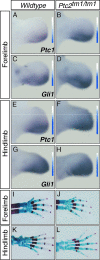
Normal limb development in Ptc2-deficient mice. (A to H) Whole-mount in situ hybridization of E11.5 limb buds. Expression of Ptc1 (A, B, E, and F) and Gli1 (C, D, G, and H) in forelimb (A to D) and hind limb (E to H) buds of wild type (A, C, E, and G) and Ptc2tm1/tm1 (B, D, F, and H) embryos is shown. Bars containing graded shading indicate increased expression of Ptc1 and Gli1 in Ptc2tm1/tm1 forelimb and hind limb buds. (I to L) Alcian blue and alizarin red staining of E18.5 wild-type and Ptc2tm1/tm1 limbs revealed no significant difference in skeletal patterning and development.
FIG. 4.
FIG. 4.
Ptc2-deficient mice develop normal cerebellums and testes, and Ptc1 and Gli1 are not upregulated. Histological staining of cerebellums (A to D) and testes (E to H) of adult wild-type (A, C, E, and G) and Ptc2tm1/tm1 (B, D, F, and H) mice is shown. (A, B, E, and F) Low-magnification views; C, D, G, H, and I to L, high-magnification views. (I to L) In situ hybridization of Ptc1 (I and J) and Gli1 (K and L) showing normal expression in Ptc2 mutants. IGL, Internal germinal layer; Pu, Purkinje cell layer. (M) Semiquantitative RT-PCR analysis of Ptc1 and Gli1 expression levels in wild-type (wt) and Ptc2tm1/tm1 (mt) cerebellum and testis. Results obtained from RT-PCR analysis were normalized to GAPDH (internal control) and are represented as expression detected in wild-type samples compared to mutant samples.
FIG. 5.
FIG. 5.
Ptc2 is not required for embryonic hair follicle development. Histological analysis of wild-type (A and M) and Ptc2tm1/tm1 (G and S) hair follicle development at E15.5 (A and G) and E18.5 (M and S) revealed normal differentiation and proliferation. In situ hybridization and immunohistochemistry for markers of hair follicle development at E15.5 and E18.5 are shown. Expression levels of Keratin-17 (B, H, N, and T), keratin-14 (C, I, O, and U), keratin-10 (D, J, P, and V), loricrin (E, K, Q, and W), and phospho-histone H3 (PH3) (F, L, R, and X) appear to be normal in the Ptc2tm1/tm1 epidermis. (Y) Cell proliferation is not affected in Ptc2tm1/tm1 skin at E15.5 or E18.5. Data represent the average numbers of PH3-positive cells (arrowheads in panels F, L, R, and X) obtained from counting 40 contiguous microscopic fields per sample. The error bars indicate standard deviations, and a t test revealed that the differences are not statistically significant. Dashed lines indicate epidermis-dermis boundaries. ep, epidermis; de, dermis; bl, basal layer; sbl, suprabasal layer; cl, cornified layer. Scale bar: 50 μM.
FIG. 6.
FIG. 6.
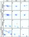
Ptc1 is upregulated in Ptc2tm1/tm1 embryonic skin. Shown is expression of Ptc1 (A, D, G, and J), Gli1 (B, E, H, and K), and Shh (C, F, I, and L) in wild-type (A to C and G to I) and Ptc2tm1/tm1 (D to F and J to L) embryos at E15.5 (A to F) and E18.5 (G to L). Ptc1 is slightly upregulated in E18.5 hair follicles (black arrowhead) and ectopically expressed in the interfollicular epidermis (IFE; red arrowhead) in Ptc2tm1/tm1 skin. Dashed lines indicate epidermis-dermis boundaries. ep, epidermis; de, dermis; hf, hair follicle. Scale bar: 50 μM.
FIG. 7.
FIG. 7.
Ptc2 mutant males develop alopecia and skin lesions. Ptc2tm1/tm1 males with ulceration and alopecia on back skin (B) and front paw skin (C) (arrowheads) are shown compared to a normal wild-type male (A). (D and G) Histological staining of wild-type back skin (D) and front paw skin (G). (E and F) Epidermal hyperplasia mutant back skin (E) and front paw skin (F). (H and I) Ulceration shown in mutant back skin (H) and mutant front paw skin (I). ep, epidermis; de, dermis; hf, hair follicle; hy; hypodermis; mu; muscle; ul, ulcer. Scale bar: 50 μM.
FIG. 8.
FIG. 8.
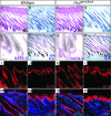
Hair loss in Ptc2 mutants is not due to aberrant hair follicle development. Shown are histological and immunohistochemical analyses of development of the differentiated layers of the hair follicle and epidermis. (A and C) Normal morphology of hair follicles. All markers analyzed are expressed in the normal pattern. (B and D) Keratin-15 expression in Henle's layer of the IRS; (E and G) GATA-3 is expressed in Huxley's layer of the IRS; (F and G) keratin-5 in the outer root sheath (ORS) and alkaline phosphatase (AP) in the dermal papillae; (M and O) keratin-6 in the companion cell layer. Epidermal development was also normal. (F, H, I, and K) Keratin-5 and keratin-14 in the basal cell layer; (J and L) keratin-10 in the suprabasal layer; (N and P), loricrin (Lor) in the cornified layer. Scale bars: 50 μM. DAPI, 4′,6′diamidino-2-phenylindole; HE, hematoxylin and eosin.
FIG. 9.
FIG. 9.
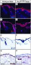
Marker analysis of skin lesions in Ptc2 mutant males. Shown are immunohistochemistry and in situ hybridization of epidermal compartments. Loricrin (Lor) expression appears to be normal (A and B). Keratin-14 expression is expanded suprabasally in ulcerated Ptc2tm1/tm1 skin (D) compared to wild-type epidermis, where it is expressed only in the basal layer (C). Keratin-17 (E and F) expression indicates expansion of the basal layer in areas of epidermal hyperplasia in affected Ptc2 mutant skin (F). Similar expression of PCNA was observed in wild-type (G) and Ptc2tm1/tm1 skin (H). Multiple layers of PCNA-expressing cells could be detected in tissue underlying ulcerated lesions (H, inset). ep, epidermis; de, dermis; hf, hair follicle; ul, ulcer. Scale bars: 50 μM.
FIG. 10.
FIG. 10.
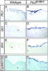
Sonic hedgehog signaling is activated in the Ptc2tm1/tm1 epidermis. Shown is marker gene analysis by in situ hybridization (A to F) and immunohistochemistry (G to H). Ptc1 (A and B), Gli1 (C and D), Shh (E and F), and cyclin D1/D2 (G to H) expression is increased in areas of epidermal hyperplasia in affected Ptc2tm1/tm1 skin. Arrowheads indicate low levels of Gli1, Shh, and cyclin D1/D2 expression in hair follicles of wild-type skin. ep, epidermis; ul, ulcer. Scale bar: 50 μM.

References

    1. Adolphe, C., M. Narang, T. Ellis, C. Wicking, P. Kaur, and B. Wainwright. 2004. An in vivo comparative study of sonic, desert and Indian hedgehog reveals that hedgehog pathway activity regulates epidermal stem cell homeostasis. Development 131:5009-5019. - PubMed
    1. Ashcroft, G. S., T. Greenwell-Wild, M. A. Horan, S. M. Wahl, and M. W. Ferguson. 1999. Topical estrogen accelerates cutaneous wound healing in aged humans associated with an altered inflammatory response. Am. J. Pathol. 155:1137-1146. - PMC - PubMed
    1. Ashcroft, G. S., M. A. Horan, and M. W. Ferguson. 1998. Aging alters the inflammatory and endothelial cell adhesion molecule profiles during human cutaneous wound healing. Lab Investig. 78:47-58. - PubMed
    1. Ashcroft, G. S., and S. J. Mills. 2002. Androgen receptor-mediated inhibition of cutaneous wound healing. J. Clin. Investig. 110:615-624. - PMC - PubMed
    1. Aszterbaum, M., A. Rothman, R. L. Johnson, M. Fisher, J. Xie, J. M. Bonifas, X. Zhang, M. P. Scott, and E. H. Epstein, Jr. 1998. Identification of mutations in the human PATCHED gene in sporadic basal cell carcinomas and in patients with the basal cell nevus syndrome. J. Investig. Dermatol. 110:885-888. - PubMed

Publication types

MeSH terms