. 2006 Sep;26(17):6656-63.
doi: 10.1128/MCB.00091-06.

Loss of the mouse ortholog of the shwachman-diamond syndrome gene (Sbds) results in early embryonic lethality

Affiliations

Loss of the mouse ortholog of the shwachman-diamond syndrome gene (Sbds) results in early embryonic lethality

Siyi Zhang et al. Mol Cell Biol. 2006 Sep.

Abstract

Mutations in SBDS are responsible for Shwachman-Diamond syndrome (SDS), a disorder with clinical features of exocrine pancreatic insufficiency, bone marrow failure, and skeletal abnormalities. SBDS is a highly conserved protein whose function remains largely unknown. We identified and investigated the expression pattern of the murine ortholog. Variation in levels was observed, but Sbds was found to be expressed in all embryonic stages and most adult tissues. Higher expression levels were associated with rapid proliferation. A targeted disruption of Sbds was generated in order to understand the consequences of its loss in an in vivo model. Consistent with recessive disease inheritance for SDS, Sbds(+/-) mice have normal phenotypes, indistinguishable from those of their wild-type littermates. However, the development of Sbds(-/-) embryos arrests prior to embryonic day 6.5, with muted epiblast formation leading to early lethality. This finding is consistent with the absence of patients who are homozygous for early truncating mutations. Sbds is an essential gene for early mammalian development, with an expression pattern consistent with a critical role in cell proliferation.


Figures

FIG. 1.
FIG. 1.
Expression profiles of Sbds mRNA and protein in adult mouse organs. (A) Multitissue Northern blot. Poly(A+) mRNA of the adult mouse tissues indicated hybridized with Sbds (upper panel) and Gapd (lower panel) cDNA probes. (B) Multitissue Western blot of mouse tissues with affinity-purified polyclonal anti-SBDS (upper panel) and control anti-mouse Gapd (lower panel) (Abcam) antibodies.
FIG. 2.
FIG. 2.
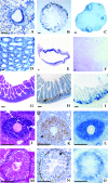
Sbds is highly expressed in rapidly proliferating tissues. In situ hybridization with a Sbds cDNA probe is shown. High expression was found in bronchiole epithelium in lung (A), small intestine epithelium (B), ovary follicles (C), testis seminiferous tube (D), and mucosal epithelium of stomach (E). In contrast, the expression in adult brain (F) is low. Increased Sbds expression was correlated with rapidly proliferating cells in small intestine epithelium (G, H, and I), ovary follicles (J, K, and L) and testis seminiferous tubes (M, N, and O). (G, J, and M) Hematoxylin and eosin staining of intestine epithelium, ovary follicles, and testis seminiferous tubes, respectively. (I, L, and O) In situ hybridization of adjacent tissue sections, respectively, with a cDNA probe consisting of the 5′ UTR and exons 1 to 3 of Sbds. (H, K, and N) Adjacent sections, respectively, were stained with an anti-BrdU antibody. Arrows mark examples of cells with incorporation of BrdU. Bars, 100 μm.
FIG. 3.
FIG. 3.
Expression of Sbds during mouse embryonic development. (A and B) Mouse embryo multiple-tissue Northern blot. Poly(A+) mRNA from whole mouse embryo at the stages indicated was hybridized with Sbds cDNA (A) and Gapd cDNA (B) probes.
FIG. 4.
FIG. 4.
Targeted disruption of the mouse Sbds gene. (A) Genomic organization of the mouse Sbds gene. The targeting vector and the desired homologous recombination outcome are illustrated. Black boxes represent Sbds exons. Probes A and B correspond to 5′ and 3′ external probes used for Southern blot-based genotyping; arrows a, b, and c correspond to oligonucleotide primers for PCR genotyping. Relevant restriction sites are BamHI (B), BssSI (S), and XbaI (X). (B) Representative Southern blot analysis of neomycin-resistant ES cells. Genomic DNA was digested with BamHI and hybridized with probe B. The 15-kb and 10-kb bands represent the wild-type (lanes 1, 3, 4, and 6) and targeted (lanes 2 and 5) alleles, respectively. (C) Representative PCR genotyping with primers a, b, and c. The observed PCR products derived from wild-type (+) and targeted (−) alleles are 354 and 184 bp, respectively. (D) Sbds expression was evaluated in wild-type and heterozygous embryonic fibroblasts by Western blotting using the anti-SBDS antibody (upper panel), with comparison to α-tubulin as a control for loading as detected by anti-α-tubulin antibody (lower panel) (Molecular Probes).
FIG. 5.
FIG. 5.
Failed development of Sbds−/− embryos. (A and B) Sbds+/− (A) and Sbds/− (B) blastocysts at E3.5 appear normal. (C to F) Sagittal sections of hematoxylin- and eosin-stained Sbds+/− (C and E) and Sbds−/− (D and F) embryos at E6.5 and E7.5; disorganization of the null embryo was apparent at E6.5 (D), with subsequent degradation clearly evident by E7.5 (F). (G to L) In situ hybridization of Sbds cDNA with exons 3 and 4 and Oct4 cDNA and Bmp4 cDNA probes on heterozygous (G, I, and K) and null (H, J, and L) E6.5 embryos. (M and N) Sbds+/− blastocysts (M) were able to outgrow after 4 days of culture. The growing ICM cells are surrounded by trophoblast giant cells (TG). By contrast, Sbds−/− blastocysts (N) retain trophoblast cells but no ICM growth. ac, amniotic cavity; ep, ectoplacental cone; em, embryonic portion; ex, extraembryonic portion; exc, exocoelom; pac, proamniotic cavity. Bars, 50 μm (A, B, M, and N) and 100 μm (C to L).

References

    1. Aggett, P. J., N. P. Cavanagh, D. J. Matthew, J. R. Pincott, J. Sutcliffe, and J. T. Harries. 1980. Shwachman's syndrome. A review of 21 cases. Arch. Dis. Child. 55:331-347. - PMC - PubMed
    1. Aggett, P. J., J. T. Harries, B. A. Harvey, and J. F. Soothill. 1979. An inherited defect of neutrophil mobility in Shwachman syndrome. J. Pediatr. 94:391-394. - PubMed
    1. Austin, K. M., R. J. Leary, and A. Shimamura. 2005. The Shwachman-Diamond SBDS protein localizes to the nucleolus. Blood 106:1253-1258. - PMC - PubMed
    1. Bodian, M., W. Sheldon, and R. Lightwood. 1964. Congenital hypoplasia of the exocrine pancreas. Acta Paediatr. 53:282-293. - PubMed
    1. Boocock, G. R., J. A. Morrison, M. Popovic, N. Richards, L. Ellis, P. R. Durie, and J. M. Rommens. 2003. Mutations in SBDS are associated with Shwachman-Diamond syndrome. Nat. Genet. 33:97-101. - PubMed

Publication types

MeSH terms