Review
. 2006 Sep;74(7):326-48.
doi: 10.1111/j.1432-0436.2006.00095.x.

From fate to function: the Drosophila trachea and salivary gland as models for tubulogenesis

Affiliations
Review

From fate to function: the Drosophila trachea and salivary gland as models for tubulogenesis

Bilal E Kerman et al. Differentiation. 2006 Sep.

Abstract

Tube formation is a ubiquitous process required to sustain life in multicellular organisms. The tubular organs of adult mammals include the lungs, vasculature, digestive and excretory systems, as well as secretory organs such as the pancreas, salivary, prostate, and mammary glands. Other tissues, including the embryonic heart and neural tube, have requisite stages of tubular organization early in development. To learn the molecular and cellular basis of how epithelial cells are organized into tubular organs of various shapes and sizes, investigators have focused on the Drosophila trachea and salivary gland as model genetic systems for branched and unbranched tubes, respectively. Both organs begin as polarized epithelial placodes, which through coordinated cell shape changes, cell rearrangement, and cell migration form elongated tubes. Here, we discuss what has been discovered regarding the details of cell fate specification and tube formation in the two organs; these discoveries reveal significant conservation in the cellular and molecular events of tubulogenesis.


Figures
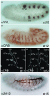
Fig. 1
Fig. 1
Tracheal morphogenesis in Drosophila embryos is largely complete by the end of embryogenesis. (A) A lateral view of a stage 10 embryo stained with antiserum to Ventral veins lacking (Vvl) reveals the ten tracheal placodes (Tr1–Tr10), which are present on both sides of the embryo. Lower levels of Vvl staining are also visible in corresponding regions in the two segments anterior to Tr1 and in one segment posterior to Tr10. (B) A lateral view of a stage 12 embryo stained with an antibody to the apical membrane protein Crumbs (Crb) reveals the primary branches of the internalized trachea. (C) A close up of tracheal metamere 4 (tr4) from a stage 11 embryo stained with Crb shows the different primary branches. (D) A close up of tracheal metamere 4 (Tr4) from a stage 12 embryo stained with Crb shows the different primary branches and their proximity to the branches from the adjacent metameres, Tr3 and Tr5. (E) A lateral view of a stage 15 embryo stained with the luminal marker protein 2A12 shows the trachea after the dorsal trunk (DT) has fused. DB, dorsal branch; DTa, DT anterior; DTp, DT posterior; GB, ganglionic branch, LT, lateral trunk; LTa, LT anterior; LTp, LT posterior; TC, transverse connectives; Tr1–10, tracheal metameres 1–10; VB, visceral branch.
Fig. 2
Fig. 2
A cartoon representation shows the types of tubes that form in the Drosophila trachea, indicating which type of tube is found in each branch and the approximate diameter of the tracheal lumen. In the cartoons, the lumen is shown in pink, with the apical membrane in red. The adherens junctions are indicated in black with the remaining lateral membrane indicated in white. The basal surface is dark blue and the cytoplasm is lighter blue. Abbreviations are as described in the legend of Fig. 1.
Fig. 3
Fig. 3
Specification of the Drosophila tracheal primordia is controlled by global patterning genes. The Salm gene limits tracheal formation to the trunk region of the embryo, from the second thoracic (T2) segment, where the first tracheal metamere forms (Tr1), through abdominal segment 8 (A8), where the last tracheal metamere forms (Tr10). Wg signaling limits tracheal formation to a subset of cells within a limited anterior–posterior portion of each segment. Dpp blocks tracheal formation in the dorsal ectoderm, whereas the ventral patterning genes are predicted to block tracheal formation in the ventral ectoderm.
Fig. 4
Fig. 4
Several signaling pathways have been implicated in tracheal migration, including endothelial growth factor (EGF), fibroblast growth factor (FGF), Dpp, and Wg. EGF signaling is localized by the expression of rho, which encodes a protease essential for the processing of the EGF ligand. EGF signaling is required for the complete internalization of the tracheal primordia. FGF signaling is limited by the expression of the ligand Bnl in the tissues toward which the tracheal branches migrate. FGF signaling is required for all migration. Dpp signaling is limited by the expression of the ligand Dpp in cells dorsal and ventral to the trachea. Dpp signaling is required for migration of the dorsal branch, lateral trunk, and ganglionic branch. Wg signaling is limited by the localization of the Wg signal in ectodermal cells near the tracheal primordia. Wg signaling is required to maintain salm expression in the dorsal trunk (DT) and to prevent the DT from migrating with the visceral branch. Green indicates the spatial position of the signal relative to the trachea. Blue indicates the tracheal primordia in the drawing along the top. Blue filled circles indicate that the cells have migrated to their correct positions in wild type and in the various signaling pathway mutants. White circles indicate that cells are missing in particular branches in the mutants.
Fig. 5
Fig. 5
The formation of type I from type II tubes requires the formation of autocellular junctions from intercellular junctions. Formation of type II tubes requires that paired cells within the type I tube reach around and contact themselves at each end to initiate the formation of an autocellular junction. The autocellular junctions then zip up from the sites of initation as the two cells slide past each other. Zipping terminates at the point where the two cells contact each other only at their ends. Apical proteins such as Pio and Dp are important in preventing the zippering from continuing to the point where the two cells would separate.
Fig. 6
Fig. 6
Different types of specialized cells form within each tracheal metamere. (A) This diagram shows the different cell types that form within a typical tracheal metamere; the cell types include stalk cells (type I and type II tubes, blue circles), fusion cells (type III tubes, red circles) and terminal cells (type IV tubes, green circles). (B) Cartoon diagram of branch fusion, showing as an example fusion of the dorsal trunk posterior and dorsal trunk anterior. The fusion cells (red) seek each other out and make contact along their basal surfaces. During initiation, the fusion cells localize E-cadherin (light blue) and other cytoskeletal proteins (yellow) typical of adherens junctions to the basal surface at the site of contact between the fusion cells. Subsequently, an actin-rich structure bridging between the apical surfaces of the two fusion cells is formed. This actin-rich structure is proposed to either physically bring the two apical surfaces together for fusion or to act as a scaffold for the accumulation of apical membrane vesicles, which will eventually fuse to create the apical surfaces connecting the two tracheal metameres. After fusion, a continuous adherens junction containing proteins such as E-cadherin is found near the apical surfaces of the two fusion cells.
Fig. 7
Fig. 7
CrebA is expressed to very high levels in the secretory cells of the salivary gland. (A) and (E) Staining of CrebA, and other early salivary gland proteins, is first detectable during embryonic stage 10, when the cells are on the surface of the embryo. (B) Beginning with cells in a dorsal–posterior domain of the placode, the cells change shape and internalize during embryonic stage 11. (C) By early stage 12, the most distal cells in the secretory tube have contacted the layer of cells known as the visceral mesoderm (VM) and have initiated posterior migration. (D) As new parts of the tube contact the VM during later stage 12, these cells also turn and migrate posteriorly. (F) The secretory tubes are fully internalized by embryonic stage 13. (G) The distal ends of the tube contact anterior portions of the midgut, which will form the gastric caeca (GC), whereas the sides of the tube make direct contact with the somatic muscle (SM) underlying the epidermis. (H) The secretory tubes maintain contact with the GC and SM at later embryonic stages.
Fig. 8
Fig. 8
The duct forms from the most ventral cells of the salivary gland primordia and internalizes to form tubes after the secretory tubes have formed. (AD) A P-element insertion in the trh gene drives β-gal expression throughout the salivary gland primordia in both duct and secretory (sec) cells. (A) Expression of the insertion is first visible when the salivary gland precursors are still on the surface of the embryo. (B) The more lateral secretory cells internalize to form tubes during embryonic stages 11 and 12. (C) The individual duct (id) tubes, which form from the more posterior duct primordia, form during embryonic stage 13 after the secretory cells have internalized and formed tubes. (D) The common duct (cd) tube forms from more anterior duct primordia and forms during embryonic stage 14.
Fig. 9
Fig. 9
Global patterning genes specify the salivary gland and cell types within the salivary gland. (A) The homeotic gene Scr is expressed in parasegment 2, where salivary glands normally form. Salivary glands form from ventral Scr-expressing ectodermal cells that do not express Dpp. Ectopic expression of Scr can result in additional salivary glands forming in segments that do not also express either tsh (in PS3–13) or Abd-B (PS14). Within the gland primordia, epithelial growth factor (EGF) signaling along the ventral midline specifies salivary duct by shutting off expression of secretory genes, such as fkh. In turn, Fkh blocks expression of duct-specific genes, and with other secretory-specific proteins, promotes development of the secretory tubes. (B and C) The duct-specific protein Ser signals its lateral neighbors to form imaginal ring cells, the precursors to the adult salivary gland. (D) In the fully formed embryonic salivary gland, the imaginal ring cells are located between the secretory tubes and the distal ends of the individual ducts.
Fig. 10
Fig. 10
Genes that specify the salivary glands disappear early through a negative feedback loop. The Scr/Exd/Hth complex induces expression of several early-expressed transcription factors (fkh, hkb, CrebA, and sage, and a splicing factor, pasilla). The complex also represses expression of hth, either directly or indirectly. As Hth is required for nuclear entry of Exd, repression of hth results in a loss of Hth and nuclear Exd. As Hth and Exd maintain Scr expression, Scr also disappears in the early salivary gland.
Fig. 11
Fig. 11
Scr/Exd/Hth complex induces expression of several early transcription factor genes, including sage, fkh, CrebA, and Hkb. Some of these transcription factors regulate their own expression as well as the expression of other early transcription factor genes. They also regulate downstream factors involved in morphogenesis and physiological function. Fkh is required to maintain its own expression as well as the expression of Sage and CrebA. Fkh regulates unknown targets to mediate the cell shape changes required for secretory cells to internalize and form tubes. It also regulates genes required for secretory cells to survive. Sage works with Fkh to maintain its own expression and the expression of two downstream enzyme-encoding genes, PH4αSG1 (indirectly) and PH4αSG2 (directly). These enzymes are required to modify target proteins that form the luminal fibrillar matrix that keeps the lumen open and uniform. CrebA regulates expression of the secretory pathway component genes (SPCGs), which are expressed to high levels in the salivary gland. High-level expression of the SPCGs mediates the high level of secretory activity characteristic of this tissue. Hkb regulates klar and crb to facilitate the apical membrane expansion required for tube elongation. Black arrows indicate regulatory interactions that are known or likely to be primarily transcriptional. Black dashed arrows indicate interactions that are primarily post-transcriptional.
Fig. 12
Fig. 12
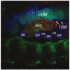
During its posterior migration, the secretory tube (sec; red from PH4αSG1 staining) directly contacts a number of other cell types, including the circular VM (cVM; blue green from overlapping α-FasIII and α-Titin staining), longitudinal VM (lVM; blue from α-Titin staining), somatic mesoderm (SM; blue from α-Titin staining) and fat body (FB; dark, unstained areas between the SM clusters).
Fig. 13
Fig. 13
Quality and quantity of luminal secretions are altered in embryos missing PH4αSG1 and PH4αSG2. (A and B) A thin sagittal section from a wild-type salivary gland (wt) reveals a relatively large and uniform luminal space filled with fibrillar material (asterisks). As the salivary tube is bent, the thin section catches the apical surface near the middle of the lumen; the bend in the tube also makes the lumen appear slightly narrower near the middle. (C and D) A thin sagittal section of a Df(3R)Exel6216 salivary gland (Df) reveals irregular, small luminal spaces with an electron dense matrix (asterisks). The number, distribution and electron density of the secretory vesicles are not different in the wild type versus deficiency salivary glands, suggesting that the differences in luminal content arise post-secretion. The reduced volume and increased electron density of the salivary secretions from the deficiency glands correlate with the luminal abnormalities seen in confocal images, with regions of tube dilation, constriction and closure. The Df(3R)Exel6216 lumenal abnormalities are rescued by salivary gland specific expression of either PH4αSG1 or PH4αSG2.

References

    1. Abrams EW, Andrew DJ. Prolyl 4-hydroxylase alpha-related proteins in Drosophila melanogaster: tissue-specific embryonic expression of the 99F8-9 cluster. Mech Dev. 2002;112:165–171. - PubMed
    1. Abrams EW, Andrew DJ. CrebA regulates secretory activity in the Drosophila salivary gland and epidermis. Development. 2005;132:2743–2758. - PubMed
    1. Abrams EW, Mihoulides WK, Andrew DJ. Fork head and Sage maintain a uniform and patent salivary gland lumen through regulation of two downstream target genes, PH4αSG1 and PH4αSG2. Development. 2006 in press. - PubMed
    1. Abrams EW, Vining MS, Andrew DJ. Constructing an organ: the Drosophila salivary gland as a model for tube formation. Trends Cell Biol. 2003;13:247–254. - PubMed
    1. Affolter M, Montagne J, Walldorf U, Groppe J, Kloter U, LaRosa M, Gehring WJ. The Drosophila SRF homolog is expressed in a subset of tracheal cells and maps within a genomic region required for tracheal development. Development. 1994b;120:743–753. - PubMed

MeSH terms

LinkOut - more resources