. 2006 Aug;23(4):561-74.
doi: 10.1016/j.molcel.2006.07.015.

Raf kinase inhibitory protein regulates aurora B kinase and the spindle checkpoint

Affiliations

Raf kinase inhibitory protein regulates aurora B kinase and the spindle checkpoint

Eva M Eves et al. Mol Cell. 2006 Aug.

Abstract

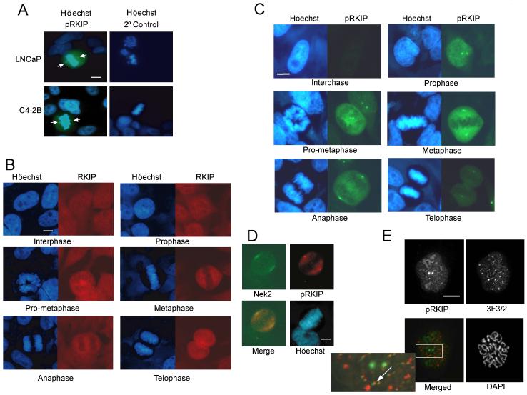
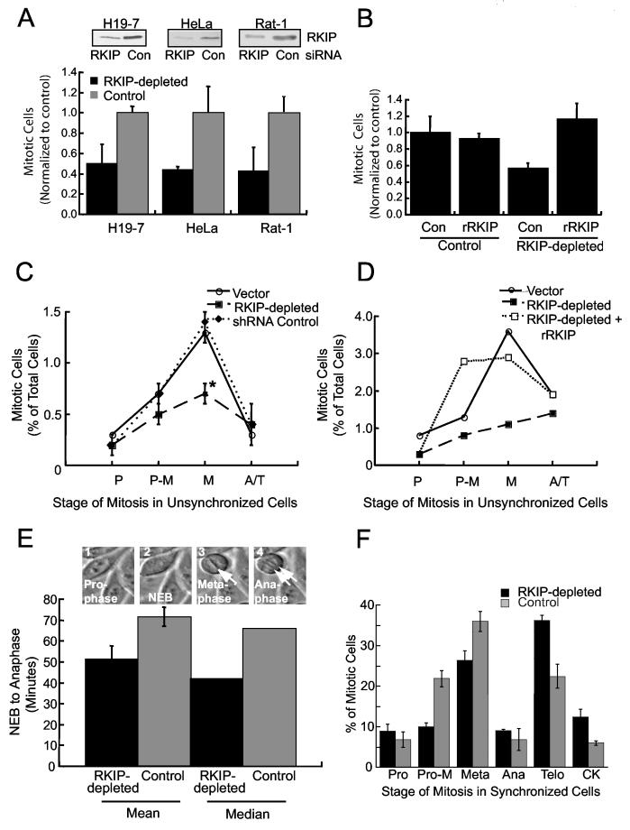
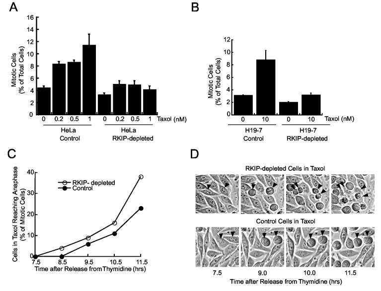
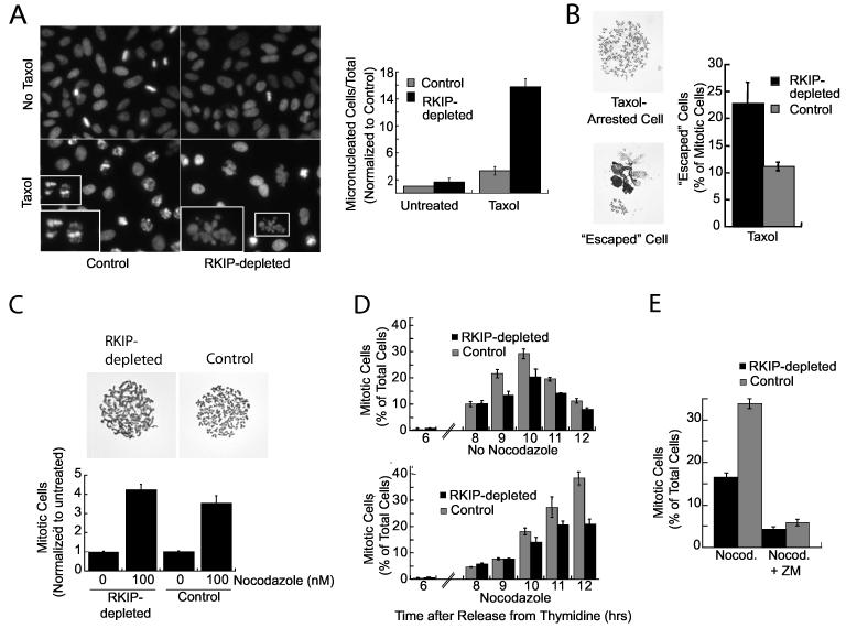
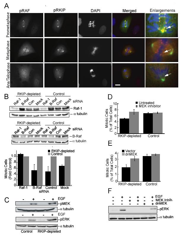
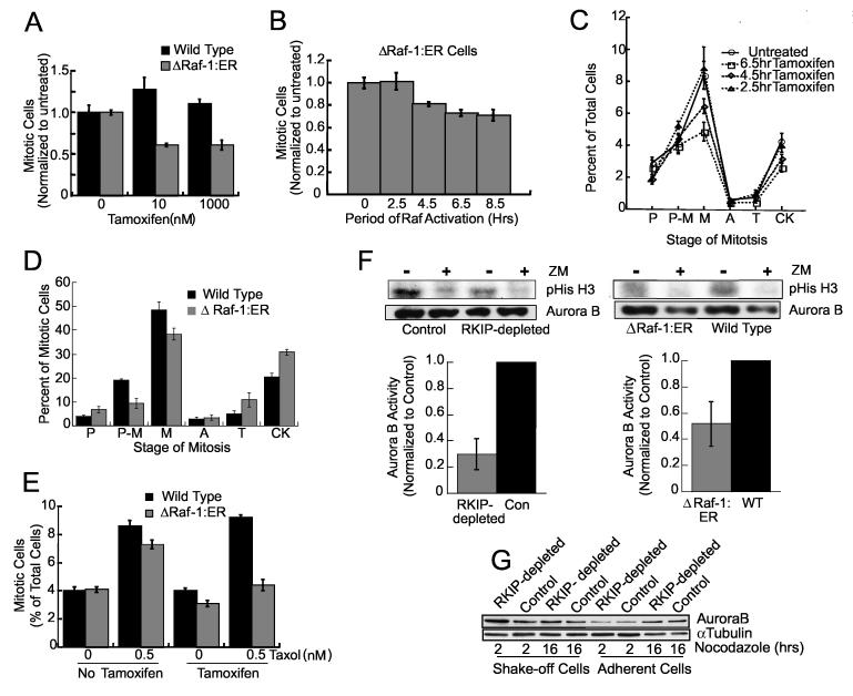
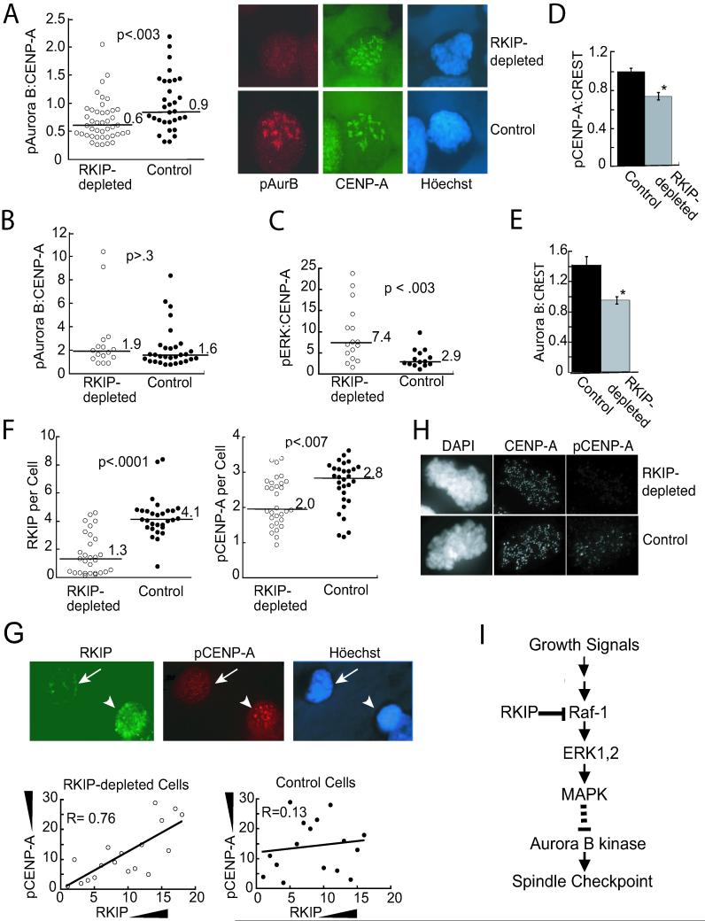
Raf kinase inhibitory protein (RKIP or PEBP) is an inhibitor of the Raf/MEK/MAP kinase signaling cascade and a suppressor of cancer metastasis. We now show that RKIP associates with centrosomes and kinetochores and regulates the spindle checkpoint in mammalian cells. RKIP depletion causes decreases in the mitotic index, the number of metaphase cells, and traversal times from nuclear envelope breakdown to anaphase, and an override of mitotic checkpoints induced by spindle poisons. Raf-1 depletion or MEK inhibition reverses the reduction in the mitotic index, whereas hyperactivation of Raf mimics the RKIP-depletion phenotype. Finally, RKIP depletion or Raf hyperactivation reduces kinetochore localization and kinase activity of Aurora B, a regulator of the spindle checkpoint. These results indicate that RKIP regulates Aurora B kinase and the spindle checkpoint via the Raf-1/MEK/ERK cascade and demonstrate that small changes in the MAP kinase (MAPK) pathway can profoundly impact the fidelity of the cell cycle.


Figures

Figure 1
Figure 1
pRKIP is Localized at Centrosomes and Kinetochores in Mitotic Cells (A) pRKIP in human prostate tumor cell lines. Arrows indicate centrosomes. (B) RKIP (Texas Red) and (C) pRKIP (FITC) in HeLa cells at different mitotic stages. (D) pRKIP (Texas Red) and Nek2 (FITC) in metaphase HeLa cell. (E) pRKIP (FITC) and 3F3/2 (rhodamine) in prometaphase Ptk-1 cell. Arrow: co-stained kinetochore. Scale bars, 10 μm.
Figure 2
Figure 2
RKIP Depletion in Rat or Human Cells Reduces Mitotic Index and Metaphase Cells, and Regulates Mitotic Progression (A) RKIP was depleted by siRNA transient transfection of H19-7, HeLa or Rat-1 cells or stable shRNA (hRKIP) transfection of HeLa cells. Mitotic cell counts were normalized to cultures transfected with control siRNA (human lamin) or shRNA (rRKIP). Means +/- SE are from multiple Rat-1(2), H19-7(4), and HeLa(5) experiments. Upper panels are RKIP immunoblots from one experiment per cell type. (B) RKIP-depleted or control HeLa cells were co-transfected with GFP and HA-tagged rRKIP or control vector. Two days later GFP+ mitotic cells were counted. (C) RKIP-depleted, control, or empty vector HeLa cells were stained with Höechst. P = prophase, P-M = prometaphase, M = metaphase, and A/T = anaphase and telophase. *p=.01 by Student t-Test. (D) RKIP-depleted HeLa cells were transfected as in (B). Two days later GFP+ cells at different stages of mitosis were counted. (E) RKIP-depleted or control HeLa cells were synchronized. The interval from NEB to anaphase was determined by time-lapse photomicroscopy. (RKIP-depleted, n=11; Control, n=30) (F) RKIP-depleted or control HeLa cells were synchronized. Nine hours after release, cells at different mitotic stages were counted. All error bars are +/-SE.
Figure 3
Figure 3
RKIP Depletion Decreases Cell Arrest in Taxol (A) RKIP-depleted or control HeLa cells were treated with Taxol for 5 hrs and mitotic cells counted. (B) RKIP-depleted or control H19-7 cells were Taxol-treated for 7 hrs and mitotic cells counted. Error bars are +/-SE. (C) RKIP-depleted or control HeLa cells were synchronized, treated with 1 nM Taxol at 2 hrs after release, and imaged beginning 5 hrs later. Mitotic cells reaching anaphase by indicated times are plotted. (D) Photomicrographs from (C). Arrowheads: RKIP-depleted and control cells that began mitosis at same time.
Figure 4
Figure 4
RKIP Depletion Enhances Taxol-Induced Chromatin Damage and Decreases Mitotic Arrest in Nocodazole(A) RKIP-depleted or control HeLa cells were untreated or treated for 12 hrs with 10 μM Taxol, stained with Höechst, and micronucleated cells counted. Insets: chromatin of Taxol-arrested cells (left), or a Taxol-escaped micronucleated cell (right). (B) RKIP-depleted or control HeLa cells were treated with 10 μM Taxol for 6 hrs, and chromosome spreads from Taxol-arrested or escaped cells were counted. (C) RKIP-depleted or control HeLa cells were treated with 0 or 100 nM nocodazole for 6 hrs and mitotic cells counted. Chromosome spreads are shown. (D) RKIP-depleted or control HeLa cells were synchronized and released for indicated times into medium +/-200 ng/ml nocodazole and mitotic cells counted. (E) HeLa RKIP-depleted or control cells were treated with 200 ng/ml nocodazole for 5 hrs. Arrested cells were shaken off and replated in nocodazole +/-5 μM Aurora B kinase inhibitor (ZM) for 4 hrs. PhosphoHistone H3+ (mitotic) cells were counted. All error bars are +/-SE.
Figure 5
Figure 5
pRKIP Co-Localizes with Activated Raf-1, and Raf-1 Depletion or MEK Inhibition Reverses the Effect of RKIP Depletion on Mitotic Index(A) Localization of pS338 Raf-1 (rhodamine) and pRKIP (FITC) in prometaphase, metaphase, and anaphase/telophase Ptk-1 cells. Scale bar, 20 μm. (B) RKIP-depleted and control HeLa cells transfected with Raf-1, B-Raf, or control siRNA for 48 hrs, were immunoblotted for Raf-1 or B-Raf and mitotic cells counted. (C) Starved RKIP-depleted or control HeLa cells were untreated or treated with 100 ng/ml EGF for 5 min and immunoblotted for pERK and pMEK. (D) After synchronization RKIP-depleted or control HeLa cells were untreated or treated with 10 μM PD098059 for 4 hrs and mitotic cells counted. (E) RKIP-depleted and control HeLa cells were infected with dnMEK1 lentivirus or control lentivirus and mitotic cells counted. (F) RKIP-depleted HeLa cells were treated with 10 μM U0126, or infected with dnMEK1 lentivirus or control lentivirus, and stimulated with EGF as in (C). Lysates were immunoblotted for pERK. α-tubulin was the loading control (B, C, F). All error bars are +/-SE.
Figure 6
Figure 6
Raf-1 Activation Mimics RKIP-Depletion (A) Wild type or HeLa cells stably expressing ΔRaf-1:ER were treated for 24 hrs with tamoxifen and mitotic cells counted. (B) 1 μM tamoxifen was added to synchronized ΔRaf-1:ER HeLa cells at .5, 2.5, 4.5, 6.5, or 9 hrs after release. At 9 hrs mitotic cells were counted. (C) Mitotic stages of ΔRaf-1:ER HeLa cells treated as in (B). P = prophase, P-M = prometaphase, M = metaphase, A = anaphase, T = telophase, and CK = cytokinesis. P<.03 by Student t-Test for 6.5 hr tamoxifen treatment. (D) Synchronized wild type or ΔRaf-1:ER HeLa cells were incubated for 2.5 hrs, 1 μM tamoxifen added for 6.5 hrs and mitotic cells counted. (E) Wild type or ΔRaf-1:ER HeLa cells were treated with 0 or 1 μM tamoxifen for 19 hrs, 0 or 0.5 nM Taxol added for 5 hrs and mitotic cells counted. Error bars are +/-SE. (F) RKIP-depleted and control (left), or tamoxifen-treated ΔRaf-1:ER and wild type (right) HeLa cells were analyzed for Aurora B kinase activity. ZM: 1 μM Aurora kinase inhibitor. Error bars are +/-SD (left) or range (right). (G) Synchronized RKIP-depleted or control HeLa cells were released into 200 ng/ml nocodazole. Arrested (shake-off) or adherent cells were immunoblotted for Aurora B.
Figure 7
Figure 7
RKIP Depletion Decreases Phosphorylated Aurora B Kinase and Phosphorylated CENP-A at Kinetochores(A) Synchronized RKIP-depleted or control HeLa cells were released into 200 ng/ml nocodazole for 9 hrs. After deconvolution microscopy, the ratio of pAurora B (Texas Red) to CENP-A (FITC) per cell was plotted. (B) Cells were treated as in (A) with 10 μM PD098059 for 7 hrs. (C) Synchronized RKIP-depleted or control HeLa cells were released for 9 hrs.. After deconvolution the ratio of pERK to CENP-A per prophase cell was plotted. (D) In nocodazole-treated (100ng/ml, 2hrs) RKIP-depleted or control HeLa cells pCENP-A and CREST were quantified at individual kinetochores. Data from 3 experiments with 19 Control and 21 RKIP-depleted cells (6-8 kinetochores per cell), *p<.02. (E) Cells treated as in (D) were immunostained for Aurora B and CREST. Data from 3 experiments with 19 Control and 16 RKIP-depleted cells (5-8 kinetochores per cell) were normalized to the control mean for each experiment, *p<.0003. Error bars are +/-SE (D,E). (F) In nocodazole-treated (200 ng/ml, 9hrs) RKIP-depleted or control HeLa cells, RKIP and pCENP-A were quantified by deconvolution microscopy. Y-axes: total pixel intensity x10-5. Lines and numbers are medians of cell populations (A,B, C, and F). For A to F, data were analyzed by the Wilcoxon Two-Sample test. (G) Low RKIP (arrow) and high RKIP (arrowhead) (FITC) cells in the RKIP-depleted population and their pCENP-A (Texas Red) immunoreactivities. RKIP-depleted and control cells below the RKIP median in (F) were ranked for RKIP and pCENP-A levels. Rankings were plotted for each cell. R is the coefficient of correlation. (H) Nocodazole-treated (100 ng/ml, 2hrs) HeLa RKIP-depleted and control cells were immunostained for pCENP-A and CENP-A. (I) Model for RKIP regulation of Aurora B kinase and the spindle checkpoint.

References

    1. Bharadwaj R, Yu H. The spindle checkpoint, aneuploidy, and cancer. Oncogene. 2004;23:2016–2027. - PubMed
    1. Campbell MS, Gorbsky GJ. Microinjection of mitotic cells with the 3F3/2 anti-phosphoepitope antibody delays the onset of anaphase. J Cell Biol. 1995;129:1195–1204. - PMC - PubMed
    1. Chatterjee D, Bai Y, Wang Z, Beach S, Mott S, Roy R, Braastad C, Sun Y, Mukhopadhyay A, Aggarwal BB, et al. RKIP sensitizes prostate and breast cancer cells to drug-induced apoptosis. J Biol Chem. 2004;279:17515–17523. - PubMed
    1. Corbit KC, Trakul N, Eves EM, Diaz B, Marshall M, Rosner MR. Activation of Raf-1 Signaling by Protein Kinase C through a Mechanism Involving Raf Kinase Inhibitory Protein. J Biol Chem. 2003;278:13061–13068. - PubMed
    1. Dhillon AS, Kolch W. Untying the regulation of the Raf-1 kinase. Arch Biochem Biophys. 2002;404:3–9. - PubMed

Publication types

MeSH terms

LinkOut - more resources