

Spinal dorsal horn neuronal responses to myelinated versus unmyelinated heat nociceptors and their modulation by activation of the periaqueductal grey in the rat
- PMID: 16916903
- PMCID: PMC1890363
- DOI: 10.1113/jphysiol.2006.117754
Spinal dorsal horn neuronal responses to myelinated versus unmyelinated heat nociceptors and their modulation by activation of the periaqueductal grey in the rat
Abstract
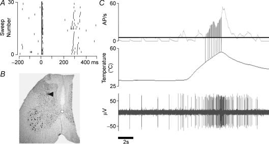
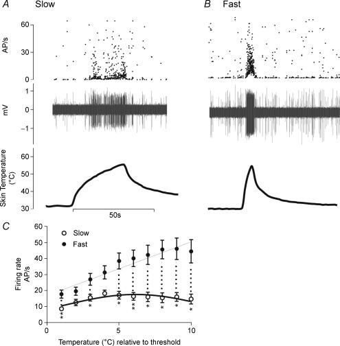
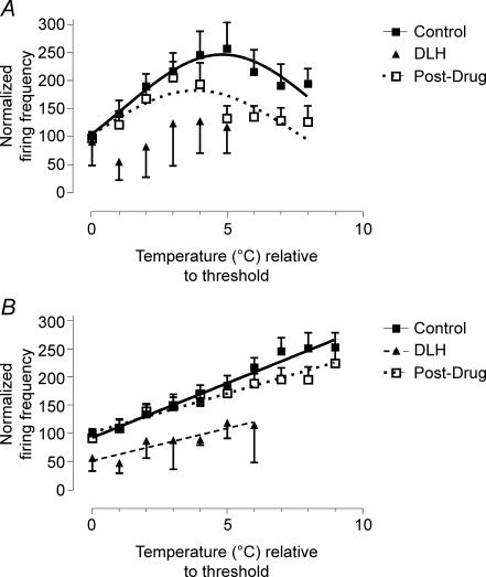
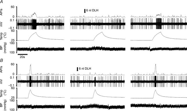
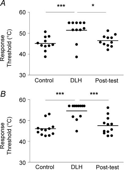
The aim of this study was to further understand the central processing of inputs arising from unmyelinated and myelinated nociceptors by (i) determining the response characteristics of Class 2 dorsal horn neurones to preferential activation of C- and A-fibre heat nociceptors, and (ii) investigating the control exerted by the dorsolateral/lateral region of the midbrain periaqueductal grey (DL/L-PAG) on C- and A-fibre-evoked responses of these neurones. The use of different rates of skin heating to preferentially activate unmyelinated (C-fibre; 2.5 degrees C s(-1)) versus myelinated (A-fibre; 7.5 degrees C s(-1)) heat nociceptors revealed that, in response to C-nociceptor activation, Class 2 neurones encode well only over the first 5 degrees C above threshold, and that at higher temperatures responses decline. In contrast, responses to A-nociceptor activation are linear and encode skin temperature over more than 10 degrees C, and almost certainly into the tissue-damaging range. PAG stimulation raised thresholds and decreased significantly the magnitude of responses to A- and C-nociceptor activation. However, differences were revealed in the effects of descending control on the relationships between skin temperature and neuronal firing rate; the linear relationship that occurred over the first 5 degrees C of slow rates of skin heating was no longer evident, whereas that to fast rates of skin heating was maintained over the entire range, albeit shifted to the right. These data indicate that the sensori-discriminative information conveyed in A-fibre nociceptors is maintained and that the information from C-nociceptors is lost in the presence of descending control from the DL/L-PAG. The data are discussed in relation to the role of the DL/L-PAG in mediating active coping strategies.
Figures
Similar articles
-
Laminar organization of spinal dorsal horn neurones activated by C- vs. A-heat nociceptors and their descending control from the periaqueductal grey in the rat.Eur J Neurosci. 2007 Aug;26(4):943-52. doi: 10.1111/j.1460-9568.2007.05716.x. Eur J Neurosci. 2007. PMID: 17714188 Free PMC article.
-
Midbrain control of spinal nociception discriminates between responses evoked by myelinated and unmyelinated heat nociceptors in the rat.Pain. 2006 Sep;124(1-2):59-68. doi: 10.1016/j.pain.2006.03.015. Epub 2006 May 2. Pain. 2006. PMID: 16650581
-
Periaqueductal grey cyclooxygenase-dependent facilitation of C-nociceptive drive and encoding in dorsal horn neurons in the rat.J Physiol. 2014 Nov 15;592(22):5093-107. doi: 10.1113/jphysiol.2014.275909. Epub 2014 Sep 19. J Physiol. 2014. PMID: 25239460 Free PMC article.
-
Inescapable and escapable pain is represented in distinct hypothalamic-midbrain circuits: specific roles for Adelta- and C-nociceptors.Exp Physiol. 2002 Mar;87(2):281-6. doi: 10.1113/eph8702356. Exp Physiol. 2002. PMID: 11856975 Review.
-
Supraspinal morphine and descending inhibitions acting on the dorsal horn of the rat.J Physiol. 1987 Mar;384:81-107. doi: 10.1113/jphysiol.1987.sp016444. J Physiol. 1987. PMID: 3309265 Free PMC article. Review.
Cited by
-
Effects of stimulation area and temperature rates on offset analgesia.Pain Rep. 2022 Oct 18;7(6):e1043. doi: 10.1097/PR9.0000000000001043. eCollection 2022 Nov-Dec. Pain Rep. 2022. PMID: 36284798 Free PMC article.
-
Spinal processing of noxious and innocuous cold information: differential modulation by the periaqueductal gray.J Neurosci. 2010 Apr 7;30(14):4933-42. doi: 10.1523/JNEUROSCI.0122-10.2010. J Neurosci. 2010. PMID: 20371814 Free PMC article.
-
Mapping the structural connectivity between the periaqueductal gray and the cerebellum in humans.Brain Struct Funct. 2019 Jul;224(6):2153-2165. doi: 10.1007/s00429-019-01893-x. Epub 2019 Jun 5. Brain Struct Funct. 2019. PMID: 31165919 Free PMC article.
-
Somatosensory Nerve Fibers Mediated Generation of De-qi in Manual Acupuncture and Local Moxibustion-Like Stimuli-Modulated Gastric Motility in Rats.Evid Based Complement Alternat Med. 2014;2014:673239. doi: 10.1155/2014/673239. Epub 2014 Apr 30. Evid Based Complement Alternat Med. 2014. PMID: 24876876 Free PMC article.
-
Periaqueductal Grey EP3 Receptors Facilitate Spinal Nociception in Arthritic Secondary Hypersensitivity.J Neurosci. 2016 Aug 31;36(35):9026-40. doi: 10.1523/JNEUROSCI.4393-15.2016. J Neurosci. 2016. PMID: 27581447 Free PMC article.
References
-
- Andrew D, Greenspan JD. Mechanical and heat sensitization of cutaneous nociceptors after peripheral inflammation in the rat. J Neurophysiol. 1999;82:2649–2656. - PubMed
-
- Budai D, Fields HL. Endogenous opioid peptides acting at mu-opioid receptors in the dorsal horn contribute to midbrain modulation of spinal nociceptive neurons. J Neurophysiol. 1998;79:677–687. - PubMed
-
- Cain DM, Khasabov SG, Simone DA. Response properties of mechanoreceptors and nociceptors in mouse glabrous skin: an in vivo study. J Neurophysiol. 2001;85:1561–1574. - PubMed
-
- Carstens E, Campell IG. Responses of motor units during the hind limb flexion withdrawal reflex evoked by noxious skin heating: phasic and prolonged suppression by midbrain stimulation and comparison with simultaneously recorded dorsal horn units. Pain. 1992;48:215–226. - PubMed
-
- Carstens E, Douglass DK. Midbrain suppression of limb withdrawal and tail flick reflexes in the rat: correlates with descending inhibition of sacral spinal neurons. J Neurophysiol. 1995;73:2179–2194. - PubMed
Publication types
MeSH terms
Grants and funding
LinkOut - more resources
Full Text Sources