. 2007 Jan;36(1):114-21.
doi: 10.1165/rcmb.2006-0174OC. Epub 2006 Aug 17.

GM-CSF regulates a PU.1-dependent transcriptional program determining the pulmonary response to LPS

Affiliations

GM-CSF regulates a PU.1-dependent transcriptional program determining the pulmonary response to LPS

Pierre-Yves Berclaz et al. Am J Respir Cell Mol Biol. 2007 Jan.

Abstract

Alveolar macrophages (AMs) normally respond to lipopolysaccharide (LPS) by activating Toll-like receptor (TLR)-4 signaling, a mechanism critical to lung host defense against gram-negative bacteria such as Pseudomonas aeruginosa. Because granulocyte macrophage colony-stimulating factor (GM-CSF)-deficient (GM(-/-)) mice are hyporesponsive to LPS, we evaluated the role of GM-CSF in TLR-4 signaling in AMs. Pulmonary TNF-alpha levels and neutrophil recruitment 4 h after intratracheal administration of Pseudomonas LPS were reduced in GM(-/-) compared with wild-type (GM(+/+)) mice. Secretion of TNF-alpha by AMs exposed to LPS ex vivo was also reduced in GM(-/-) mice and restored in mice expressing GM-CSF specifically in the lungs (SPC-GM(+/+)/GM(-/-) mice). LPS-dependent NF-kappaB promoter activity, TNF-alpha secretion, and neutrophil chemokine release were reduced in AM cell lines derived from GM(-/-) mice (mAM) compared with GM(+/+) (MH-S). Retroviral expression of PU.1 in mAM cells, which normally lack PU.1, rescued all of these AM defects. To determine whether GM-CSF, via PU.1, regulated expression of TLR-4 pathway components, mRNA and protein levels for key components were evaluated in MH-S cells (GM(+/+), PU.1(Positive)), mAM cells (GM(-/-), PU.1(Negative)), and mAMPU.1+ cells (GM(-/-), PU.1(Positive)). Cluster of differentiation antigen-14, radioprotective 105, IL-1 receptor-associated kinase (IRAK)-M mRNA, and protein were dependent upon GM-CSF and restored by expression of PU.1. In contrast, expression of other TLR-4 pathway components (myeloid differentiation-2, TLR-4, IRAK-1, IRAK-2, Toll/IL-1 receptor domain containing adapter protein/MyD88 adaptor-like, myeloid differentiation primary-response protein 88, IRAK-4, TNF receptor-associated factor-6, NF-kappaB, inhibitor of NF-kappaB kinase) were not GM-CSF or PU.1-dependent. These results show that GM-CSF, via PU.1, enables AM responses to P. aeruginosa LPS by regulating expression of a specific subset of components of the TLR-4 signaling pathway.


Figures

<b>Figure 1.</b>
Figure 1.
GM-CSF deficiency impairs LPS-stimulated pulmonary neutrophil recruitment. Four hours after intratracheal Pseudomonas LPS administration, pulmonary neutrophil influx determined by differential cytometry was reduced in GM-CSF–deficient (GM−/−) mice compared with wild-type (GM+/+) controls (n = 10, P < 0.001 [ANOVA]).
<b>Figure 2.</b>
Figure 2.
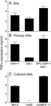
GM-CSF deficiency impairs Pseudomonas LPS-stimulated TNF-α release by AMs in vivo and in vitro. (A) Levels of TNF-α in the BAL fluid recovered 4 h after pulmonary administration of LPS were reduced in GM−/− mice compared with GM+/+ controls and were restored to supranormal levels in BAL from GM−/− mice carrying a transgene overexpressing GM-CSF in the lungs (SPC-GM+/+/GM−/− mice; n = 5, P < 0.001 [ANOVA]). (B) TNF-α secretion by primary AMs after recovery from BAL fluid, adherence to plastic for 1 h and exposure to LPS for 4 h. TNF-α secretion was reduced in AMs from GM−/− mice compared with GM+/+ controls and was restored to supranormal levels in cells from SPC-GM+/+/GM−/− mice (n = 5, P = 0.001 [ANOVA]). (C) TNF-α secretion by cultured murine AM cell lines after exposure to LPS for 4 h. TNF-α secretion was readily detectable in MH-S (GM+/+) macrophages, absent in mAM (GM−/−) macrophages (asterisk) and was restored to supranormal levels in mAMPU.1+ (GM−/−, PU.1Positive) macrophages by retroviral-mediated overexpression of the transcription factor PU.1 (n = 5, P < 0.001 [Kruskal-Wallis ANOVA on ranks]).
<b>Figure 3.</b>
Figure 3.
Neutrophil chemotaxin release from Pseudomonas LPS-stimulated AMs is regulated by GM-CSF, via PU.1. (A) Quantification of neutrophil chemotactic activity for wild type neutrophils in a Boyden chamber. AM cell lines were exposed to LPS for 24 h and culture supernatant was collected and evaluated for the presence of neutrophil chemotactic activity. LPS-exposure stimulated secretion of neutrophil chemotactic activity from MH-S cells (GM+/+) but not mAM cells (GM−/−) and was restored in mAMPU.1+ cells (GM−/−, retroviral PU.1+) (n = 9/group; P < 0.001 [Kruskal-Wallis ANOVA on ranks]). As controls, fMLP stimulated readily increased neutrophil chemotaxis compared with media alone (hatched and open bars, respectively; n = 3/group, P < 0.001). (B) Quantification of LPS-stimulated MIP-1α secretion. Cell lines were exposed to LPS as above and MIP-1α was quantified in culture supernatant by ELISA. LPS stimulated significantly increased MIP-1α secretion in MH-S and mAMPU.1+ cells, but did not stimulate any increase in mAM cells (n = 4/group, P < 0.001 [Kruskal-Wallis ANOVA on ranks]). * indicates “not detected”.
<b>Figure 4.</b>
Figure 4.
GM-CSF regulates Pseudomonas LPS-stimulated neutrophil recruitment via the TLR-4 signaling pathway. (A) The percentage of neutrophils in cells recovered by BAL 4 h after pulmonary administration of LPS was reduced in HEJ mice (TLR-4 deficient) compared with SNJ (TLR-4-replete) (n = 9, P < 0.001 [ANOVA]). (B) Levels of TNF-α in the BAL fluid recovered 4 h after pulmonary administration of LPS were reduced in HEJ mice compared with SNJ controls (n = 15, P < 0.001 [ANOVA]). (C) Neutrophil chemotaxin release by primary AMs after exposure to LPS for 24 h. Neutrophil chemotaxin release by macrophages from MyD88-deficient (MyD88−/−) mice was reduced compared with wild-type controls (MyD88+/+) (n = 3, P < 0.001 [ANOVA]) as measured by the Boyden chamber assay. As a positive control, fMLP stimulated neutrophil chemotaxis (n = 3, hatched bars). (D) LPS-stimulated NF-κB activity was markedly reduced in cultured alveolar macrophages from GM−/− mice (mAM) compared with GM+/+ mice (MH-S) and was restored by expression of PU.1 in GM−/− cells (mAMPU1+) (n = 20, P < 0.001 [ANOVA]).
<b>Figure 5.</b>
Figure 5.
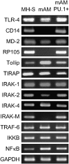
GM-CSF, via PU.1, regulates mRNA levels for multiple components of the TLR-4 signaling pathway in AMs. mRNA transcripts encoding both positive and negative signaling components of the TLR-4 signaling pathway in cultured alveolar macrophages were detected by RT-PCR. Note the absence of transcripts for CD14, RP105, and IRAK-M in mAM cells.
<b>Figure 6.</b>
Figure 6.
GM-CSF, via PU.1, regulates multiple components of TLR-4 signaling pathway in AMs. (A) Evaluation of RP105 on cultured alveolar macrophage cell lines by flow cytometry. Cells were immunostained with PE-labeled, anti-RP105 (black line) or isotype control (gray shading) antibodies and evaluated by flow cytometry. (B) RP105 levels on AMs were quantified by determining the difference between the mean fluorescence of respective cell lines immunostained with anti-RP105 antibody minus the isotype control antibody. RP105 was readily detectible on MH-S and mAMPU.1+ cells and absent on mAM cells (n = 3 determinations/cell line; P < 0.001 [Kruskal-Wallis ANOVA on ranks]). (C) Evaluation of CD14 and IRAK-M on cultured alveolar macrophage cell lines by Western blot analysis.
<b>Figure 7.</b>
Figure 7.
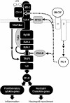
Proposed mechanism by which GM-CSF regulates AM-mediated proinflammatory cytokine signaling and neutrophil recruitment upon exposure to Pseudomonas LPS. GM-CSF receptor binding stimulates increased levels of the transcription factor, PU.1, in AMs (see Ref. 6). In turn, PU.1 stimulates expression of TLR-4 signaling pathway components (arrows). Expression of other TLR-4 components in alveolar macrophages is unaffected by GM-CSF or PU.1 (unmarked).

Similar articles

Cited by

References

    1. Goldstein E, Lippert W, Warshauer D. Pulmonary alveolar macrophage, defender against bacterial infecion of the lung. J Clin Invest 1974;54:519–528. - PMC - PubMed
    1. Poltorak A, He X, Smirnova I, Liu MY, Van Huffel C, Du X, Birdwell D, Alejos E, Silva M, Galanos C, et al. Defective LPS signaling in C3H/HeJ and C57BL/10ScCr mice: mutations in Tlr4 gene. Science 1998;282:2085–2088. - PubMed
    1. Medzhitov R, Preston-Hurlburt P, Kopp E, Stadlen A, Chen C, Ghosh S, Janeway CA Jr. MyD88 is an adaptor protein in the hToll/IL-1 receptor family signaling pathways. Mol Cell 1998;2:253–258. - PubMed
    1. Akira S, Takeda K. Toll-like receptor signalling. Nat Rev Immunol 2004;4:499–511. - PubMed
    1. Kobayashi K, Hernandez LD, Galan JE, Janeway CA Jr, Medzhitov R, Flavell RA. IRAK-M is a negative regulator of Toll-like receptor signaling. Cell 2002;110:191–202. - PubMed

Publication types

MeSH terms