Review
. 2006;16(1-2):1-22.

The vestibular-related frontal cortex and its role in smooth-pursuit eye movements and vestibular-pursuit interactions

Affiliations
Review

The vestibular-related frontal cortex and its role in smooth-pursuit eye movements and vestibular-pursuit interactions

Junko Fukushima et al. J Vestib Res. 2006.

Abstract

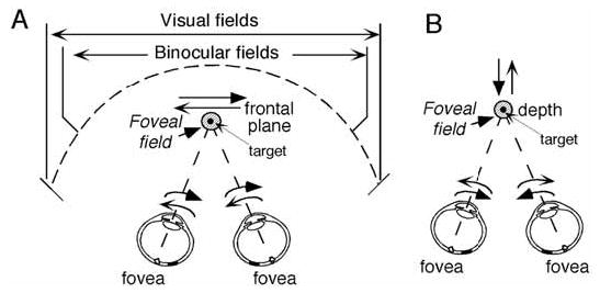
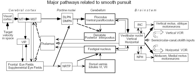
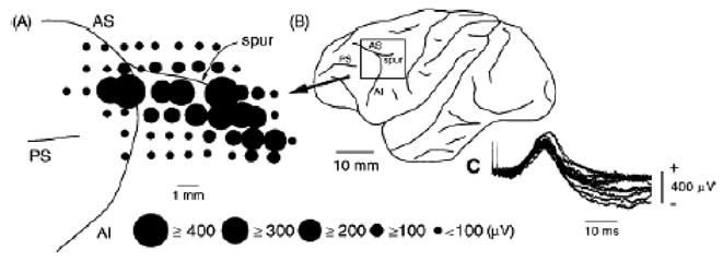
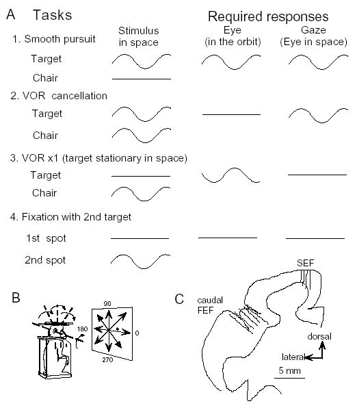
In order to see clearly when a target is moving slowly, primates with high acuity foveae use smooth-pursuit and vergence eye movements. The former rotates both eyes in the same direction to track target motion in frontal planes, while the latter rotates left and right eyes in opposite directions to track target motion in depth. Together, these two systems pursue targets precisely and maintain their images on the foveae of both eyes. During head movements, both systems must interact with the vestibular system to minimize slip of the retinal images. The primate frontal cortex contains two pursuit-related areas; the caudal part of the frontal eye fields (FEF) and supplementary eye fields (SEF). Evoked potential studies have demonstrated vestibular projections to both areas and pursuit neurons in both areas respond to vestibular stimulation. The majority of FEF pursuit neurons code parameters of pursuit such as pursuit and vergence eye velocity, gaze velocity, and retinal image motion for target velocity in frontal and depth planes. Moreover, vestibular inputs contribute to the predictive pursuit responses of FEF neurons. In contrast, the majority of SEF pursuit neurons do not code pursuit metrics and many SEF neurons are reported to be active in more complex tasks. These results suggest that FEF- and SEF-pursuit neurons are involved in different aspects of vestibular-pursuit interactions and that eye velocity coding of SEF pursuit neurons is specialized for the task condition.


Figures

Fig. 1
Fig. 1
Foveal field and frontal pursuit (A) and vergence pursuit (B). Schematic top views of visual fields and foveal field projection (shaded circles) in 3D space and target (small dot within the foveal field). For frontal target motion (A), left and right eyes rotate in the same directions. For target motion in depth (B), both eyes rotate in the opposite directions.
Fig. 2
Fig. 2
Major pathways related to frontal pursuit and vestibular inputs. Thick lines are proposed main smooth-pursuit pathways [55]. Dashed lines from the thalamus to the cortex indicate that the nature of thalamic pursuit signals are still unknown. Modified from ref. .
Fig. 3
Fig. 3
Vestibular-evoked potentials recorded on the surface of the periarcuate cortex of the monkey. A, distribution of vestibular-evoked positive potentials on the periarcuate cortex. The diameter of the circle is proportional to the amplitude of the positive peak of a vestibular-evoked potential at the center of the circle. AI, inferior ramus of the arcuate sulcus; AS, superior ramus of the arcuate sulcus; PS, principal sulcus. B, schematic drawing of the left cerebral cortex showing the recorded periarcuate area in A (boxed area). C, typical surface potentials evoked by stimulation of the contralateral labyrinth with two stimuli at 500 mA. Reproduced from ref. with permission.
Fig. 4
Fig. 4
Behavioral tasks used to dissociate eye movements in the orbit from those in space (A,B) and recording locations (C). A and B, Each task (rows) shows the idealized, intended movement of the target and chair-fixed head (first column), the eye (second column) and gaze (third column). C, Recording locations in caudal FEF and SEF [28,34].
Fig. 5
Fig. 5
Discharge characteristics of FEF pursuit neurons. A1–A2, discharge of a single neuron during vertical pursuit (A1) and pursuit during different directions (A2). B1 and B2, discharge during VOR cancellation in the pitch plane (B1) and VOR cancellation along different directions (B2). A3 and B3, polar plots of preferred directions of FEF neurons during frontal pursuit (A3) and rotational VOR cancellation (B3). A4 and B4, amplitude of discharge modulation plotted against peak eye (A4) and gaze (B4) velocity for individual neurons (Reproduced and modified from ref. with permission).
Fig. 6
Fig. 6
Discharge characteristics of a single FEF pursuit neuron during different task conditions. A–C, Responses during frontal pursuit, VOR cancellation, and VOR x1, respectively. D, Response during chair rotation in complete darkness. Eye-velocity and gaze-velocity are clipped.
Fig. 7
Fig. 7
Discharge modulation of FEF and SEF pursuit neurons during frontal-pursuit, VOR cancellation and VOR x1. A and D compare preferred directions during smooth-pursuit and VOR cancellation for FEF and SEF neurons, respectively. Dashed and straight line slopes in A and D = one. B and E compares sensitivity (re stimulus velocity) during smooth-pursuit and VOR cancellation. C and F compares sensitivity (re stimulus velocity) during smooth-pursuit and VOR x1. Open and filled squares in A, B, C are gaze-velocity and eye/head velocity FEF neurons. Open squares and dots in D, E, F are gaze-velocity and pursuit+vestibular SEF neurons, respectively. Reproduced and modified from refs. , with permission.
Fig. 8
Fig. 8
Discharge of a representative pursuit plus vestibular SEF neuron. Discharge during horizontal smooth-pursuit (A), yaw VOR cancellation (B), yaw VOR x1 (C), yaw rotation in complete darkness (D), and pitch VOR cancellation (E). Each section shows stimulus velocity, “de-saccaded” and superimposed horizontal eye-velocity (H Ė or vertical eye velocity (V Ė), spikes rasters, and histograms of neuron discharge with superimposed fitted sine waves. Reproduced from ref. with permission.
Fig. 9
Fig. 9
Effects of muscimol injection into the caudal FEF on pursuit. Horizontal pursuit eye movements before (A) and one hour after (C) muscimol injection (15 μg) into the caudal FEF (E). Yaw VOR cancellation before (B) and after (D) muscimol infusion. Abbreviations: HE and H Ė, horizontal eye position and velocity, respectively. Pos and vel, position and velocity, respectively. Reproduced from refs. , with permission.
Fig. 10
Fig. 10
Preferred direction of an FEF pursuit neuron during passive whole body translation in complete darkness (A, B) and discharge modulation of another neuron during right/left translation with a target (C). Linear motion (0.3 Hz, ±10 cm) was given along the same earth-horizontal direction in complete darkness while the orientation of the monkeys’ whole body was changed as indicated in A. B, preferred linear motion direction of a single FEF pursuit neuron. In C, linear motion was applied along left/right direction while the target moved with the monkey (LVOR cancellation) and while the target stayed stationary in space during translation (LVOR x1). Reproduced from ref. with permission.
Fig. 11
Fig. 11
Comparison of vergence pursuit modulation of FEF and MST neurons. A and B, representative discharge modulation of FEF (A) and MST (B) pursuit neurons during sinusoidal vergence pursuit at different frequencies (±5°) as indicated. C compares mean (±SD) phase differences of group A and B FEF pursuit neurons at different frequencies relative to the values at 0.5 Hz. D is a similar plot for MST pursuit neurons and simultaneously recorded eye movement responses. Phase shifts in C and D were calculated by fitting a sinusoid using a least-squared error algorithm in all traces. Notice a distortion manifested in vergence eye velocity at 1.0 Hz (bottom traces in A and B indicated by open and filled arrowheads). Open arrowheads indicate actual peak convergence eye velocity. Filled arrowheads indicate the peak of the fitted function. They are clearly different at 1.0 Hz but virtually identical at 0.5 Hz (also ref. 87). Actual peak convergence eye velocity exhibited phase lag (relative to the value at 0.5 Hz) of less than 10° in A and B at 1.0 Hz that are within the error bars of FEF group A neurons in C. Open triangles in C and D indicate a model that contains a delay of only 200 ms. Reproduced from refs. , with permission.
Fig. 12
Fig. 12
Visual responses of a representative FEF pursuit neuron. For all traces, the monkey fixated a stationary spot while the second test spot moved sinusoidally along different directions (C). Upper panels for A-B are 1st target, horizontal and vertical eye position (HE, VE), second target velocity, and rasters and histograms of neuron responses when the second target was continuously visible. In the lower panels, the second spot was extinguished for more than half of each cycle as indicated (OFF). C shows directional tuning of this neuron with (open circles) and without (filled circles) blanking the second target. Reproduced from ref. with permission.
Fig. 13
Fig. 13
Stimulus trajectory for cross-axis vestibular-pursuit training and vertical pursuit eye movements after training. A, stimulus trajectory. Inter-trial intervals for chair motion were random (top trace). Chair was rotated in the yaw plane at 20°/s for 1 s. Delay between the onset of chair motion and target motion onset is marked by vertical dashed lines. B shows de-saccaded mean ± SD vertical eye velocity. Dashed line in B indicates the onset of chair motion. Vertical bars with rightward arrows in B indicate the onset of actual target motion at different delays. Upward arrows in B indicate onset of vertical smooth eye movements. All traces are aligned on the onset of chair motion. C plots mean ± SD latencies against the delays between the onset of chair and target motion for cross-axis training in two monkeys. Open diamonds and filled circles are values with and without blanking the target, respectively. 7–10 different recording sessions were combined to calculate mean and SD which was smaller than the symbol size in most cases. Only one plus SD is shown for means with blanking and one minus SD is shown for means without blanking. Linear regressions are shown for filled circles (i.e., without blanking). Reproduced from ref. with permission.
Fig. 14
Fig. 14
Responses of a caudal FEF pursuit neuron during adaptive pursuit induced by cross-axis vestibular-pursuit training. Superimposed traces of chair/target position and eye position and velocity before training to yaw rotation in complete darkness (A), to downward spot movement (B), and combined presentation of vertical target and yaw rotation after 30 min of similar training (C). Upward arrows on chair/target traces indicate onset of leftward chair rotation (A, C) and downward spot movement (B, C). HE, VE, and VĖ indicate horizontal eye position, vertical eye position and vertical eye velocity, respectively. Reproduced from ref. with permission.
Fig. 15
Fig. 15
Effects of muscimol infusion into SEF (A–E) for vertical pursuit and pitch VOR cancellation. All eye velocity records in A–E were de-saccaded and averaged. Arrows in B and E indicate impaired smooth eye movements. Reproduced from ref. with permission.

Similar articles

Cited by

References

    1. Akao T, Kurkin S, Fukushima K. Latency of adaptive vergence eye movements induced by vergence-vestibular interaction training in monkeys. Exp Brain Res. 2004;158:129–132. - PubMed
    1. Akao T, Kurkin S, Fukushima J, Fukushima K. Visual and vergence eye movement related responses of pursuit neurons in the caudal frontal eye fields to motion-in-depth stimuli. Exp Brain Res. 2005;164:92–108. - PubMed
    1. Akao T, Mustari MJ, Fukushima J, Kurkin S, Fukushima K. Discharge characteristics of pursuit neurons in MST during vergence eye movements. J Neurophysiol. 2005;93:2415–2434. - PubMed
    1. Akao T, Kasahara S, Kurkin S, Fukushima K. Coordinate frames in representing pursuit signals in simian frontal eye fields (FEF) J Physiol Sci. 2006;56(Suppl):S187. (Abstr).
    1. Amador N, Schlag-Rey M, Schlag J. Reward-predicting and reward-detecting neuronal activity in the primate supplementary eye field. J Neurophysiol. 2000;84:2166–2170. - PubMed

Publication types

LinkOut - more resources