. 2006 Sep 6;25(17):3934-42.
doi: 10.1038/sj.emboj.7601281. Epub 2006 Aug 17.

Identification of CD63 as a tissue inhibitor of metalloproteinase-1 interacting cell surface protein

Affiliations

Identification of CD63 as a tissue inhibitor of metalloproteinase-1 interacting cell surface protein

Ki-Kyung Jung et al. EMBO J. .

Abstract

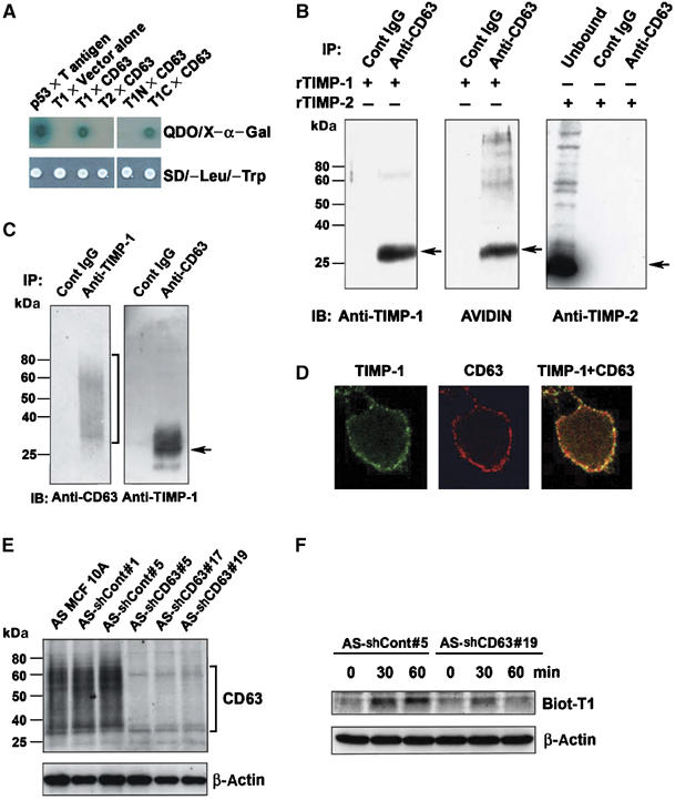
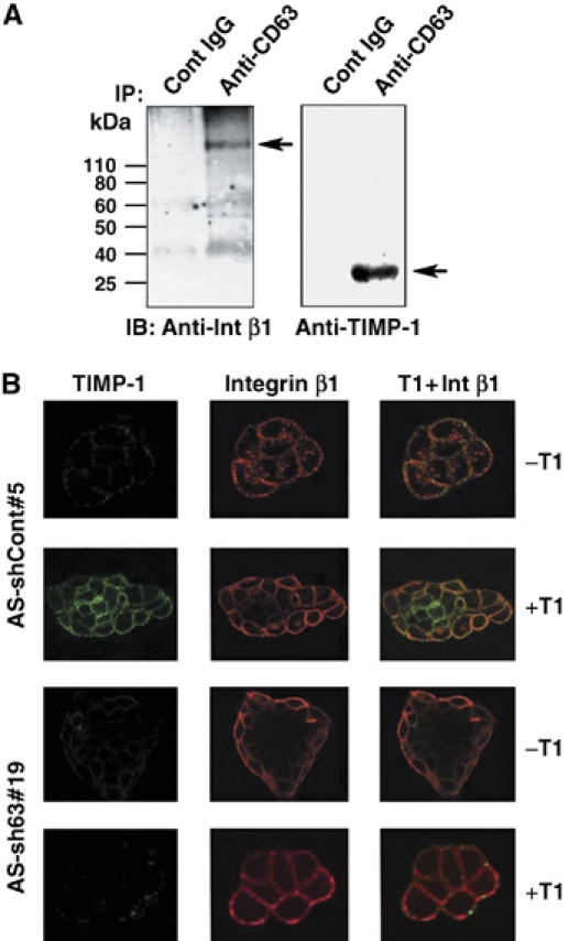
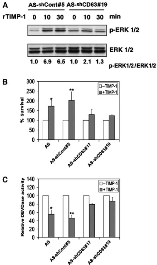
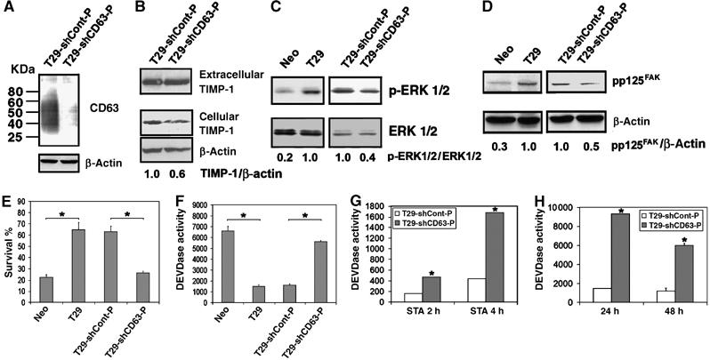
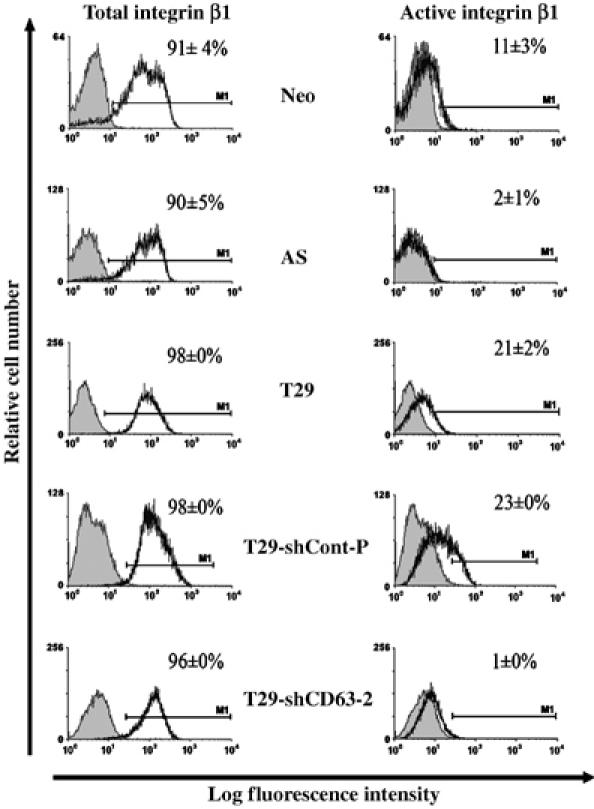
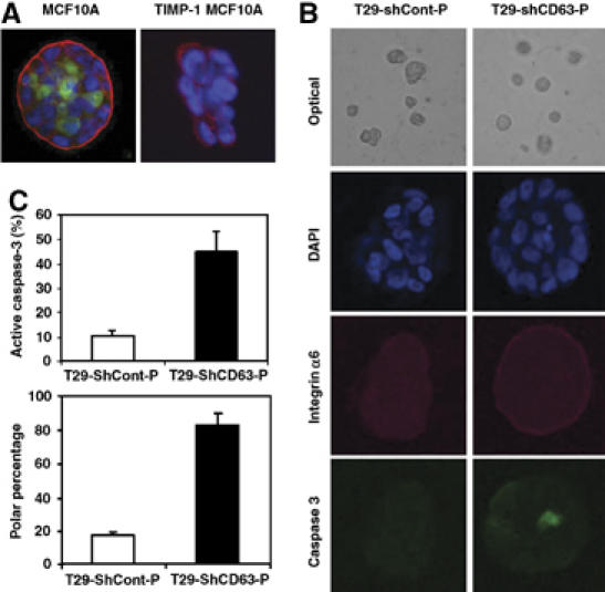
This study identified CD63, a member of the tetraspanin family, as a TIMP-1 interacting protein by yeast two-hybrid screening. Immunoprecipitation and confocal microscopic analysis confirmed CD63 interactions with TIMP-1, integrin beta1, and their co-localizations on the cell surface of human breast epithelial MCF10A cells. TIMP-1 expression correlated with the level of active integrin beta1 on the cell surface independent of cell adhesion. While MCF10A cells within a three-dimensional (3D) matrigel matrix form polarized acinar-like structures, TIMP-1 overexpression disrupted breast epithelial cell polarization and inhibited caspase-mediated apoptosis in centrally located cells, necessary for the formation and maintenance of the hollow acinar-like structures. Small hairpin RNA (shRNA)-mediated CD63 downregulation effectively reduced TIMP-1 binding to the cell surface, TIMP-1 co-localization with integrin beta1, and consequently reversed TIMP-1-mediated integrin beta1 activation, cell survival signaling and apoptosis inhibition. CD63 downregulation also restored polarization and apoptosis of TIMP-1 overexpressing MCF10A cells within a 3D-matrigel matrix. Taken together, the present study identified CD63 as a cell surface binding partner for TIMP-1, regulating cell survival and polarization via TIMP-1 modulation of tetraspanin/integrin signaling complex.


Figures

Figure 1
Figure 1
TIMP-1-specific interaction with CD63. (A) Yeast AH109 expressing p53 bait (as a positive control), full-length TIMP-1 (amino acids 1–184) lacking signal peptide (T1), full-length TIMP-2 (T2), N-terminal domain (amino acids 1–125) of TIMP-1 (T1N), or C-terminal domain (amino acids 126–184) of TIMP-1 (T1C) was mated with Y187 expressing large T antigen prey (as a positive control), vector alone (as a negative control), or full-length CD63. Diploids grow on SD/−Leu/−Trp selective media. The specific interaction between bait and prey proteins was detected on quadruple dropout (QDO) media SD/−Leu/−Trp/−His/−Ade containing X-α-Gal. (B) Anti-CD63 immunoprecipitates of MCF10A cell lysates in the presence of 500 ng/ml biotinylated rTIMP-1 (left and middle panel) or rTIMP-2 (right panel) were analyzed by Western blot analysis with anti-TIMP-1 monoclonal antibody, avidin-HRP, and anti-TIMP-2 polyclonal antibody. (C) TIMP-1 overexpressing MCF10A (T29) cell lysates were immunoprecipitated with anti-TIMP-1 polyclonal antibody or anti-CD63 monoclonal antibody, followed by immunoblot analysis using anti-CD63 under nonreducing condition or anti-TIMP-1 antibody under reducing condition, respectively. (D) MCF10A cells were grown on the coverslips overnight, blocked with PBS containing 10% horse serum 1% BSA, and co-stained with anti-CD63 Ab/Texas red conjugated secondary Ab (red staining), or with anti-TIMP-1 Ab/FITC conjugated secondary Ab (green staining). Co-localization of TIMP-1 and CD63 is shown as yellow staining (live cell staining). (E) Cell lysates of AS MCF10A clones stably transfected with control vector (AS-shCont#1 and AS-shCont#5) or with shRNA vector targeting CD63 (AS-shCD63#5, AS-shCD63#17 and AS-shCD63#19) were subjected to immunoblot analysis with an anti-CD63 antibody in a nonreducing condition. The bottom panel shows the β-actin levels of the same blot reprobed with an anti-human β-actin antibody. (F) AS-shCont#5 and AS-shCD63#19 cells were incubated with biotinylated TIMP-1 (500 ng/ml) for 0, 30, and 60 min at 37°C. For the 0 min time point, cells were washed right after the addition of biotinylated TIMP-1. Cells lysates (20 μg/lane) were subjected to immunoblot analysis with avidin-HRP.
Figure 2
Figure 2
CD63 mediates TIMP-1 binding to cell surface and TIMP-1 co-localization with integrin β1 in human breast epithelial cells. (A) MCF10A cell lysates were immunoprecipitated with anti-CD63 antibody, followed by immunoblotting with anti-integrin β1 or anti-TIMP-1 monoclonal antibody. (B) AS-shCont#5 and AS-shCD63#19 cells were cultured on the coverslips overnight and incubated with or without 500 ng/ml TIMP-1 for 30 min. Live cells were co-stained with anti-integrin β1 Ab/Texas red conjugated secondary Ab (red staining) and anti-TIMP-1 Ab/FITC conjugated secondary Ab (green staining). Colocalization of TIMP-1 and integrin β1 is shown as yellow staining.
Figure 3
Figure 3
CD63 is required for exogenous TIMP-1-mediated ERKs activation and inhibition of apoptosis. (A) AS-shCont#5 and AS-shCD63#19 cells were cultured in serum-free medium for 48 h and then incubated with 500 ng/ml TIMP-1 for 10 and 30 min. Cell lysates (20 μg/lane) were subjected to immunoblot analysis with anti-active ERKs (pERK1/2) and anti-ERK1/2 antibodies. Densitometry analysis of the signals between pERK1/2 and ERK1/2 were presented by normalizing to the signal ratio in AS-shCont#5 or AS-shCD63#19 cells at 0 min. (B) AS MCF10A, AS-shCont#5, AS-shCD63#17, and AS-shCD63#19 were cultured in serum-free medium for 48 h in the presence and absence of 500 ng/ml TIMP-1. The percentage of cell survival was determined by WST-1 assay and normalized to the respective cells cultured in serum containing medium. Shown are the means±s.e. of the sextuple experiments. *P<0.05 versus AS MCF10A cells without TIMP-1 treatment (t-test); **P<0.05 versus AS-shCont#5 cells without TIMP-1 treatment (t-test). (C) AS MCF10A, AS-shCont#5, AS-shCD63#17, and AS-shCD63#19 were cultured in serum-free medium for 24 h in the presence and absence of 500 ng/ml TIMP-1. The cells were then subjected to DEVDase activity assay. Shown are the means±s.e. of the sextuple experiments. *P<0.05 versus AS cells without TIMP-1 treatment (t-test); **P<0.001 versus AS-shCont#5 cells without TIMP-1 treatment (t-test).
Figure 4
Figure 4
Downregulation of CD63 expression reverses TIMP-1mediated antiapoptotic activity. (A) T29-shCont-P and T29-shCD63-P cells were subjected to immunoblot analysis with an anti-CD63 antibody in a nonreducing condition. The bottom panel shows the β-actin levels of the same blot reprobed with an anti-human β-actin antibody. (B) Conditioned medium and cell lysates of T29-shCont-P and T29-shCD63-P cells were subjected to immunoblot analysis with an anti-TIMP-1 antibody. The bottom panel shows the β-actin levels of the same blot reprobed with an anti-human β-actin antibody. Densitometry analysis of the signals between cellular TIMP-1 and β-actin was presented by normalizing to the signal ratio in T29-ShCont-P cells. (C) Cell lysates (40 μg/lane) of 48 h serum-starved MCF10Aneo (Neo), T29, T29-shCont-P, and T29-shCD63-P cells were subjected to immunoblot analysis with anti-active ERKs (pERK1/2) and anti-ERK1/2 antibodies. Densitometry analysis of the signals between pERK1/2 and ERK1/2 were presented by normalizing to the signal ratio in T29 (left panel) or T29-shCont-P cells (right panel). (D) Cell lysates (40 μg/lane) of 48 h serum-starved Neo, T29, T29-shCont-P, and T29-shCD63-P cells were subjected to immunoblot analysis with anti-FAK (pY397) and anti-FAK antibodies. Densitometry analysis of the signals between anti-FAK (pY397) and anti-FAK were presented by normalizing to the signal ratio in T29 (left panel) or T29-shCont-P cells (right panel). (E) MCF10Aneo (Neo), T29, T29-shCont-P, and T29-shCD63-P cells were cultured in serum-free medium for 48 h. The percentage of cell survival was determined by WST-1 assay, and normalized to the respective cells cultured in serum containing medium. Three independent experiments were performed and the error bars represent standard deviation of the mean of sextuplicates. Asterisks depict statistically significant differences between Neo and T29 cells and between T29-shCont-P and T29-shCD63-P cells, by an unpaired, unequal t-test (*P<0.001). (F) MCF10Aneo (Neo), T29, T29-shCont-P, and T29-shCD63-P cells were cultured in serum-free medium for 48 h. The cells were then washed with PBS and lysed with 200 μl caspase lysis buffer as previously described (Liu et al, 2003, 2005). After lysates were centrifuged at 16 000 g for 10 min, DEVDase activity in 50 μl cytosol was assayed and the activity was normalized per μg protein. Three independent experiments were performed and the error bars represent standard deviation of the mean of sextuplicates. Asterisks depict statistically significant differences between Neo and T29 cells and between T29-shCont-P and T29-shCD63-P cells, by an unpaired, unequal t-test (*P<0.001). (G, H) Apoptosis was induced in T29-shCont-P and T29-shCD63-P cells by treatment with 0.5 μM staurosporine (G) or by culturing on polyHEMA-coated dishes (H). At indicated time points, the cells were washed with PBS and lysed with 200 μl caspase lysis buffer and DEVDase activity was measured. Three independent experiments were performed and the error bars represent standard deviation of the mean of triplicates. *P<0.05 versus T29-shCont-P cells at the respective time points (t-test).
Figure 5
Figure 5
TIMP-1 enhances the level of integrin β1 in activated conformation on the cell surface. MCF10Aneo (Neo), AS TIMP-1 MCF10A (AS), TIMP-1 overexpressing MCF10A (T29), T29-shCont-P and T29-shCD63 cells were plated on polyHEMA coated tissue culture plates for 24 h and stained indirectly with either anti integrin β1 antibody (left panels) or an LIBS antibody recognizing only the active integrin β1 (right panels) followed by incubation with a FITC conjugated secondary antibody. Fluorescence was measured using a FACSCalibur machine. The percentage of gated cells stained for active or total integrin β1 (solid black line) was normalized to the percentage of gated cells stained with the FITC-secondary antibody only (negative control, shaded area). About 20 000 cells per experimental condition were analyzed in duplicates and the means±s.d. were shown.
Figure 6
Figure 6
Downregulation of CD63 expression results in phenotypic reversion of TIMP-1 overexpressing MCF10A cells to the parental MCF10A cells in 3D culture. (A, B) MCF10A and TIMP-1 overexpressing MCF10A (T29) cells (A) and T29-shCont-P and T29-shCD63-P cells (B) were cultured in GFR matrigel as described in the Materials and methods (see Supplemental data). At 8 days, confocal microscopic images of cross-sections through the middle of developing acini are shown. Cells were stained with anti-integrin α6 Ab/Texas red conjugated secondary Ab (red staining) to delineate the basement membrane, anti-active caspase-3 Ab/FITC conjugated secondary Ab (green staining) to detect apoptotic cells, and with DAPI (blue) to counterstain the nuclei. (C) Quantitative analysis of the percentage of spheroids including active caspase-3 and polarity are shown in the right panel. (About 30 spheroids were analyzed for each condition from three independent experiments and the means±s.d. were shown).

Similar articles

Cited by

References

    1. Airola K, Karonen T, Vaalamo M, Lehti K, Lohi J, Kariniemi AL, Keski-Oja J, Saarialho-Kere UK (1999) Expression of collagenases-1 and -3 and their inhibitors TIMP-1 and -3 correlates with the level of invasion in malignant melanomas. Br J Cancer 80: 733–743 - PMC - PubMed
    1. Avalos BR, Kaufman SE, Tomonaga M, Williams RE, Golde DW, Gasson JC (1988) K562 cells produce and respond to human erythroid-potentiating activity. Blood 71: 1720–1725 - PubMed
    1. Berditchevski F (2001) Complexes of tetraspanins with integrins: more than meets the eye. J Cell Sci 114: 4143–4151 - PubMed
    1. Berditchevski F, Bazzoni G, Hemler ME (1995) Specific association of CD63 with the VLA-3 and VLA-6 integrins. J Biol Chem 270: 17784–17790 - PubMed
    1. Berditchevski F, Odintsova E (1999) Characterization of integrin–tetraspanin adhesion complexes: role of tetraspanins in integrin signaling. J Cell Biol 146: 477–492 - PMC - PubMed

Publication types

MeSH terms