. 2006 Sep 6;25(17):3955-65.
doi: 10.1038/sj.emboj.7601273. Epub 2006 Aug 17.

Roles of HIPK1 and HIPK2 in AML1- and p300-dependent transcription, hematopoiesis and blood vessel formation

Affiliations

Roles of HIPK1 and HIPK2 in AML1- and p300-dependent transcription, hematopoiesis and blood vessel formation

Yukiko Aikawa et al. EMBO J. .

Abstract

Histone acetyltransferases (HATs) p300 and CREB-binding protein (CBP) function as co-activators for a variety of sequence-specific transcription factors, including AML1. Here, we report that homeodomain-interacting protein kinase-2 (HIPK2) forms a complex with AML1 and p300, and phosphorylates both AML1 and p300 to stimulate transcription activation as well as HAT activities. Phosphorylation of p300 is triggered by phosphorylated AML1 as well as by PU.1, c-MYB, c-JUN and c-FOS, and is inhibited by dominant-negative HIPK2. Phosphorylation of p300 and AML1 is impaired in Hipk1/2 double-deficient mouse embryos. Double-deficient mice exhibit defects in primitive/definitive hematopoiesis, vasculogenesis, angiogenesis and neural tube closure. These phenotypes are in part similar to those observed in p300- and CBP-deficient mice. HIPK2 also phosphorylates another co-activator, MOZ, in an AML1-dependent manner. We discuss a possible mechanism by which transcription factors could regulate local histone acetylation and transcription of their target genes.


Figures

Figure 1
Figure 1
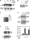
AML1 is phosphorylated at Ser249 and Ser276 by HIPK2. (A) Phosphorylation sites are evolutionarily conserved. (B) Migration of WT and mutant AML1 in L-G cells. (C) Phosphatase treatment of AML1. Purified WT and S249/276A mutant AML1b were treated with (+) or without (−) calf intestine alkaline phosphatase (CIAP) and analyzed by immunobloting with anti-AML1 antibody. (D) AML1 complex contains HIPK2. Purified AML1 complexes were analyzed by immunobloting with anti-HIPK2 antibody. (E) Interaction of AML1 with HIPK2. 293T cells were transfected with HA-AML1b together with either FLAG-tagged WT or KD HIPK2. Immunoprecipitates with anti-FLAG were analyzed by immunoblotting with anti-HA antibody. (F) Phosphorylation of AML1 by HIPK2. Cell extracts from 293T cells expressing WT or mutants of HA-tagged AML1 and HIPK2 were subjected to immunoblot analysis with anti-HA antibody. (G) Phosphorylation of AML1 in vitro. Purified WT or mutant AML1b proteins were tested for in vitro kinase activity. (H) HIPK2 activates AML1-mediated transcription. SaOS2 cells were transfected with 50 ng of MPO-luc, 200 ng of LNCX-AML1b, 250 ng of MOZ or p300, 250 ng of WT or KD mutant of HIPK2 and 2 ng of phRL-cmv. Cell lysates were analyzed for luciferase activity at 24 h after transfection.
Figure 2
Figure 2
HIPK2 mediates AML1-dependent phosphorylation of p300. (A) AML1 induces phosphorylation of p300. 293T cells were transfected with FLAG-AML1b, HA-p300 and HIPK2. Cell lysates were immunoprecipitated with anti-FLAG antibodies. Cell lysates (1%) and immunoprecipitates were then analyzed by immunoblotting with anti-HA antibody. (B) CIAP-treatment of AML1–p300 complex. 293T cells were transfected with HA-p300 and FLAG-AML1b. After immunoprecipitation with anti-HA antibodies, immunoprecipitates were treated with CIAP and analyzed by immunoblotting with anti-HA antibody. (C) Phosphorylation of p300 in vitro. Purified WT p300 protein was incubated with or without HIPK2 in the presence of [γ32P]ATP and was separated by 7% SDS–PAGE. Gels were dried and subjected to autoradiography. (D) Phosphorylation of AML1 is required for AML1-induced phosphorylation of p300. 293T cells were transfected with HA-p300 and FLAG-tagged WT, S249/276A (2A) or S249/276A (2D) mutant AML1b. After immunoprecipitation with anti-FLAG antibodies, cell lysates (1%) and immunoprecipitates were analyzed by immunoblotting with anti-HA antibody. (E) HIPK2 and AML1 stimulates HAT activity of p300. 293T cells were transfected with FLAG-p300 together with either WT or KD HIPK2, or AML1b. After immunoprecipitation with anti-FLAG antibodies, immunoprecipitates were analyzed for HAT activity. (F) HIPK2 and AML1 stimulate transcription activation by a Gal4-p300 fusion protein. 293T cells were transfected with 500 ng of pFR-luc(Gal4-luc), 100 ng of Gal4-p300, 400 ng of WT or KD HIPK2, or AML1b and 2 ng of phRL-cmv. Cell lysates were analyzed for luciferase activity at 24 h after transfection.
Figure 3
Figure 3
Phosphorylation sites of p300 by HIPK2. (A) Schematic representation of p300 mutants used for determining HIPK2-induced phosphorylation sites. (B) Amino-acid sequence of putative HIPK2 phosphorylation sites of p300. Serines or threonines shown in red were substituted with alanines. (C) HIPK2-induced activation of transcription by Gal4-p300 mutants. 293T cells were transfected with 500 ng of pFR-luc(Gal4-luc), 100 ng of WT or mutant Gal4-p300, 400 ng of HIPK2 and 2 ng of phRL-cmv. Cell lysates were prepared at 24 h after transfection and were analyzed for luciferase activity. (D) Phosphorylation of p300 mutants by HIPK2. 293T cells were transfected with WT or mutants of Gal4-p300 together with HIPK2. Cell lysates were subjected to immunoblot analysis with anti-Gal4 antibody.
Figure 4
Figure 4
Interaction of p300 and HIPK2. (A) Schematic representation of p300 deletion mutants used for determining HIPK2-binding domains. (B) Interaction of p300 with HIPK2. 293T cells were transfected with p300 together with FLAG-tagged WT or KD HIPK2, or AML1b. (B–D) Cell lysates (1%) and immunoprecipitates with anti-FLAG antibodies were analyzed by immunoblotting with anti-p300, anti-HA or anti-FLAG antibodies. (C) Reciprocal coimmunoprecipitation of HIPK2 and p300. (D) Interaction of p300 mutants with HIPK2 and AML1. 293T cells were transfected with p300 mutants as shown together with KD HIPK2 or AML1b. The lower panel represents a longer exposure of the same membrane shown in the middle panel. (E) HIPK2 translated in vitro and labelled with 35S was tested for binding to bacterially produced GST, GST-AML1b and GST-p300(HAT) proteins as shown. The input lane represents 10% of the material used for binding to the GST fusion protein.
Figure 5
Figure 5
HIPK2 mediates phosphorylation of p300 induced by a set of transcription factors. (A) Phosphorylation of p300 by various transcription factors. 293T cells were transfected with HA-p300 together with various transcription factors. Cell lysates were subjected to immunoblot analysis with anti-HA antibody. (B) Effects of WT and KD HIPK2 on transcription factor-induced phosphorylation of p300. 293T cells were transfected with HA-p300 and the transcription factors indicated together with WT and KD HIPK2. (C) Effects of KD HIPK2 on phosphorylation of p300. 293T cells were transfected with HA-p300 (0.2 μg) and either C/EBPɛ, C/EBPα or AML1b (0.4 μg) together with increasing amounts (0.2 and 0.4 μg) of KD HIPK2.
Figure 6
Figure 6
HIPK1 and HIPK2 are essential for phosphorylation of p300 and AML1. (A) Phosphorylation of p300 in HIPK1- and/or HIPK2-deficient embryos. Proteins were extracted from WT and mutant E10.0 embryos and were subjected to immunoblot analysis with anti-p300 antibody. (B) Expression of HIPK2 and phosphorylation of p300 during differentiation of ES cells. WT and Hipk1/2 double-deficient ES cells were exposed to 1 μM retinoic acid for 2 and 4 days. The cell lysates were subjected to immunoblot analysis with anti-HIPK2 and anti-p300 antibodies. (C) Expression of HIPK2 and phosphorylation of p300 during differentiation of myeloid cells. 32Dcl3 cells were exposed to 10 ng G-CSF for indicated number of days. (D) Phosphorylation of AML1 in HIPK1- and/or HIPK2-deficient yolk sac. Yolk sac cells from E10.0 embryos were cultured on OP9 cells for 5 days. Cell lysates were subjected to immunoblot analysis with anti-AML1 and phospho-S249-specific AML1 antibodies. (E) Phosphorylation of p300 and AML1 by HIPK1 and HIPK2. 293T cells were transfected with HA-p300 or AML1 (0.2 μg) together with increasing amounts (0.05, 0.1, 0.2 μg) of HIPK1 or HIPK2. Cell lysates were subjected to immunoblot analysis with anti-HA antibody. (F) 293T cells were transfected with WT or mutant Gal4-p300 or HA-AML1 together with HIPK1 or HIPK2. Cell lysates were subjected to immunoblot analysis with anti-Gal4 or anti-HA antibody.
Figure 7
Figure 7
HIPK1 and HIPK2 are involved in p300- and AML1-mediated transcription. (A) 293T cells were transfected with 500 ng of pFR-luc(Gal4-luc), 100 ng of Gal4-p300, 400 ng of HIPK1 or HIPK2, and 2 ng of phRL-cmv. (B) SaOS2 cells were transfected with 50 ng of MPO-luc, 200 ng of LNCX-AML1b, 250 ng of MOZ or p300, 250 ng of HIPK1 or HIPK2, and 2 ng of phRL-cmv. (C) WT and HIPK1/2 double-deficient MEFs were transfected with 50 ng of MPO-luc, 200 ng of LNCX-AML1b, 500 ng of MOZ or p300, and 2 ng of phRL-cmv. Cell lysates were analyzed for luciferase activity at 24 h after transfection.
Figure 8
Figure 8
Hematopoiesis in HIPK1/2 mutant embryos. (A) Yolk sac cells from embryos at E9.5 were dispersed into single-cell suspensions and the numbers of nucleated erythroid cells were counted. (B) Colony-forming cells in P-Sp cultures. P-Sp explants derived from E9.5 embryos obtained by intercrossing HIPK1−/−,HIPK2+/− mice were cultured on OP9 stromal cells for 7 days and were subjected to colony-formation assay. Dot-filled and oblique-line-filled boxes represent the number of erythroid and myeloid colonies, respectively. (C) Colony-forming cells in yolk sac. Yolk sac cells were directly subjected to colony-formation assay.
Figure 9
Figure 9
HIPK1/2 mutant embryos showed defective vasculogenesis and angiogenesis. (A, B) Phenotypic comparison of E10.0 Hipk1−/−, Hipk2+/+ and Hipk1−/−,Hipk2−/− embryos with (A) and without (B) yolk sac. (C) Whole-mount immunohistostaining with anti-CD31 (PECAM-1) mAb of E9.5 Hipk1−/−,Hipk2+/+ and Hipk1−/−,Hipk2−/− embryos. (D) Primary vascular plexus observed in mutant heads. (E) Migration of endothelial cells forming intersomitic vessels. (FH) Vasculo-angiogenesis in P-Sp culture. P-Sp explants derived from E8.75 embryos obtained by intercrossing Hipk1−/−,Hipk2+/− mice were cultured on OP9 stromal cells and were stained with anti-CD31 mAb. Representative results are shown in (F). The average values of areas of vascular sheet formation and vascular network formation are shown in (G) and (H), respectively.

References

    1. Auerbach W, Dunmore JH, Fairchild-Huntress V, Fang Q, Auerbach AB, Huszar D, Joyner AL (2000) Establishment and chimera analysis of 129/SvEv- and C57BL/6-derived mouse embryonic stem cell lines. Biotechniques 29: 1024–1028, 1030, 1032 - PubMed
    1. Britos-Bray M, Friedman AD (1997) Core binding factor cannot synergistically activate the myeloperoxidase proximal enhancer in immature myeloid cells without c-Myb. Mol Cell Biol 17: 5127–5135 - PMC - PubMed
    1. Chen CJ, Deng Z, Kim AY, Blobel GA, Lieberman PM (2001) Stimulation of CREB binding protein nucleosomal histone acetyltransferase activity by a class of transcriptional activators. Mol Cell Biol 21: 476–487 - PMC - PubMed
    1. Choi CY, Kim YH, Kim YO, Park SJ, Kim EA, Riemenschneider W, Gajewski K, Schulz RA, Kim Y (2005) Phosphorylation by the DHIPK2 protein kinase modulates the corepressor activity of Groucho. J Biol Chem 280: 21427–21436 - PubMed
    1. Choi CY, Kim YH, Kwon HJ, Kim Y (1999) The homeodomain protein NK-3 recruits Groucho and a histone deacetylase complex to repress transcription. J Biol Chem 274: 33194–33197 - PubMed

Publication types

MeSH terms