. 2006 Sep;116(9):2532-42.
doi: 10.1172/JCI28054. Epub 2006 Aug 17.

Sepsis-induced suppression of lung innate immunity is mediated by IRAK-M

Affiliations

Sepsis-induced suppression of lung innate immunity is mediated by IRAK-M

Jane C Deng et al. J Clin Invest. 2006 Sep.

Abstract

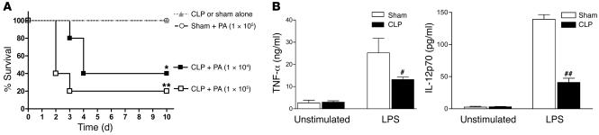
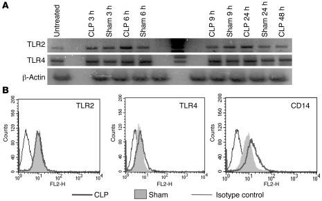
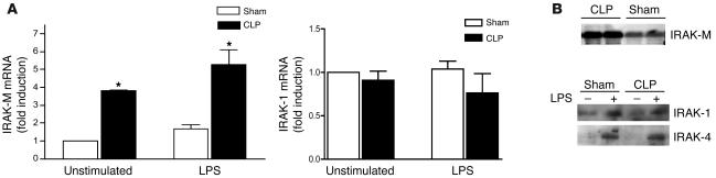
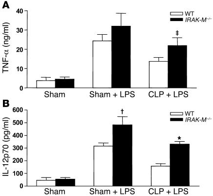
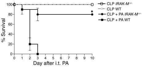
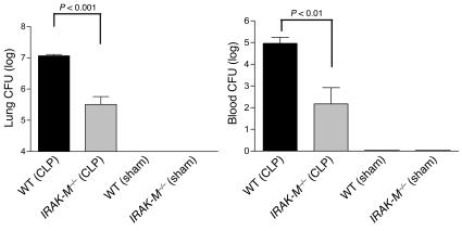
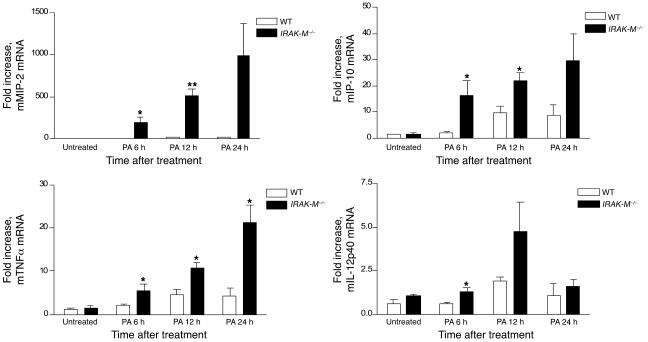
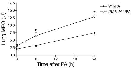
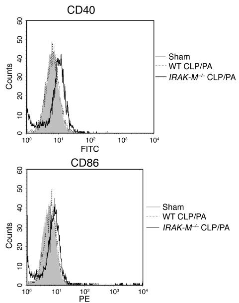
Sepsis results in a state of relative immunosuppression, rendering critically ill patients susceptible to secondary infections and increased mortality. Monocytes isolated from septic patients and experimental animals display a "deactivated" phenotype, characterized by impaired inflammatory and antimicrobial responses, including hyporesponsiveness to LPS. We investigated the role of the LPS/TLR4 axis and its inhibitor, IL-1 receptor-associated kinase-M (IRAK-M), in modulating the immunosuppression of sepsis using a murine model of peritonitis-induced sepsis followed by secondary challenge by intratracheal Pseudomonasaeruginosa. Septic mice demonstrated impaired alveolar macrophage function and increased mortality when challenged with intratracheal Pseudomonas as compared with nonseptic controls. TLR2 and TLR4 expression was unchanged in the lung following sepsis, whereas levels of IRAK-M were upregulated. Macrophages from IRAK-M-deficient septic mice produced higher levels of proinflammatory cytokines ex vivo and greater costimulatory molecule expression in vivo as compared with those of their WT counterparts. Following sepsis and secondary intrapulmonary bacterial challenge, IRAK-M(-/-) animals had higher survival rates and improved bacterial clearance from lung and blood compared with WT mice. In addition, increased pulmonary chemokine and inflammatory cytokine production was observed in IRAK-M(-/-) animals, leading to enhanced neutrophil recruitment to airspaces. Collectively, these findings indicate that IRAK-M mediates critical aspects of innate immunity that result in an immunocompromised state during sepsis.


Figures

Figure 1
Figure 1. Effect of abdominal sepsis on survival following secondary i.t. Pseudomonas challenge (A) and AM cytokine production in response to LPS (B).
(A) Wild-type C57BL/6 mice underwent either CLP with a 26-gauge needle or sham surgery. Twenty-four hours later, mice were administered i.t. P. aeruginosa (PA) at the indicated doses and monitored for 10 days after challenge for survival. *P < 0.01; **P < 0.001 compared with CLP; n = 10/group; data representative of experiments performed in duplicate. (B) At 24 hours following CLP or sham surgery, mice were sacrificed for the isolation of AMs by BAL. AMs were adherence purified and then stimulated ex vivo with LPS (1 μg/ml) in media or with media alone (unstimulated) for 16 hours. Cell supernatants were collected for determination of TNF-α and IL-12p70 levels by ELISA. #P < 0.05; ##P < 0.01 as compared with corresponding sham; n = 5/group; experiments were performed in duplicate.
Figure 2
Figure 2. Changes in TLR expression in lung after CLP or sham surgery.
(A) Time-dependent expression of TLR2 and TLR4 mRNA in lung after sham surgery or CLP. TLR mRNA was isolated from whole lung at the times noted and mRNA levels determined by RT-PCR. n = 3 animals/time point combined; experiments were performed in duplicate. (B) Cell-surface expression of TLR2, TLR4, and CD14 by AMs after CLP or sham surgery. At 24 hours after CLP or sham surgery, AMs were isolated by BAL and stained with anti-TLR2, -TLR4, or -CD14 antibodies. Surface expression of these markers was determined by flow cytometry. Histograms are representative of experiments performed in duplicate, with AMs combined from 3 animals per group.
Figure 3
Figure 3. Induction of IRAK-M mRNA (A) or protein (B) in pulmonary macrophages after CLP.
At 24 hours after CLP or sham surgery, lungs were harvested and digested with collagenase to obtain single-cell suspensions. Pulmonary macrophages were obtained by adherence purification for 2 hours. The cells were unstimulated or incubated with LPS (1 μg/ml) ex vivo for 6 hours for RNA analysis or incubated with LPS (1 μg/ml) for 16 hours for protein analysis. (A) Expression of IRAK-1 and IRAK-M mRNA was determined by quantitative PCR. Fold increase represents that over expression level from unstimulated macrophages isolated from sham-operated mice. *P < 0.05 as compared with sham control. Each condition represents pulmonary macrophages from 3–5 animals combined. (B) Protein levels of IRAK-1, IRAK-4, and IRAK-M were determined by Western immunoblotting.
Figure 4
Figure 4. TNF-α (A) and IL-12p70 (B) production by AMs isolated from sham- and CLP-operated WT and IRAK-M–/– mice following ex vivo stimulation with LPS.
AMs were isolated from WT and IRAK-M–/– mice 24 hours after either sham surgery or CLP and cultured at a concentration of 5 × 10⁵/ml in the presence or absence of LPS (100 ng/ml) for 16 hours. *P < 0.05 and P = 0.05 compared with cytokine production from LPS-stimulated AMs from WT mice isolated 24 hours after CLP. P < 0.05 compared with cytokine production from LPS-stimulated AMs from WT mice isolated 24 hours after sham surgery. Data represent combined results from 2 independent experiments; n = 5–16 per condition.
Figure 5
Figure 5. Survival following CLP and i.t. Pseudomonas infection in WT and IRAK-M–/– mice.
At 24 hours after CLP or sham surgery, WT and IRAK-M–/– mice were i.t. challenged with P. aeruginosa (1 × 10⁵ CFU/mouse). All sham-operated groups had 100% survival at 10 days (data not shown), as did the groups that underwent CLP alone. Data are representative of experiments performed in triplicate. *P < 0.001, IRAK-M–/– versus WT after CLP and i.t. Pseudomonas challenge (n = 10/group).
Figure 6
Figure 6. Lung and blood CFU following CLP and i.t. P. aeruginosa administration in WT and IRAK-M–/– mice.
WT and IRAK-M–/– mice underwent CLP or sham surgery on day 0 and i.t. Pseudomonas administration on day 1. Lung and blood were collected for CFU determination 24 hours after administration of bacteria. Both WT and IRAK-M–/– sham-operated groups had undetectable bacterial CFU in lung and blood following i.t. Pseudomonas administration. n = 5–6/group; experiments were performed in duplicate.
Figure 7
Figure 7. Lung cytokine mRNA expression following CLP and i.t. Pseudomonas challenge in WT and IRAK-M–/– mice.
WT and IRAK-M–/– mice underwent CLP followed 24 hours later by i.t. P. aeruginosa administration. At various time points following Pseudomonas administration, lungs were harvested for determination of cytokine mRNA levels by real-time quantitative PCR. Data are presented as fold increase in mRNA levels over WT untreated. *P < 0.05; **P < 0.001; n = 3–4 animals/condition combined.
Figure 8
Figure 8. Total lung MPO activity following CLP and i.t. P. aeruginosa challenge in WT and IRAK-M–/– mice.
n = 3 animals per group for time 0; n = 8 animals per group for the 6 and 24 hour time points. *P < 0.01 compared with WT mice.
Figure 9
Figure 9. Cell-surface expression of CD40 and CD86 by AMs after CLP or sham surgery followed by i.t. P. aeruginosa administration.
At 24 hours after CLP or sham surgery (time 0) or 6 hours after P. aeruginosa administration, AMs were isolated by BAL and stained with anti-CD40, -CD80, or -CD86 antibodies. Surface expression of these markers was determined by flow cytometry gating on macrophages based on size and surface complexity characteristics. Histograms are representative of experiments performed in duplicate, with AMs combined from 3 animals per group. Shaded histogram represents AMs from mice 24 hours after sham surgery.

References

    1. Jacobs R.F., Kiel D.P., Balk R.A. Alveolar macrophage function in a canine model of endotoxin-induced lung injury. Am. Rev. Respir. Dis. 1986;134:745–751. - PubMed
    1. Munoz C., et al. Dysregulation of in vitro cytokine production by monocytes during sepsis. J. Clin. Invest. 1991;88:1747–1754. - PMC - PubMed
    1. Nelson S., Chidiac C., Bagby G., Summer W.R. Endotoxin-induced suppression of lung host defenses. J. Med. 1990;21:85–103. - PubMed
    1. Volk H.D., et al. Monocyte deactivation — rationale for a new therapeutic strategy in sepsis. Intensive Care Med. 1996;22(Suppl. 4):S474–S481. - PubMed
    1. Volk, H.D., et al. 1993. Alterations in function and phenotype of monocytes from patients with septic disease: predictive value and new therapeutic strategies. In Host defense dysfunction in trauma, shock, and sepsis. E. Faist, J.L. Meakins, and F.W. Schildberg, editors. Springer–Verlag. Berlin/Heidelberg, Germany. 365–371.

Publication types

MeSH terms

Substances